Confocal microscopy reveals persisting stromal changes after myopic photorefractive keratectomy in zero haze corneas
- PMID: 9930270
- PMCID: PMC1722439
- DOI: 10.1136/bjo.82.12.1393
Confocal microscopy reveals persisting stromal changes after myopic photorefractive keratectomy in zero haze corneas
Abstract
Aims: Micromorphological examination of the central cornea in myopic patients 8-43 months after excimer laser photorefractive keratectomy (PRK), using the slit scanning confocal microscope.
Methods: Patients were selected from a larger cohort of individuals on the basis of full corneal clarity (haze grading 0 to +1; mean 0.3) and their willingness to participate in the study. 15 eyes of 10 patients with myopic PRK (-4 to -11 D; mean 6.7) and an uneventful postoperative interval of 8-43 months (mean 26) were examined. Contact lenses had been worn by eight of the 10 patients for 4-11 years (mean 6.7) before surgery. Controls included the five untreated fellow eyes of PRK patients, 10 healthy, age matched volunteers without a history of ocular inflammation or contact lens wear, and 20 patients who had worn rigid gas permeable (n = 10) or soft contact lenses (n = 10) for 2-11 years. Subjects were examined with a real time flying slit, scanning confocal microscope using x25 and x50 objectives.
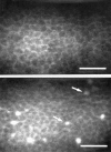
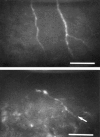
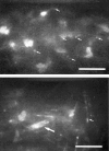
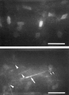
Results: In PRK treated patients and contact lens wearers, basal layer epithelial cells sporadically displayed enhanced reflectivity. The subepithelial nerve plexus was observed in all individuals, but was usually less well contrasted in the PRK group, owing to the presence of a very discrete layer of subepithelial scar tissue, which patchily enhanced background reflectivity. Within all layers of the stroma, two distinct types of abnormal reflective bodies were observed in all PRK treated eyes, but in none of the controls. One had the appearance of long (> = 50 microns), slender (2-8 microns in diameter) dimly reflective rods, which sometimes contained bright, punctate, crystal-like inclusions, arranged linearly and at irregular intervals. The other was shorter (< 25 microns), more slender in form (< 1 micron in diameter), and highly reflective; these so called needles were composed of crystal-like granules in linear array, with an individual appearance similar to the bright punctate inclusions seen in rods, but densely packed. Both of these unusual structures were confined, laterally, to the ablated area, but were otherwise distributed throughout all stromal layers, with a clear predominance in the anterior ones. These rods and needles were observed in all PRK treated corneas, irrespective of previous contact lens wear. On the basis of qualitative inspection, the incidence of rods and needles did not appear to correlate with either the volume of tissue ablated or the length of the postoperative interval. In contact lens wearing controls, highly reflective granules, reminiscent of those from which the needles were composed, were found scattered as isolated entities throughout the entire depth and lateral extent of the corneal stroma, but rods and needles were never encountered. The corneal endothelium exhibited no obvious abnormalities.
Conclusion: Confocal microscopy 8-43 months after PRK revealed belated changes in the corneal stroma. These were manifested as two distinct types of abnormal reflective bodies, which had persisted beyond the stage when acute wound healing would have been expected to be complete. The clinical significance of these findings in the context of contrast visual acuity and long term status of the cornea is, as yet, unknown.
Figures


References
MeSH terms
LinkOut - more resources
Full Text Sources
