. 1998 Dec;82(12):1407-11.
doi: 10.1136/bjo.82.12.1407.

The conjunctiva in corneal epithelial wound healing

Affiliations

The conjunctiva in corneal epithelial wound healing

H S Dua. Br J Ophthalmol. 1998 Dec.

Abstract

Background/aims: During the healing of corneal epithelial wounds with limbal involvement, conjunctival epithelium often migrates across the denuded limbus to cover the corneal surface. It is believed that, over a period of time, conjunctival epithelium covering the cornea assumes characteristics of corneal epithelium by a process referred to as conjunctival transdifferentiation. The purpose of this study was to examine, clinically, the fate of conjunctival epithelial cells covering the cornea and to assess the healing of corneal epithelial wounds when the conjunctival epithelium was removed or actively prevented from crossing the limbus and extending onto the cornea.

Methods: 10 patients with conjunctivalisation of the cornea were followed for an average of 7.5 months. Five patients in this group had their conjunctival epithelium removed from the corneal surface and allowed to heal from the remaining intact corneal epithelium. In another four patients with corneal epithelial defects, the conjunctival epithelium was actively prevented from crossing the limbus by mechanically scraping it off.

Results: The area of cornea covered by conjunctival epithelium appeared thin, irregular, attracted new vessels and was prone to recurrent erosions. Conjunctivalisation of the visual axis affected vision. Removal of conjunctival epithelium from the cornea allowed cells of corneal epithelial phenotype to cover the denuded area with alleviation of symptoms and improvement of vision. It was also established that migration of conjunctival epithelium onto corneal surface could be anticipated by close monitoring of the healing of corneal epithelial wounds, and prevented by scraping off conjunctival epithelium before it reached the limbus.

Conclusion: This study shows that there is little clinical evidence to support the concept that conjunctival transdifferentiation per se, occurs in humans. "Replacement" of conjunctival epithelium by corneal epithelial cells may be an important mechanism by which conjunctival "transdifferentiation" may occur. In patients with partial stem cell deficiency this approach can be a useful and effective alternative to partial limbal transplantation, as is currently practised.


Figures

Figure 1
Figure 1
Slit lamp diffuse view of a fluorescein stained cornea of a patient with a corneal graft showing a clear demarcation between corneal and conjunctival epithelial phenotypes. The pupillary area is covered by conjunctival epithelium. Tiny "buds" of corneal epithelium can be seen along the line of contact between corneal and conjunctival epithelium (arrowheads) (×10).
Figure 2
Figure 2
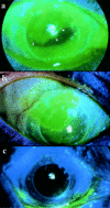
Slit lamp diffuse view of a fluorescein stained cornea of a patient with a corneal graft showing a clear demarcation between corneal and conjunctival epithelial phenotypes. The conjunctival epithelium shows light staining with fluorescein and blood vessels can be seen extending on the conjunctivalised epithelium. Tiny "buds" of corneal epithelium can be seen along the line of contact between corneal and conjunctival epithelium (arrowheads). The pupillary area is covered by corneal epithelium with is "sustained" by two clock hours of intact limbus (between large arrows). The best corrected visual acuity was 6/12. Scraping of conjunctival epithelium is not usually necessary in such cases (×10).
Figure 3
Figure 3
Slit lamp diffuse view of a fluorescein stained cornea of a patient with corneal surface problems following superficial chemical injury. The corneal surface shows a clear demarcation between corneal and conjunctival epithelial phenotypes. The conjunctival epithelium shows light staining with fluorescein. The pupillary area is covered by corneal epithelium with is "sustained" by four clock hours of intact limbus (between arrows). The best corrected visual acuity was 6/9. Scraping of conjunctival epithelium is not usually necessary in such cases (×10).
Figure 4
Figure 4
(a) Broad beam slit lamp photograph of a patient with extensive conjunctivalisation of the cornea. Only a small area of the corneal surface, corresponding to one and a half clock hours of the inferotemporal limbus, is covered by corneal epithelium. Note the well defined line of demarcation (arrowheads) between corneal and conjunctival epithelium. The patient's vision was 6/18 (×10). (b) Fluorescein stained picture of the cornea immediately after removal of conjunctival epithelium over and around the pupillary area (×6). (c) The same eye on day 2 following removal of conjunctival epithelium. Note that the pupillary area is being covered by migration of corneal epithelium from the small area of preserved corneal epithelium illustrated in (a) (×6). (d) On day 5 following removal of conjunctival epithelium, healing is almost complete and the pupillary area is covered by healthy corneal epithelium. The visual acuity improved to 6/9 (×6).
Figure 5
Figure 5
(a) Fluorescein stained photograph of a patient with a corneal graft showing just over 25% of the graft surface covered by conjunctival epithelium (arrowheads). The area appears irregular and shows light fluorescein staining. The patient was symptomatic and vision was a blurry 6/24 (×10). (b) The same eye immediately following removal of the conjunctival epithelium (×10). (c) After complete healing had occurred on day 5, the graft surface was covered by healthy corneal epithelium and the best corrected vision improved to 6/12 with alleviation of symptoms (×6).
Figure 6
Figure 6
(a) Fluorescein stained photograph of a patient with Stevens-Johnson syndrome who had over 50% of the superior corneal surface covered with conjunctival epithelium. The line of demarcation between corneal and conjunctival epithelium was not very well defined (arrowheads). The patient's vision was 6/12 and her main complaint was that of irritation and intermittent sharp pains due to recurrent erosions (×10). (b) The same eye with the patient looking down to show that the superior conjunctiva too was showing coarse punctate staining with fluorescein and had injected vessels (×10). (c) The eye after removal of the conjunctival epithelium from the pupillary area and superiorly. Note that the removal of epithelium was not extended across the limbus (×10). (d) Six days following removal of conjunctival epithelium, the pupillary area and a part of the superior cornea is covered by healthy corneal epithelium. The demarcation between conjunctival epithelium and corneal epithelium is now well defined (arrowheads). The patient's vision improved to 6/5 but she was more impressed by the reduction in her symptoms (×10).
Figure 7
Figure 7
(a) Fluorescein stained photograph of the cornea of a patient who had presented several months after a chemical injury, showing a clear demarcation between corneal and conjunctival epithelial phenotypes (arrowheads). The pupillary area is almost entirely covered by conjunctival epithelium. Tiny "buds" of corneal epithelium can be seen along the line of contact between corneal and conjunctival epithelium. The patient's vision was 3/18 (×10). (b) The eye after removal of all conjunctival epithelium from the corneal surface and limbus (×10). (c) The eye on day 3 following removal of conjunctival epithelium. The corneal sheet has covered the pupillary area but the conjunctival epithelium has encroached on to cornea along the temporal limbus (×10). (d) The eye after complete healing. A new line of contact is established between corneal and conjunctival epithelial phenotypes (arrowheads) but the pupillary area is covered by healthy corneal epithelium. The patient's vision improved to 6/9 (×10).

References

    1. Ophthalmic Surg. 1992 Oct;23(10):680-4 - PubMed
    1. Doc Ophthalmol. 1951;5-6:184-92 - PubMed
    1. Invest Ophthalmol Vis Sci. 1990 Sep;31(9):1903-13 - PubMed
    1. Am J Ophthalmol. 1990 Dec 15;110(6):646-56 - PubMed
    1. Invest Ophthalmol Vis Sci. 1987 Sep;28(9):1497-504 - PubMed

Publication types