

Differential expression of costimulatory molecules in chronic inflammatory periodontal disease tissue
- PMID: 9933436
- PMCID: PMC1905181
- DOI: 10.1046/j.1365-2249.1999.00763.x
Differential expression of costimulatory molecules in chronic inflammatory periodontal disease tissue
Abstract
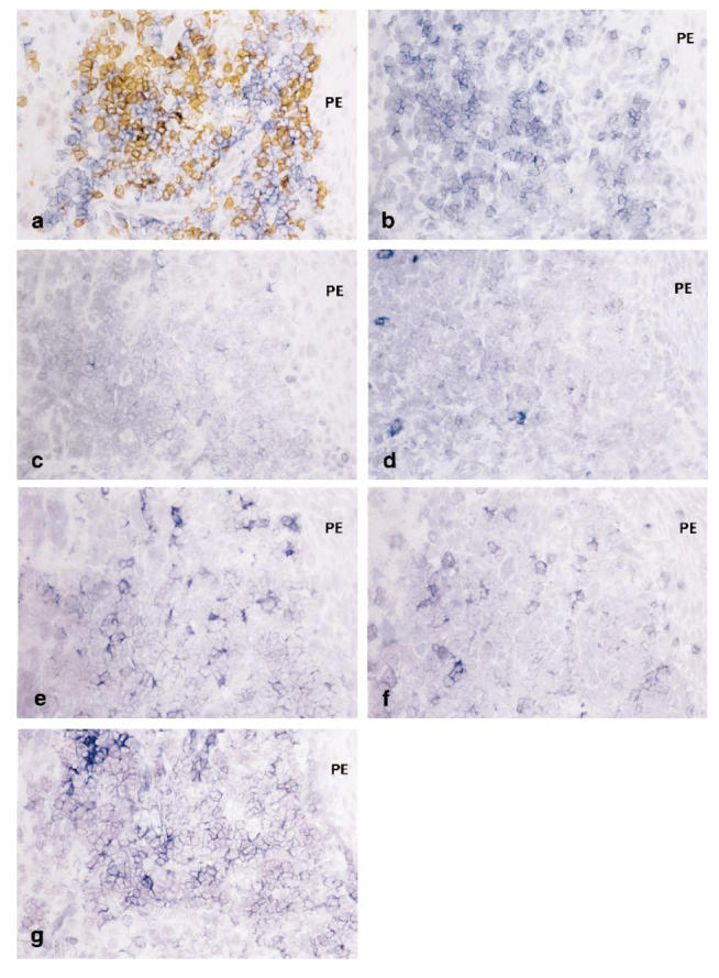
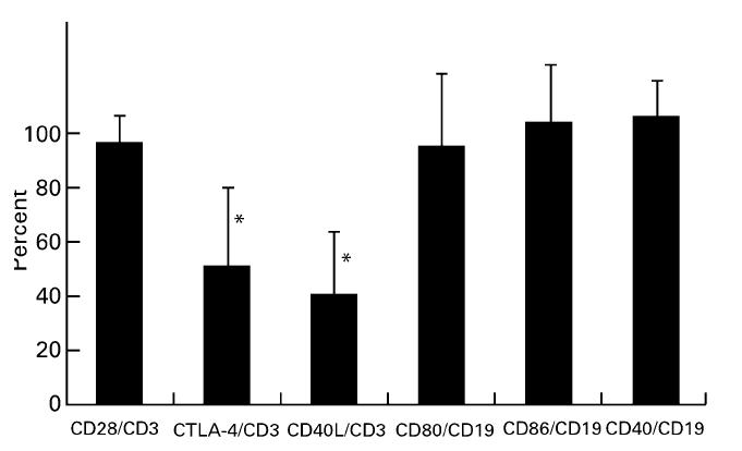
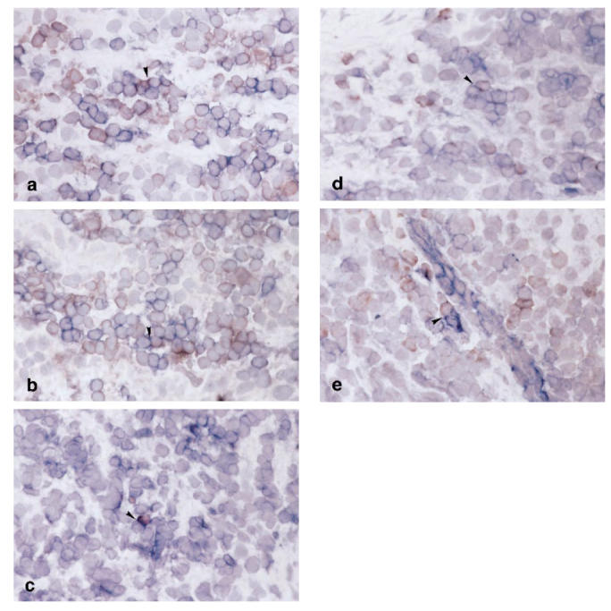
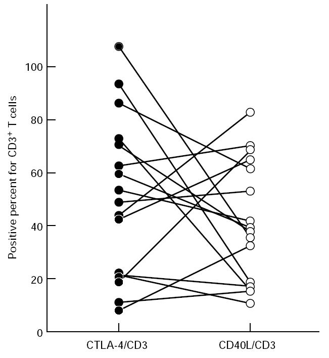
Although B cell activation and subsequent immunoglobulin production are the immunopathological features of chronic inflammatory periodontal disease, in situ expression of costimulatory molecules in humoral immunity has not been investigated. In the present study we examined the expression of CD40, CD40 ligand (CD40L), CD80, CD86, CD28 and cytolytic T lymphocyte-associated antigen-4 (CTLA-4) on lymphocytes immunohistochemically. Cryostat sections were prepared from the gingival tissue samples of 14 patients with moderate to advanced adult periodontitis. In vitro kinetics of the expression of CD40L and CTLA-4 by peripheral blood T cells and that of CD80 and CD86 by peripheral blood B cells were also investigated by flow cytometry. Positive percentage expression of CD40L, CD28 and CTLA-4, and CD40, CD80 and CD86 was calculated for the number of CD3+ and CD19+ cells, respectively. Flow cytometric analysis demonstrated that the expression of CD40L and CTLA-4 on T cells, and CD80 and CD86 on B cells of peripheral blood was up-regulated upon activation. While most T cells and B cells expressed CD28, and CD80 and CD86, respectively, in gingival tissues, the expression of CD40L and CTLA-4 was lower but highly variable between specimens. Furthermore, these two molecules seemed to be expressed reciprocally in the lesion. As both CD40L and CTLA-4 expression are induced transiently by stimulation, variability in the expression of the molecules may reflect immunological activities and participation in the regulation of B cell activation of the lesion.
Figures
Similar articles
-
Costimulatory molecules in human periodontal disease tissues.J Periodontal Res. 2001 Apr;36(2):92-100. doi: 10.1034/j.1600-0765.2001.360205.x. J Periodontal Res. 2001. PMID: 11327084
-
Localization in situ of costimulatory molecules and cytokines in B-cell non-Hodgkin's lymphoma.Immunology. 1998 Aug;94(4):580-6. doi: 10.1046/j.1365-2567.1998.00550.x. Immunology. 1998. PMID: 9767448 Free PMC article.
-
Altered expression of costimulatory molecules in myasthenia gravis.Muscle Nerve. 2000 Jun;23(6):946-53. doi: 10.1002/(sici)1097-4598(200006)23:6<946::aid-mus16>3.0.co;2-4. Muscle Nerve. 2000. PMID: 10842273
-
The role of CD40 ligand in costimulation and T-cell activation.Immunol Rev. 1996 Oct;153:85-106. doi: 10.1111/j.1600-065x.1996.tb00921.x. Immunol Rev. 1996. PMID: 9010720 Review.
-
CD28/CTLA-4 and CD80/CD86 families: signaling and function.Immunol Res. 1999;19(1):1-24. doi: 10.1007/BF02786473. Immunol Res. 1999. PMID: 10374692 Review.
Cited by
-
Activation of Langerhans-Type Dendritic Cells Alters Human Cytomegalovirus Infection and Reactivation in a Stimulus-Dependent Manner.Front Microbiol. 2016 Sep 14;7:1445. doi: 10.3389/fmicb.2016.01445. eCollection 2016. Front Microbiol. 2016. PMID: 27683575 Free PMC article.
-
Elevated CTLA-4 expression on CD4 T cells from periodontitis patients stimulated with Porphyromonas gingivalis outer membrane antigen.Clin Exp Immunol. 2000 Feb;119(2):280-6. doi: 10.1046/j.1365-2249.2000.01126.x. Clin Exp Immunol. 2000. PMID: 10632663 Free PMC article.
-
Effects of periodontitis on cancer outcomes in the era of immunotherapy.Lancet Healthy Longev. 2023 Apr;4(4):e166-e175. doi: 10.1016/S2666-7568(23)00021-1. Lancet Healthy Longev. 2023. PMID: 37003275 Free PMC article. Review.
-
Elevated humoral immune response to heat shock protein 60 (hsp60) family in periodontitis patients.Clin Exp Immunol. 2000 May;120(2):285-93. doi: 10.1046/j.1365-2249.2000.01216.x. Clin Exp Immunol. 2000. PMID: 10792378 Free PMC article.
-
The Murine Oral Metatranscriptome Reveals Microbial and Host Signatures of Periodontal Disease.J Dent Res. 2023 May;102(5):565-573. doi: 10.1177/00220345221149675. Epub 2023 Mar 8. J Dent Res. 2023. PMID: 36883648 Free PMC article.
References
-
- Mackler BF, Frostad KB, Robertson PB, Levy BM. Immunoglobulin bearing lymphocytes and plasma cells in human periodontal disease. J Periodont Res. 1977;12:37–45. - PubMed
-
- Seymour GJ, Greenspan JS. The phenotypic characterization of lymphocyte subpopulations in established human periodontal disease. J Periodont Res. 1979;14:39–46. - PubMed
-
- Gemmell E, Feldner B, Seymour GJ. CD45RA and CD45RO positive CD4 cells in human peripheral blood and periodontal disease tissue before and after stimulation with periodontopathic bacteria. Oral Microbiol Immunol. 1992;7:84–88. - PubMed
-
- Yamazaki K, Nakajima T, Aoyagi T, Hara K. Immunohistological analysis of memory T lymphocytes and activated B lymphocytes in tissues with periodontal disease. J Periodont Res. 1993;28:324–34. - PubMed
-
- Takeuchi Y, Yoshie H, Hara K. Expression of interleukin-2 receptor and HLA-DR on lymphocyte subsets of gingival crevicular fluid in patients with periodontitis. J Periodont Res. 1991;26:502–10. - PubMed
Publication types
MeSH terms
Substances
LinkOut - more resources
Full Text Sources
Research Materials
