. 1999 Feb;10(2):329-44.
doi: 10.1091/mbc.10.2.329.

Detection of transient in vivo interactions between substrate and transporter during protein translocation into the endoplasmic reticulum

Affiliations
Free PMC article

Detection of transient in vivo interactions between substrate and transporter during protein translocation into the endoplasmic reticulum

M Dünnwald et al. Mol Biol Cell. 1999 Feb.
Free PMC article

Abstract

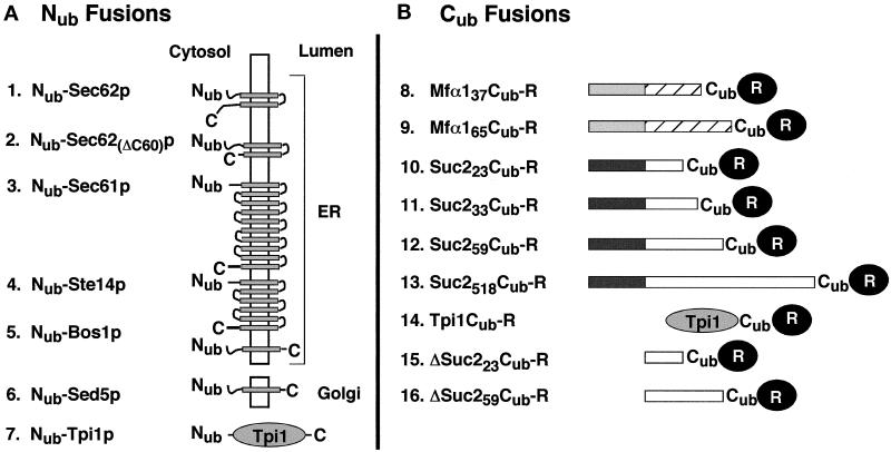
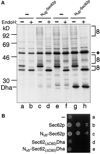
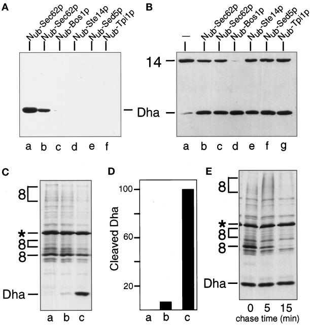
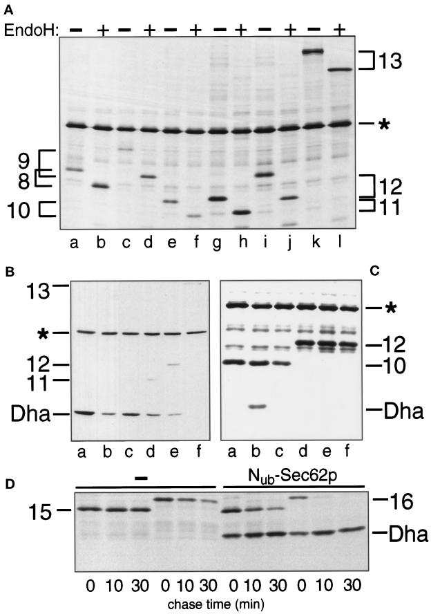
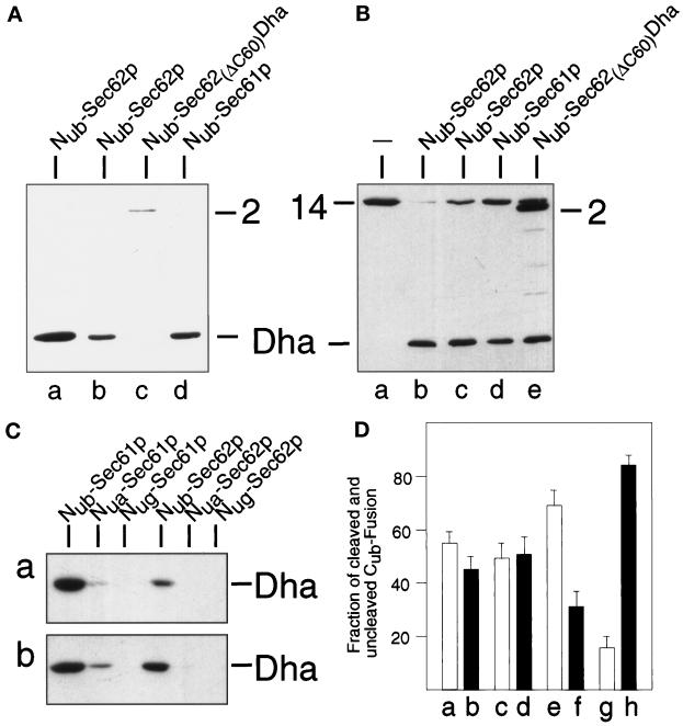
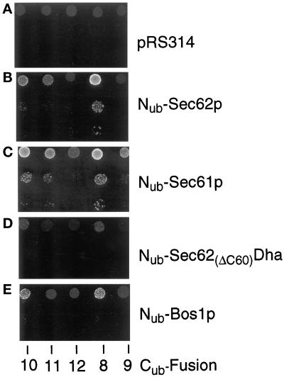
The split-ubiquitin technique was used to detect transient protein interactions in living cells. Nub, the N-terminal half of ubiquitin (Ub), was fused to Sec62p, a component of the protein translocation machinery in the endoplasmic reticulum of Saccharomyces cerevisiae. Cub, the C-terminal half of Ub, was fused to the C terminus of a signal sequence. The reconstitution of a quasi-native Ub structure from the two halves of Ub, and the resulting cleavage by Ub-specific proteases at the C terminus of Cub, serve as a gauge of proximity between the two test proteins linked to Nub and Cub. Using this assay, we show that Sec62p is spatially close to the signal sequence of the prepro-alpha-factor in vivo. This proximity is confined to the nascent polypeptide chain immediately following the signal sequence. In addition, the extent of proximity depends on the nature of the signal sequence. Cub fusions that bore the signal sequence of invertase resulted in a much lower Ub reconstitution with Nub-Sec62p than otherwise identical test proteins bearing the signal sequence of prepro-alpha-factor. An inactive derivative of Sec62p failed to interact with signal sequences in this assay. These in vivo findings are consistent with Sec62p being part of a signal sequence-binding complex.


Figures

Figure 1
Figure 1
The split-Ub technique and its application to the analysis of protein translocation. (A) Nub and Cub are linked to the interacting proteins X and Y. The X–Y complex brings Nub and Cub into close proximity. Nub and Cub reconstitute a quasi-native Ub moiety, which is cleaved by the UBPs, yielding the free reporter R (Johnsson and Varshavsky, 1994a). (B) Using split-Ub to monitor the proximity between a secretory protein and a component of the translocation machinery. A signal sequence-bearing Cub fusion (SS-Y-Cub-R) and a Nub fusion (Nub-X) are coexpressed in a cell. Pathway 1: when Nub is linked to a protein not involved in the targeting for translocation, the uncleaved (except for the signal sequence SS) Y-Cub-R enters the ER. Pathway 2: when Nub is linked to a protein involved in the targeting for translocation the signal sequence of the SS-Y-Cub-R brings Nub and Cub into close contact. As a result, some of the SS-Y-Cub-R and Nub-X molecules interact to form the quasi-native Ub, yielding the free reporter R in the cytosol.
Figure 2
Figure 2
Nub and Cub test fusions. (A) Nub (residues 1–36 of Ub) was fused to the N terminus of either a transmembrane protein (constructs 1–6) or a cytosolic protein (construct 7). The N termini of all proteins are located in the cytosol. The orientations and the numbers of membrane-spanning regions (shaded boxes) were derived from the published studies of these proteins, except for Ste14p, for which the exact number of the domains and the localization of the C terminus are not yet known. The Nub fusions 1–5 are located in the membrane of the ER; the Nub fusion 6 resides in the membrane of the early Golgi. The Nub fusion 2 is a Sec62p derivative lacking the C-terminal 60 residues. The Nub fusion 7 contains the full-length triosephosphate isomerase (Nub-Tpi1p). (B) Cub fusions. The Cub fusions 8 and 9 contain the signal sequence of the prepro-α-factor (shaded boxes), followed by either 37 (construct 8) or 65 residues (construct 9) of the mature α-factor sequence (striped boxes) and a 7-residue linker sequence (not shown). Cub fusions 10–13 contain the signal sequence of the Suc2p invertase (dark boxes) followed by 23 (construct 10), 33 (construct 11), 59 (construct 12), or 518 residues (construct 13) of the mature sequence of invertase (open boxes) and a 7-residue linker sequence (not shown). The Cub fusion 14 contains the complete sequence of S. cerevisiae triosephosphate isomerase (Tpi1p) followed by a 17-residue linker peptide and Cub. The Cub fusions 15 and 16 are the signal sequence-lacking counterparts of the fusions 10 and 12. Cub is always followed by a reporter protein. The reporter is DHFR-ha or Ura3p for the Cub fusions 8–13, and DHFR-ha for the Cub fusions 14, 15, and 16.
Figure 3
Figure 3
Sec62p is close to the signal sequence of the α-factor precursor. (A) S. cerevisiae cells expressing Mfα37-Cub-Dha (construct 8; Figure 2) were labeled for 5 min with 35S-methionine. The extracted proteins were immunoprecipitated with anti-ha antibody, followed by a mock treatment (lanes a, c, e, and g) or the treatment with EndoH (lanes b, d, f, and h), and SDS-PAGE. The results with cells coexpressing Nug- or Nub-Sec62p are shown in lanes c and d and g and h, respectively. The analysis was performed with Nug-Sec62p in the S. cerevisiae mutant RSY529 carrying a ts allele of SEC62 (lanes a–d) or with Nub-Sec62p in the wild-type yeast (lanes e–h). Number 8 (following the numbering of the constructs in Figure 2) on the right indicates the positions of uncleaved Mfα37-Cub-Dha and its glycosylated forms. An asterisk denotes an unrelated yeast protein that cross-reacts with the anti-ha antibody. (B) Nub-Sec62p encodes a functionally active protein. RSY529 cells carrying an empty plasmid (a), Sec62p (b), Nub-Sec62p (c), Sec62(ΔC60)Dha (d), or Nub-Sec62(ΔC60)Dha (e) were spotted on minimal media and grown for 2 d at 30°C (semipermissive temperature for unmodified RSY529).
Figure 4
Figure 4
The in vivo proximity between Sec62p and Mfα37-Cub-Dha is transient and specific. (A) Immunoblot analysis of extracts of S. cerevisiae coexpressing the Mfα37-Cub-Dha (construct 8; Figure 2) and one of the following constructs: Nub-Sec62p, integrated (lane a) or plasmid-borne (lane b); Nub-Bos1p (lane c); Nub-Ste14p (lane d); Nub-Sed5p(lane e); and Nub-Tpi1p (lane f). (B) Immunoblot analysis of extracts of S. cerevisiae expressing the Tpi1p-Cub-Dha fusion (construct 14; Figure 2) alone (lane a) or together with one of the following constructs: Nub-Sec62p, either integrated (lane b) or plasmid-borne (lane c); Nub-Bos1p (lane d); Nub-Ste14p (lane e); Nub-Sed5p(lane f); and Nub-Tpi1p (lane g). Number 14 on the left indicates the position of uncleaved Tpi1p-Cub-Dha. (C) S. cerevisiae cells expressing Mfα37-Cub-Dha (construct 8; Figure 2) together with either the vector (lane a), Nub-Bos1p (lane b), or Nub-Sec62p (lane c) were labeled for 5 min with 35S-methionine. The extracted proteins were immunoprecipitated with anti-ha antibody and analyzed by SDS-PAGE. (D) Quantitation of the pulse-labeling experiment (C) using Phosphor-Imager. The extent of Dha release in the presence of Nub-Sec62p was arbitrarily set at 100. The averages of three experiments are shown. Lanes a, b, and c are the same as in panel C. (E) S. cerevisiae cells expressing Mfα37-Cub-Dha together with Nub-Sec62p were labeled for 5 min with 35S-methionine and chased for 5 and 15 min, followed by extraction of proteins, immunoprecipitation with anti-ha antibody, and SDS-PAGE.
Figure 5
Figure 5
The nature of the signal sequence and its distance from Cub determine the extent of cleavage of Cub-R in the presence of Nub-Sec62p. (A) S. cerevisiae expressing Mfα37-Cub-Dha (construct 8; Figure 2) (lanes a and b), Mfα65-Cub-Dha (construct 9) (lanes c and d), Suc223-Cub-Dha (construct 10) (lanes e and f), Suc233-Cub-Dha (construct 11) (lanes g and h), Suc259-Cub-Dha (construct 12) (lanes i and j) and Suc2518-Cub-Dha (construct 13) (lanes k and l) were labeled with 35S-methionine for 5 min. The extracted proteins were either mock-treated (lanes a, c, e, g, i, and k) or treated with EndoH (lanes b, d, f, h, j, and l), followed by immunoprecipitation with anti-ha antibody and SDS-PAGE. (B) Same as panel A but the cells also contained Nub-Sec62p in addition to the Cub-fusions Mfα37-Cub-Dha, Mfα65-Cub-Dha, Suc223-Cub-Dha, Suc233-Cub-Dha, Suc259-Cub-Dha, Suc2518-Cub-Dha (lanes a–f). The analysis was carried out by immunoblotting whole- cell extracts with the anti-ha antibody. (C) S. cerevisiae cells expressing Suc223-Cub-Dha (construct 10; Figure 2) (lanes a–c) and Suc259-Cub-Dha (construct 12; Figure 2) (lanes d–f) together with either Nub-Sec62p (lanes b and e), Nub-Bos1p (lanes c and f) or the vector (lanes a and d) were labeled for 5 min with 35S-methionine. Whole-cell extracts were immunoprecipitated with anti-ha antibody, followed by SDS-PAGE and autoradiography. (D) S. cerevisiae cells expressing ΔSuc223-Cub-Dha (construct 15; Figure 2) or ΔSuc259-Cub-Dha (construct 16; Figure 2) together with either the vector (first six lanes) or Nub-Sec62p (last six lanes) were labeled for 5 min with 35S-methionine and chased for 10 and 30 min, followed by extraction, immunoprecipitation with anti-ha antibody, and SDS-PAGE. Numbers 15 and 16 indicate the positions of the corresponding (uncleaved) Cub fusions.
Figure 6
Figure 6
Sec61p, but not a mutant of Sec62p, are close to the nascent chain of a translocated protein. (A) These assays employed S. cerevisiae expressing Mfα37-Cub-Dha (construct 8, Figure 2) and one of the following Nub fusions (Figure 2): Nub-Sec62p, either integrated (lane a) or plasmid borne (lane b); Nub-Sec62(ΔC60)Dha (lane c); and Nub-Sec61p (lane d). Whole-cell extracts from these strains were subjected to immunoblot analysis with anti-ha antibody. (B) Same as panel A but the same Nub fusions were coexpressed with Tpi1-Cub-Dha (construct 14; Figure 2). Numbers 2 and 14 indicate the positions of the corresponding (uncleaved) fusions. (C) Lane a: S. cerevisiae expressing Suc223-Cub-Dha (construct 10; Figure 2) together with either Nub-Sec61p, Nua-Sec61p, Nug-Sec61p, Nub-Sec62p, Nua-Sec62p, or Nug-Sec62p; lane b: same as lane a but cells expressed Mfα37-Cub-Dha (construct 8; Figure 2) instead of Suc223-Cub-Dha. (D) S. cerevisiae cells expressing Mfα37-Cub-Dha together with Nub-Sec61p (a and b) or Nub-Sec62p (e and f), and cells expressing Suc223-Cub-Dha together with Nub-Sec61p (c and d) or Nub-Sec62p (g and h) were labeled for 5 min with 35S-methionine. Whole-cell extracts were immunoprecipitated with anti-ha antibody, followed by SDS-PAGE, and quantitation of the cleaved and uncleaved Cub fusions using PhosphorImager. Shown are the relative amounts of the cleaved (white bars: a, c, e, and g) and uncleaved (black bars: b, d, f, and h) Cub fusions. The sum of a cleaved and uncleaved fusion was set at 100 in each of the three independent experiments. SDs are also indicated.
Figure 7
Figure 7
The use of a metabolic marker to assess the proximity between a component of the translocation machinery and a translocated protein. S. cerevisiae expressing the Cub fusions 8–12 (Figure 2) that contained Ura3p instead of Dha (see the main text) were transformed with the vector (A) or plasmids expressing Nub-Sec62p (B), Nub-Sec61p (C), Nub-Sec62(ΔC60)Dha (D), and Nub-Bos1p (E). Cells were grown in a liquid uracil-containing SD medium, and ∼105, 103, and 102 cells were spotted onto uracil-lacking SD medium. Plates were examined after 18 h at 30°C.

References

    1. Allison DS, Young ET. Single-amino-acid substitutions within the signal sequence of yeast prepro-α-factor affect membrane translocation. Mol Cell Biol. 1988;8:1915–1922. - PMC - PubMed
    1. Aronheim A, Zandi E, Hennemann H, Elledge SJ, Karin M. Isolation of an AP-1 repressor by a novel method for detecting protein- protein interactions. Mol Cell Biol. 1997;17:3094–3102. - PMC - PubMed
    1. Banfield DK, Lewis MJ, Rabouille C, Warren G, Pelham HR. Localization of Sed5, a putative vesicle targeting molecule, to the cis- Golgi network involves both its transmembrane and cytoplasmic domains. J Cell Biol. 1994;127:357–371. - PMC - PubMed
    1. Beckmann R, Bubeck D, Grassucci R, Penczek P, Verschoor A, Blobel G, Frank J. Alignment of conduits for the nascent polypeptide chain in the ribosome-Sec61 complex. Science. 1997;278:2123–2126. - PubMed
    1. Biederer T, Volkwein C, Sommer T. Degradation of subunits of the Sec61p complex, an integral component of the ER membrane, by the ubiquitin-proteasome pathway. EMBO J. 1996;15:2069–2076. - PMC - PubMed

Publication types

MeSH terms

LinkOut - more resources