. 1999 Feb;10(2):487-99.
doi: 10.1091/mbc.10.2.487.

The stem-loop binding protein (SLBP1) is present in coiled bodies of the Xenopus germinal vesicle

Affiliations
Free PMC article

The stem-loop binding protein (SLBP1) is present in coiled bodies of the Xenopus germinal vesicle

J Abbott et al. Mol Biol Cell. 1999 Feb.
Free PMC article

Abstract

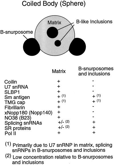
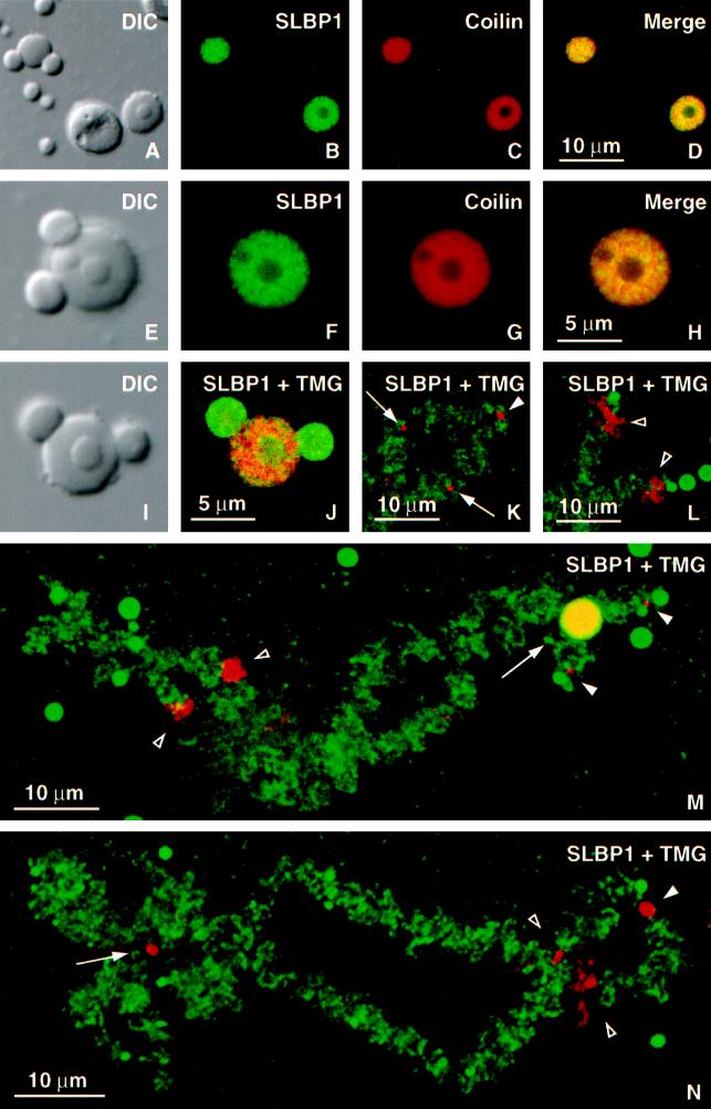
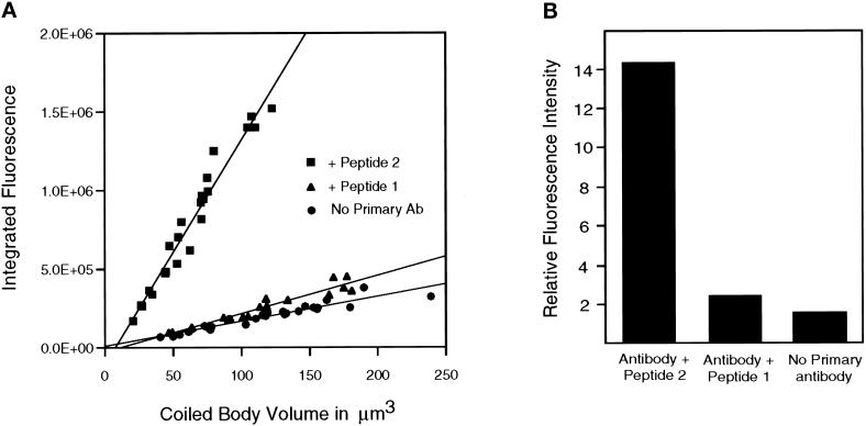
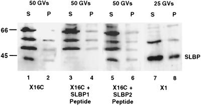
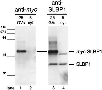
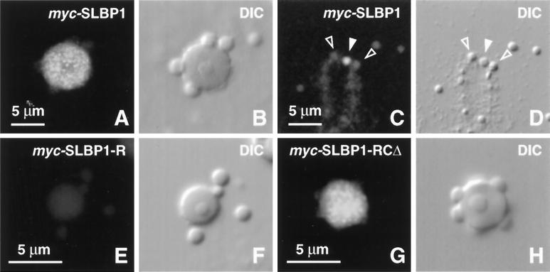
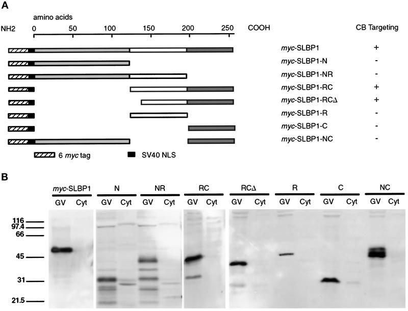
The stem-loop binding protein (SLBP1) binds the 3' stem-loop of histone pre-mRNA and is required for efficient processing of histone transcripts in the nucleus. We examined the localization of SLBP1 in the germinal vesicle of Xenopus laevis oocytes. In spread preparations of germinal vesicle contents, an anti-SLBP1 antibody stained coiled bodies and specific chromosomal loci, including terminal granules, axial granules, and some loops. After injection of myc-tagged SLBP1 transcripts into the oocyte cytoplasm, newly translated myc-SLBP1 protein was detectable in coiled bodies within 4 h and in terminal and axial granules by 8 h. To identify the region(s) of SLBP1 necessary for subnuclear localization, we subcloned various parts of the SLBP1 cDNA and injected transcripts of these into the cytoplasm of oocytes. We determined that 113 amino acids at the carboxy terminus of SLBP1 are sufficient for coiled body localization and that disruption of a previously defined RNA-binding domain did not alter this localization. Coiled bodies also contain the U7 small nuclear ribonucleoprotein particle (snRNP), which participates in cleavage of the 3' end of histone pre-mRNA. The colocalization of SLBP1 and the U7 snRNP in the coiled body suggests coordinated control of their functions, perhaps through a larger histone-processing particle. Some coiled bodies are attached to the lampbrush chromosomes at the histone gene loci, consistent with the view that coiled bodies in the oocyte recruit histone-processing factors to the sites of histone pre-mRNA transcription. The non-histone chromosomal sites at which SLBP1 is found include the genes coding for 5 S rRNA, U1 snRNA, and U2 snRNA, suggesting a wider role for SLBP1 in the biosynthesis of small non-spliced RNAs.


Figures

Figure 1
Figure 1
Diagram of a coiled body from a Xenopus GV and a list of some of its molecular components. The coiled body consists of three parts: a matrix, B-snurposomes attached to the surface, and B-like inclusions. The number of attached B-snurposomes and inclusions is variable, and they may be absent. The attached B-snurposomes are identical in all respects to the hundreds of free B-snurposomes in the nucleoplasm. The inclusions are generally smaller than B-snurposomes but are otherwise identical. The terms coiled body and sphere are used synonymously. In some previous publications (Wu et al., 1996), the term C-snurposome referred to the matrix and inclusions only.
Figure 2
Figure 2
Localization of SLBP1 in the Xenopus GV by immunofluorescence. Stained images were taken with a Leica (Heidelberg, Germany) TCS NT confocal microscope. (A–D) DIC and immunofluorescence images of two coiled bodies, a nucleolus, and several B-snurposomes double-stained with serum X16C against SLBP1 (fluorescein) and mAb H1 against Xenopus coilin (Cy3). SLBP1 and coilin are both limited to the matrix of the coiled body, being excluded from the B-snurposomes on the surface (left coiled body) and the internal B-like inclusion (right coiled body). (E–H) Higher magnification of a similar coiled body that has two B-snurposomes on the surface and two inclusions. Note the patchy distribution of SLBP1 staining and the lack of complete correspondence between the SLBP1 and coilin stains in the merged image. (I and J) Coiled body double-stained with serum X16C for SLBP1 (Cy3) and mAb K121 for the TMG cap of snRNAs (fluorescein). mAb K121 detects U7 snRNA in the matrix of the coiled body (Bellini and Gall, 1998) and splicing snRNAs in the B-snurposomes and inclusions (Wu et al., 1991). (K–N) Lampbrush chromosomes double-stained with serum X16C for SLBP1 (Cy3) and mAb K121 for TMG (fluorescein). The nascent transcripts on almost all chromosome loops are associated with splicing snRNAs and hence stain strongly for TMG. Those few loops that stain red with serum X16C do not stain with mAb K121, presumably because they lack splicing snRNAs (open arrowheads in L–N). Terminal granules stain only with X16C (filled arrowheads in K, M, and N); the same is true of axial granules (arrows in K and N). M shows chromosome 9, which has a coiled body attached at the histone gene locus (arrow). In this case the single coiled body joins the two homologous chromosomes; in other cases each homologue may have its own coiled body. The coiled body is yellow, because it stains with both antibodies.
Figure 3
Figure 3
Staining of coiled bodies with anti-SLBP1 serum X16C is reduced by treatment with peptide-1, against which the antibody was raised. (A) Images of stained coiled bodies were taken with a charge-coupled device (CCD) camera; the total fluorescence from individual coiled bodies was then measured as the sum of pixel values and plotted as a function of coiled body volume. The linear relationship shows that the amount of SLBP1 in a coiled body is proportional to its volume. Staining was markedly reduced by previous treatment of the antibody with peptide-1, whereas the unrelated peptide-2 had no effect. (B) The slopes of the curves in A were determined and compared. The slope represents the amount of stain per unit volume and is assumed to be a measure of stain per unit of SLBP1.
Figure 4
Figure 4
Western blot of GV proteins probed with antibodies against Xenopus SLBP1. GV contents were centrifuged to separate the soluble nucleoplasm (S) from the pellet (P), which contains chromosomes, nucleoli, B-snurposomes, and coiled bodies. (Lanes 1 and 2) Proteins from 50 GVs probed with antibody X16C. (Lanes 3 and 4) Proteins from 50 GVs probed with X16C in the presence of peptide-1, against which the antibody was raised. A single band with a mobility corresponding to SLBP1 (∼40 kDa) is specifically competed. (Lanes 5 and 6) Proteins from 50 GVs probed with X16C in the presence of the unrelated peptide-2. (Lanes 7 and 8) Proteins from 25 GVs probed with antibody X1, which is more specific for SLBP1 on Western blots but gives poor immunofluorescent staining. Molecular weight standards are on the left.
Figure 5
Figure 5
Transcripts of full-length myc plus NLS–tagged SLBP1 were injected into the oocyte cytoplasm. Sixteen hours later, GVs and cytoplasms (cyt) were manually isolated, and their proteins were separated on an SDS-polyacrylamide gel. Western blots were made with mAb 9E10 against the c-myc tag (lanes 1 and 2) or with antibody X1 against SLBP1 (lanes 3 and 4). All myc-tagged SLBP1 is found in the nucleus and is recognized by both antibodies at a mobility corresponding to ∼50 kDa. Antibody X1 also recognizes endogenous SLBP1 in both nucleus and cytoplasm at a mobility of ∼40 kDa. It also stains a minor cross-reacting band in both nucleus and cytoplasm just below the heavy band of myc-SLBP1.
Figure 6
Figure 6
Localization of myc plus NLS–tagged SLBP1 in GV spreads. Each pair of panels shows a DIC image and the corresponding immunofluorescent image after staining with mAb 9E10 against the c-myc tag. (A and B) A single coiled body from an oocyte injected 8 h previously with transcripts of full-length myc-SLBP1. The tagged protein accumulates in the matrix of the coiled body in a distinctly granular pattern. (C and D) End of a chromosome 8 h after a similar injection. The solid arrowhead points to a stained terminal granule; the open arrowheads point to two adjacent unstained B-snurposomes. (E and F) A single coiled body and three B-snurposomes 8 h after injection of transcripts of myc-SLBP1-R, which consists of the RNA-binding domain alone. Staining is at background level. (G and H) A single coiled body 8 h after injection of transcripts of myc-SLBP1-RCΔ. This protein accumulates in the matrix of the coiled body despite lacking a functional RNA-binding domain.
Figure 7
Figure 7
(A) Full-length myc plus NLS–tagged SLBP1 and constructs were derived by deleting the amino terminus (amino acids 1–122), the RNA-binding region (amino acids 123–195), or the carboxy terminus (amino acids 196–253), either singly or in pairs. Construct myc-SLBP1-RCΔ had a further deletion of 17 amino acids from the left end of the RNA-binding region. The table on the right shows which constructs were targeted to coiled bodies (CB). (B) In vitro transcripts were produced from clones encoding these constructs and were injected into the cytoplasm of stage V–VI oocytes. GVs and cytoplasm (Cyt) were isolated 10 h later, and expressed proteins were detected on Western blots with mAb 9E10 against the c-myc epitope. In each case a protein was detected in the GV, whereas little reactivity was seen in the cytoplasm. All constructs included an SV40 NLS to ensure import into the GV. Construct myc-SLBP1-R has an anomalously low mobility.

References

    1. Bauer DW, Gall JG. Coiled bodies without coilin. Mol Biol Cell. 1997;8:73–82. - PMC - PubMed
    1. Bauer DW, Murphy C, Wu Z, Wu C-HH, Gall JG. In vitro assembly of coiled bodies in Xenopus egg extract. Mol Biol Cell. 1994;5:633–644. - PMC - PubMed
    1. Bellini M, Gall JG. Coilin can form a complex with the U7 snRNP. Mol Biol Cell. 1998;9:2987–3001. - PMC - PubMed
    1. Birchmeier C, Schümperli D, Sconzo G, Birnstiel ML. 3′ editing of mRNAs: sequence requirements and involvement of a 60-nucleotide RNA in maturation of histone mRNA precursors. Proc Natl Acad Sci USA. 1984;81:1057–1061. - PMC - PubMed
    1. Bond UM, Yario TA, Steitz JA. Multiple processing-defective mutations in a mammalian histone pre-mRNA are suppressed by compensatory changes in U7 RNA both in vivo and in vitro. Genes Dev. 1991;5:1709–1722. - PubMed

Publication types

MeSH terms

LinkOut - more resources