. 1999 Feb 15;19(4):1165-78.
doi: 10.1523/JNEUROSCI.19-04-01165.1999.

Interactions of calmodulin and alpha-actinin with the NR1 subunit modulate Ca2+-dependent inactivation of NMDA receptors

Affiliations

Interactions of calmodulin and alpha-actinin with the NR1 subunit modulate Ca2+-dependent inactivation of NMDA receptors

J J Krupp et al. J Neurosci. .

Abstract

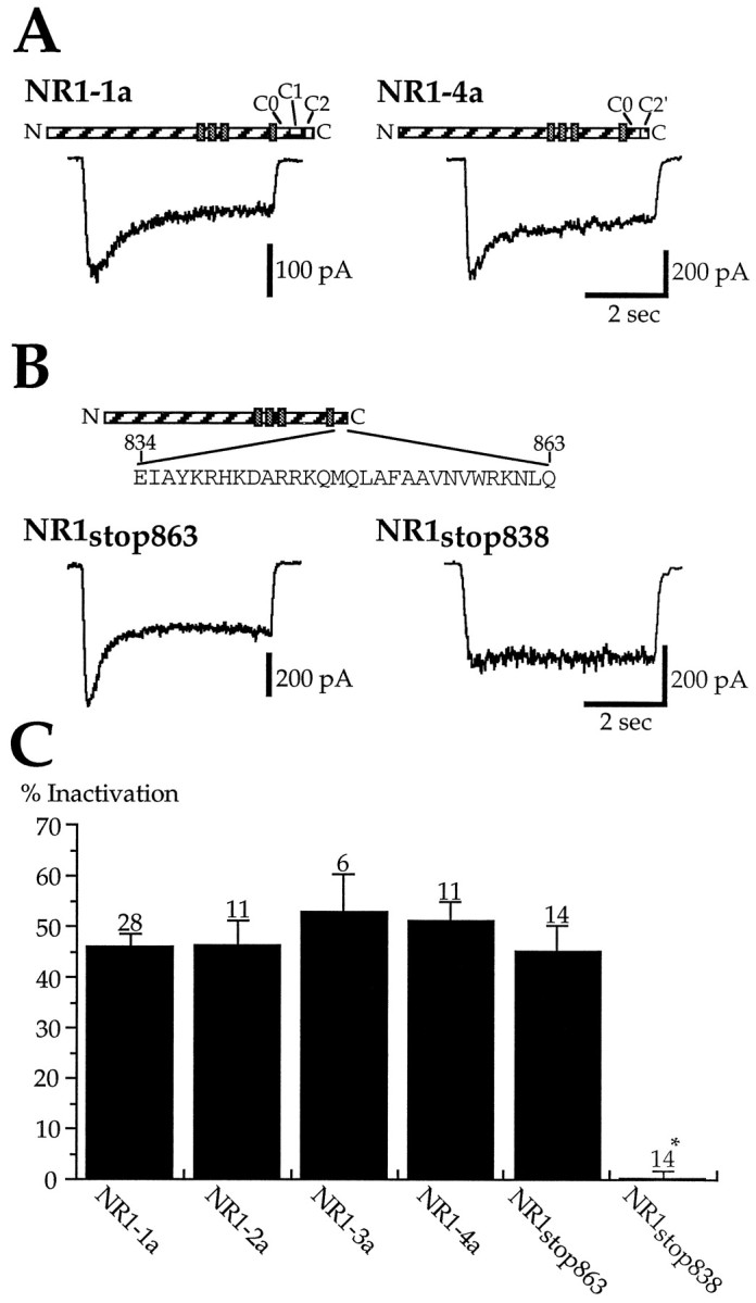
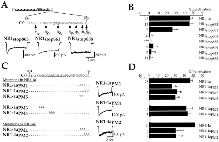
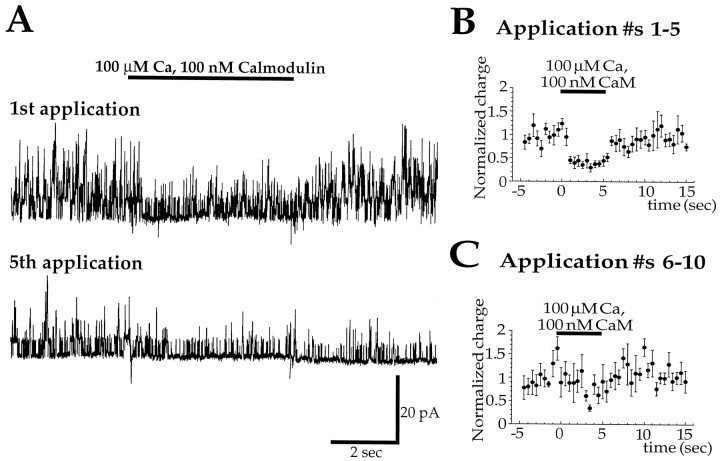
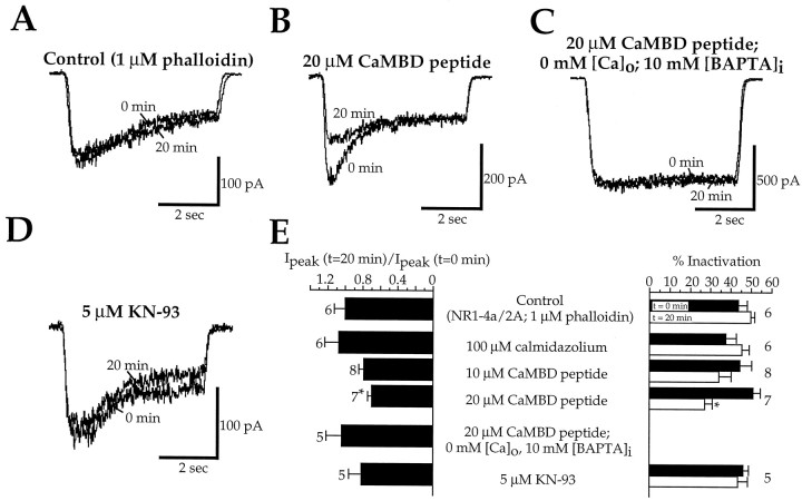
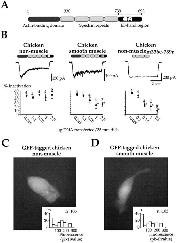
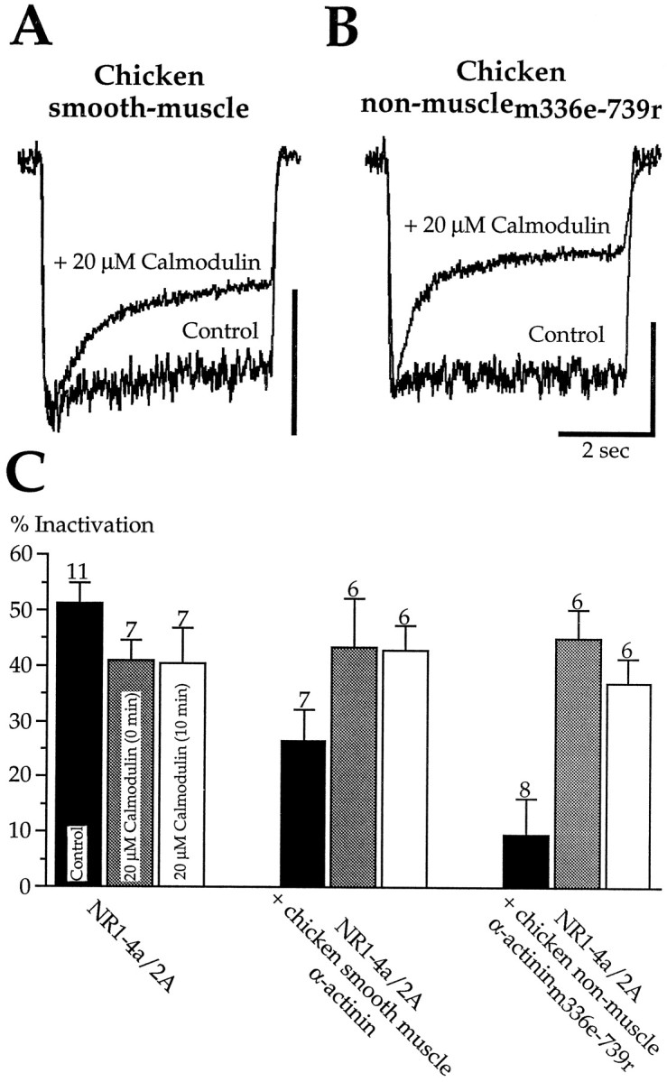
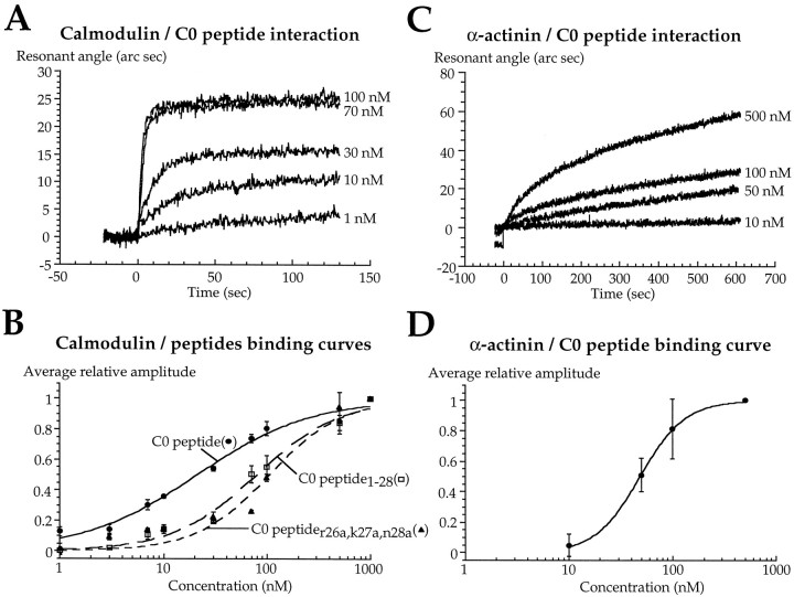
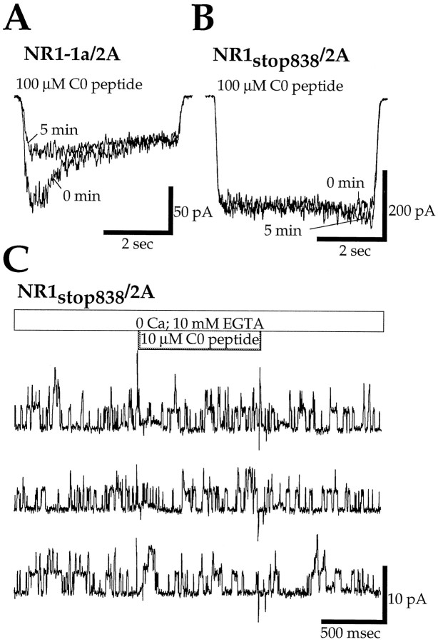
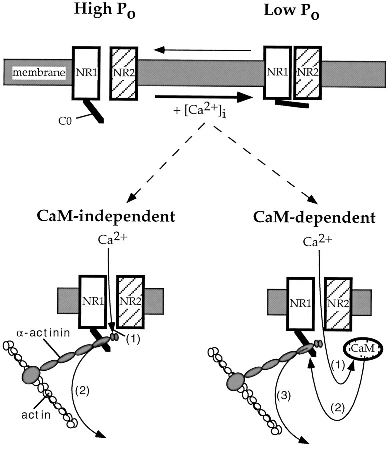
Glutamate receptors are associated with various regulatory and cytoskeletal proteins. However, an understanding of the functional significance of these interactions is still rudimentary. Studies in hippocampal neurons suggest that such interactions may be involved in calcium-induced reduction in the open probability of NMDA receptors (inactivation). Thus we examined the role of the intracellular domains of the NR1 subunit and two of its binding partners, calmodulin and alpha-actinin, on this process using NR1/NR2A heteromers expressed in human embryonic kidney (HEK) 293 cells. The presence of the first 30 residues of the intracellular C terminus of NR1 (C0 domain) was required for inactivation. Mutations in the last five residues of C0 reduced inactivation and produced parallel shifts in binding of alpha-actinin and Ca2+/calmodulin to the respective C0-derived peptides. Although calmodulin reduced channel activity in excised patches, calmodulin inhibitors did not block inactivation in whole-cell recording, suggesting that inactivation in the intact cell is more complex than binding of calmodulin to C0. Overexpression of putative Ca2+-insensitive, but not Ca2+-sensitive, forms of alpha-actinin reduced inactivation, an effect that was overcome by inclusion of calmodulin in the whole-cell pipette. The C0 domain also directly affects channel gating because NR1 subunits with truncated C0 domains that lacked calmodulin or alpha-actinin binding sites had a low open probability. We propose that inactivation can occur after C0 dissociates from alpha-actinin by two distinct but converging calcium-dependent processes: competitive displacement of alpha-actinin by calmodulin and reduction in the affinity of alpha-actinin for C0 after binding of calcium to alpha-actinin.


Figures

Fig. 1.
Fig. 1.
The C0 domain of the NR1 subunit is essential for inactivation. A, NR1 splice variants were coexpressed with NR2A in HEK293 cells. NMDA (10 μm) in the presence of 50–100 μm glycine was applied at a holding potential of −50 mV (2 mm[Ca2+]o). This protocol reveals inactivation of NMDA channels but does not induce macroscopic glycine-independent desensitization (Legendre et al., 1993; Rosenmund and Westbrook, 1993b). Full inactivation of ∼50% was observed with NR1–1a and NR1–4a. The structure of the NR1 splice variants is depicted above the current traces. The four hydrophobic domains are indicated by gray boxes. B, NR1/2A heteromers expressing an NR1 mutant truncated after the C0 domain (NR1stop863) also showed full inactivation. In contrast, inactivation was absent in heteromers with an NR1 truncation of almost the entire C terminus (NR1stop838).C, Pooled data for the degree of inactivation observed with different NR1/2A heteromers. Asterisk indicates significant difference compared with NR1–1a.
Fig. 2.
Fig. 2.
Residues 859–863 within the C0 domain of NR1 are critical for inactivation. A, B, In contrast to the full inactivation (∼50%) observed with NR1 constructs containing the entire C0 domain (NR1stop863), inactivation was completely absent if the last five residues in C0 were deleted (NR1stop858). Deletion of aa 862 and 863 produced a partial reduction of inactivation (NR1stop861). The NR1 subunit structure with the C0 amino acid sequence are indicated at thetop. Arrows point to the last amino acid of each truncation mutant. Asterisks in Bindicate significant differences compared with NR1–1a. C, D, Point mutants within the C0 domain confirmed that residues 859–863 are critical for inactivation. Triple alanine (NR1–1aPM1, NR1–1aPM2) or double glutamate (NR1–1aPM5) mutations in residues 859–863 reduced inactivation, whereas alanine mutations in C0 closer to the fourth membrane region (NR1–1aPM3, NR1–1aPM4) did not affect inactivation. The C1 domain did not affect inactivation because triple alanine mutations in NR1–4a (NR1–1aPM1, NR1–1aPM2) did not differ from the same mutations in NR1–1a.Asterisks in D indicate significant differences compared with NR1–1a for NR1–1aPM1–5 and NR1–4a for NR1–4aPM1+2.
Fig. 3.
Fig. 3.
Calmodulin rapidly and reversibly reduces the open probability of NMDA channels in “early” inside-out patches.A, The first application of 100 μm Ca plus 100 nm calmodulin to an inside-out patch from an HEK293 cell expressing NR1–4a/2A heteromers inhibited single-channel activity rapidly and reversibly (top). However, the inhibition was markedly reduced by the fifth application, made 4.5 min after excision of the patch. The overall channel activity also decreased with time of recording (rundown). Patches were exposed to a calcium-free solution containing 10 mm EGTA between calmodulin applications. B, Pooled data for the first five applications of calmodulin to a different inside-out patch than inA. Data are represented as charge per 500 msec and were normalized to the average charge in the 5 sec preceding application of calmodulin. C, Pooled data for applications 6–10 of calmodulin to the same patch as in B. The effect of calmodulin is no longer apparent. Similar results were obtained for four other patches.
Fig. 4.
Fig. 4.
Calmodulin inhibitors do not block inactivation.A, Under control conditions, both the peak amplitude and percentage inactivation of NR1–4a/2A heteromers remained constant during the first 20 min of whole-cell recording. B, Intracellular perfusion of 20 μm CaMBD peptide reduced the peak current amplitude, but had no effect on the steady-state current at the end of the agonist application. C, The effect of CaMBD peptide did not occur in calcium-free medium with 10 mm BAPTA in the whole-cell pipette. D, The CaM kinase II inhibitor KN-93 (5 μm in intracellular solution) had no effect on the response, suggesting that the CaMBD peptide did not act by inhibiting CaM kinase II. E, The ratio of the peak currents (t = 20 min/t = 0 min) are plotted at leftfor the calmodulin inhibitors and KN-93. The percentage inactivation att = 0 min (black bars) andt = 20 min (white bars) are plotted at right. Asterisks indicate significant differences to respective control.
Fig. 5.
Fig. 5.
Overexpression of putative calcium-insensitive α-actinin reduces inactivation. A, Subunits of chicken nonmuscle α-actinin are composed of an N-terminal actin-binding domain, a central rod domain of four spectrin repeats, and a C-terminal domain that contains two EF-hand motifs. B, Cotransfection of chicken nonmuscle α-actinin did not affect inactivation over a 100-fold range of cDNA amount transfected. In contrast, inactivation was reduced with 1 and 2.5 μg of DNA transfected with the chicken smooth muscle α-actinin. These two isoforms are splice variants of one gene, differing only in the predicted functionality of the first EF-hand motif. Cotransfection of a truncated α-actinin, encoding only the central rod domain, produced a dose-dependent block of inactivation. All recordings shown are from cells transfected with 2.5 μg of DNA of the respective α-actinin/35 mm dish. Asterisks indicate significant differences to cells transfected with NR1–4a/2A. C, D, The lack of effect of chicken nonmuscle α-actinin was not caused by low expression levels. C shows an HEK293 cell cotransfected with 2.5 μg DNA/35 mm dish of an N-terminal GFP-tagged chicken nonmuscle α-actinin. The bright fluorescence indicates high expression of the protein. Responses recorded from such cells showed full inactivation. Inset shows the fluorescence intensity (on a scale of 1 to 254) of randomly selected cells from a dish transfected with an N-terminal GFP-tagged chicken nonmuscle α-actinin (average pixel value: 128.0 ± 7.6;n = 106). D shows an HEK293 cell cotransfected with 2.5 μg DNA/35 mm dish of an N-terminal GFP-tagged chicken smooth muscle α-actinin. The distribution and average fluorescence intensity of cells transfected with this clone were similar to cells transfected with the GFP-tagged nonmuscle isoform (seeinset) (average pixel value: 104.3 ± 7.9;n = 102).
Fig. 6.
Fig. 6.
Calmodulin and α-actinin competitively affect calcium-dependent inactivation. A, Inclusion of calmodulin (20 μm) in the whole-cell pipette restored inactivation in HEK293 cells transfected with 2.5 μg DNA/35 mm dish of the putative calcium-insensitive chicken smooth muscle α-actinin. Recordings are from two cells. Scale bar is 100 pA for the control cell and 400 pA for the calmodulin-loaded cell. B, Calmodulin (20 μm) in the pipette also restored inactivation in HEK293 cells transfected with the central rod domain of α-actinin (2.5 μg DNA/35 mm dish). Recordings are from two cells. Scale bar is 100 for the control cell and 250 pA for the calmodulin-loaded cell.C, Pooled data are illustrated from experiments as inA and B.
Fig. 7.
Fig. 7.
The C0 peptide interacts with α-actinin as well as calmodulin in vitro. A, Protein–protein interactions were assessed by an optical method, as described in Materials and Methods, with binding detected as an increase in the resonant angle of incident light. Addition of calmodulin at the indicated concentrations ([Ca2+] = 1 mm) to C0 peptide-coated cuvettes produced dose-dependent binding to the C0 peptide. Because the change in resonant angle is linear to the mass, the change in resonant angle is a direct measure of the peptide–protein interactions. B, Dose–response curves for the interaction of three different C0 peptides with calmodulin. The amplitude of the resonant angle change was measured after the reaction had reached equilibrium (2 min after addition of calmodulin) and normalized to the response at 1 μm calmodulin. Experiments with C0 peptides corresponding to NR1 constructs producing reduced inactivation had a lower calmodulin affinity as compared with the full-length C0 peptide. C, Rabbit skeletal muscle α-actinin also bound to C0 but showed slower kinetics. D, Estimated dose–response curve for the interaction of the C0 peptide with α-actinin as normalized to the response at 500 nm, the maximal concentration of α-actinin that could be used in our experiments.
Fig. 8.
Fig. 8.
The C0 peptide reduced the peak current in whole-cell recording but did not affect channel activity in inside-out patches. A, The addition of C0 peptide (100 μm) to the pipette reduced the peak current amplitude in NR1–1a/2A heteromers (10 μm NMDA; 2 mm[Ca2+]o) after 5 min of whole-cell recording. B, In the presence of extracellular calcium, the C0 peptide (100 μm) had no effect on the peak current amplitude when NR1–1a was replaced with the C-terminal truncation mutant NR1stop838. C, Inside-out patches (Vh = +65 mV) from cells expressing NR1stop838/2A heteromers were sequentially exposed to a calcium-free solution (+10 mm EGTA) and a calcium-free solution containing 10 μm C0 peptide. The peptide had no effect on single-channel activity.
Fig. 9.
Fig. 9.
The C0 domain affects the open probability of NMDA channels and thus has an intrinsic effect on NMDA channel gating.A, Responses from two HEK293 cells expressing either NR1–4a/2A heteromers or NR1stop843/2A heteromers illustrate the protocol used to estimate the open probability (Po). Responses were evoked by a low concentration of NMDA (10 μm). To avoid contamination of the Po measurement by inactivation, recordings were made with a BAPTA-containing intracellular solution, and agonist was applied in Ca-free extracellular solution. After the responses had reached equilibrium, the open-channel blocker MK-801 (20 μm) was coapplied with NMDA. The onset of MK-801 block can be described by two exponentials, with the slower time constant reflecting channels that enter the open state during the presence of MK-801, i.e., it is proportional to Po (see Results for further details). Note that the slow component of MK-801 block is slower for NR1stop843/2A heteromers, indicating a lower Po. Currents are normalized to their steady-state amplitude. B, Semilogarithmic plot of the current decays in MK-801 for responses from NR1–4a/2A heteromers evoked under control conditions (10 μm NMDA), with a lower concentration of agonist (7.5 μm NMDA), or from cells coexpressing the central rod domain of α-actinin (α-actininm336e-739r). The coexpression of the central rod domain did not affect the slow component of MK-801 block, whereas the reduction in agonist concentration slowed the onset of MK-801 block, indicating the expected lower Po with lower agonist concentration.C, Semilogarithmic plot of the current decays in MK-801 for responses from NR1/2A heteromers containing several NR1 truncation mutants. Note that the onset of MK-801 block is slower (i.e., lowPo) with NR1stop858 and NR1stop843, which presumably do not bind calmodulin or α-actinin. The onset of MK-801 block is fast (i.e., highPo) after truncation of almost the entire C0 domain (NR1stop838). D, The ratio between the amount of charge during steady-state current (measured during the 2 sec application preceding MK-801) and the amount of charge during the application of MK-801 is the same for all conditions tested, indicating that differences in MK-801 kinetics between different conditions do not underlie the observed differences in the onset of MK-801 block. E, Pooled data for the slow time constant τslow under all conditions tested demonstrates that the presence of a C0 domain incapable of binding calmodulin and α-actinin results in a low Po.Asterisks indicate significant differences compared with control (NR1–4a/2A; 10 μm NMDA).
Fig. 10.
Fig. 10.
A molecular model for inactivation. C0 has an intrinsic effect on channel gating that can shift the open probability between high and low Po states. When the channel has a high Po (top left), C0 is probably attached to actin via α-actinin (for clarity this interaction is not shown). Influx of calcium triggers the dissociation of α-actinin from C0, leading to a lowPo (top right). This effect underlies calcium-dependent inactivation of NMDA channels and can be induced by two mechanisms: calmodulin dependent and calmodulin independent. In the calmodulin-independent mechanism (bottom left), calcium binds to the EF-hand of α-actinin (1), thereby reducing the affinity of α-actinin for C0, resulting in the dissociation of α-actinin from C0 (2). This mechanism incorporates our findings with the overexpressed α-actinins and explains the lack of effect of calmodulin inhibitors on whole-cell inactivation. In the calmodulin-dependent mechanism (bottom right), calcium binds to calmodulin (1). The activated calmodulin then competes with α-actinin for binding to C0 (2), resulting in the dissociation of α-actinin from C0 (3). This mechanism incorporates our competition experiments, in which exogenous calmodulin overcame the effects of overexpressed α-actinins.

Similar articles

Cited by

References

    1. Baron MD, Davison MD, Jones P, Critchley DR. The sequence of chick α-actinin reveals homologies to spectrin and calmodulin. J Biol Chem. 1987;262:17623–17629. - PubMed
    1. Beggs AH, Byers TJ, Knoll JHM, Boyce FM, Bruns GAP, Kunkel LM. Cloning and characterization of two human skeletal muscle α-actinin genes located on chromosomes 1 and 11. J Biol Chem. 1992;267:9281–9288. - PubMed
    1. Bennett V. Spectrin-based membrane skeleton: a multipotential adaptor between plasma membrane and cytoplasm. Physiol Rev. 1990;70:1029–1065. - PubMed
    1. Brakeman PR, Lanahan AA, O’Brien R, Roche K, Barnes CA, Huganir RL, Worley PF. Homer: a protein that selectively binds metabotropic glutamate receptors. Nature. 1997;386:284–288. - PubMed
    1. Carpén O, Pallai P, Staunton DE, Springer TA. Association of intercellular adhesion molecule-1 (ICAM-1) with actin-containing cytoskeleton and α-actinin. J Cell Biol. 1992;118:1223–1234. - PMC - PubMed

Publication types

MeSH terms

LinkOut - more resources