. 1999 Feb 15;19(4):1247-56.
doi: 10.1523/JNEUROSCI.19-04-01247.1999.

Activation of protein kinase A contributes to the expression but not the induction of long-term hyperexcitability caused by axotomy of Aplysia sensory neurons

Affiliations

Activation of protein kinase A contributes to the expression but not the induction of long-term hyperexcitability caused by axotomy of Aplysia sensory neurons

X Liao et al. J Neurosci. .

Abstract

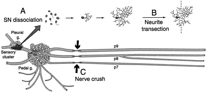
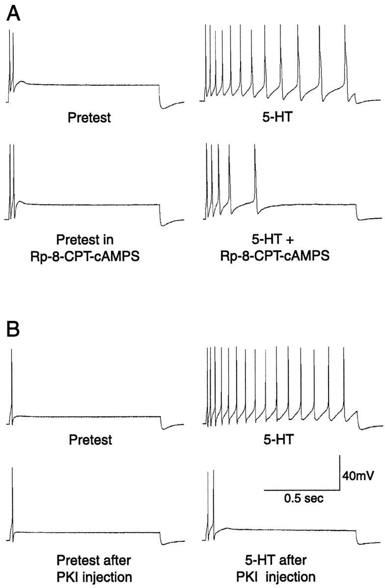
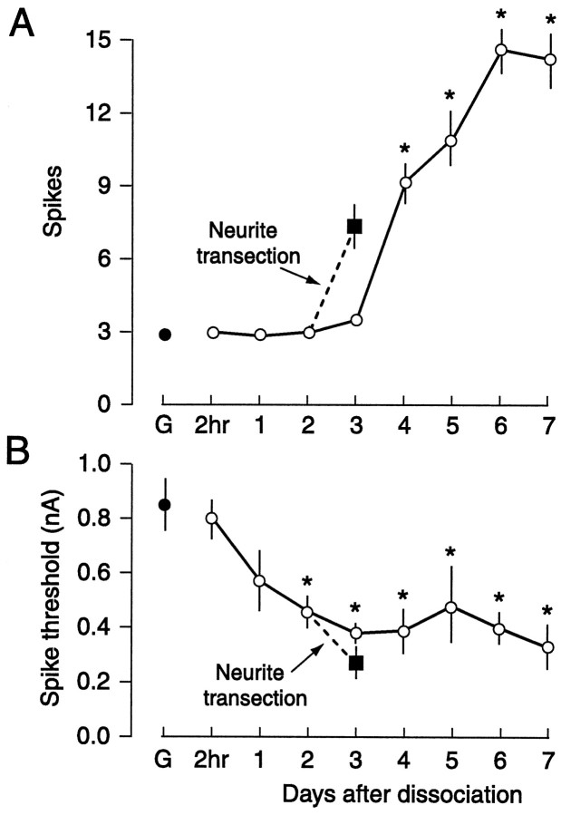
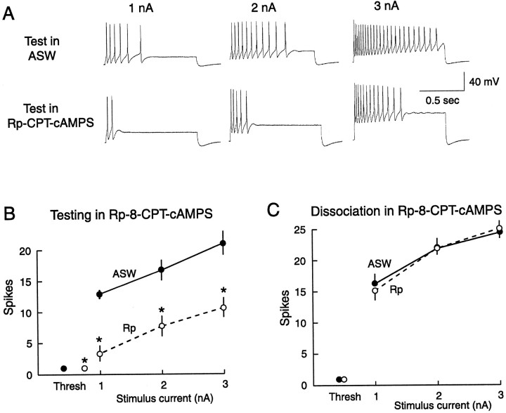
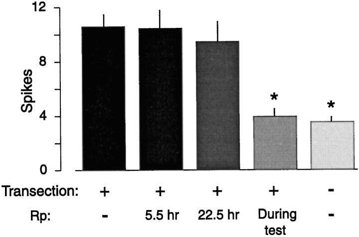
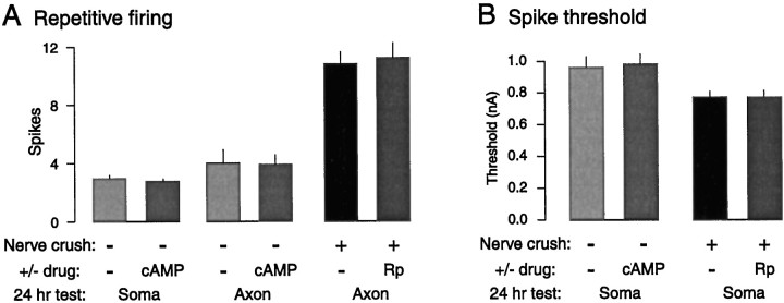
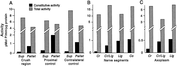
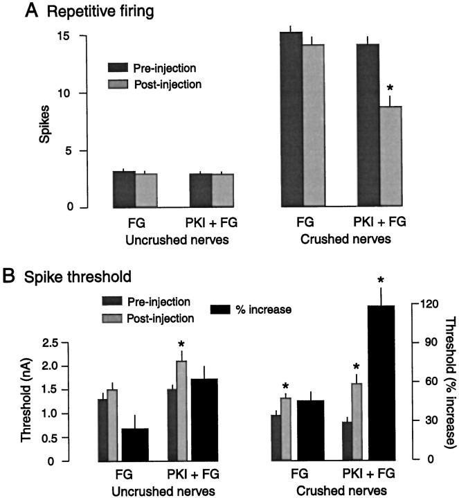
Nociceptive sensory neurons (SNs) in Aplysia provide useful models to study both memory and adaptive responses to nerve injury. Induction of long-term memory in many species, including Aplysia, is thought to depend on activation of cAMP-dependent protein kinase (PKA). Because Aplysia SNs display similar alterations in models of memory and after nerve injury, a plausible hypothesis is that axotomy triggers memory-like modifications by activating PKA in damaged axons. The present study disproves this hypothesis. SN axotomy was produced by (1) dissociation of somata from the ganglion [which is shown to induce long-term hyperexcitability (LTH)], (2) transection of neurites of dissociated SNs growing in vitro, or (3) peripheral nerve crush. Application of the competitive PKA inhibitor Rp-8-CPT-cAMPS at the time of axotomy failed to alter the induction of LTH by each form of axotomy, although the inhibitor antagonized hyperexcitability produced by 5-HT application. Strong activation of PKA in the nerve by coapplication of a membrane-permeant analog of cAMP and a phosphodiesterase inhibitor was not sufficient to induce LTH of either the SN somata or axons. Furthermore, nerve crush failed to activate axonal PKA or stimulate its retrograde transport. Therefore, PKA activation plays little if any role in the induction of LTH by axotomy. However, the expression of LTH was reduced by intracellular injection of the highly specific PKA inhibitor PKI several days after nerve crush. This suggests that long-lasting activation of PKA in or near the soma contributes to the maintenance of long-term modifications produced by nerve injury.


Figures

Fig. 1.
Fig. 1.
Three forms of sensory neuron (SN) axotomy. A, Dissociation of SN somata, which severed the major axon(s) of each SN close to the soma. Neurites began to grow within hours after plating.B, Transection close to the soma of outgrowing neurites from isolated SNs 2 d after dissociation. C, Axotomy produced by crushing pedal nerves containing SN axons, eitherin vivo or in an in vitro preparation (in which all nerves were left as long as possible). In both cases nerves innervating the midbody region and tail (notably p7, p8, and p9) were crushed ∼1 cm from the pedal ganglion. In some studies PKA activity was measured in segments of the nerve or axoplasm extruded from nerve segments (Fig. 7) after crushing pedal nerves ∼3 cm from the ganglion. Some of the nerves were ligated (data not shown) midway between the crush site and the ganglion to accumulate material transported retrogradely from the crush site (see measurement of PKA activity in Materials and Methods).
Fig. 2.
Fig. 2.
Examples of the reduction of immediate 5-HT-induced hyperexcitability by PKA antagonists. A, Bath application of 1 mm Rp-8-CPT-cAMPS reduced the increase in repetitive firing (compared with the pretest) caused by applying 5 μm 5-HT to the bath. B, Injection of PKI (1.5 mm, with 0.05% FG dye) into the SN reduced the increase in repetitive firing produced by puffing 50 μm 5-HT onto the cell soma.
Fig. 3.
Fig. 3.
Long-term hyperexcitability of SNs produced by dissociation. Repetitive firing (A) and spike threshold (B) were tested in SNs sampled at the indicated times after dissociation. G, Control cells tested in excised ganglia preparations during the same period. Data in this and other figures are shown as means ± SEM. *Significant differences (p < 0.05) compared with control SNs in ganglia. In addition to producing changes in repetitive firing and spike threshold, dissociation increasedRin (see Results). Also shown is the previously reported enhancement of excitability 24 hr later produced by transecting SN neurites 2 d after dissociation (Ambron et al., 1996).
Fig. 4.
Fig. 4.
Rp-8-CPT-cAMPS applied during dissociation does not prevent the induction of LTH, although it reduces the expression of LTH when applied later. A, Examples of repetitive firing tested at three test current intensities in the presence and absence of Rp-8-CPT-cAMPS 4–5 d after dissociation. B, Significant depression of repetitive firing and elevation of spike threshold when SNs (dissociated 4–5 d previously) were tested in the presence of Rp-8-CPT-cAMPS (Rp). Thresh, Mean threshold in nanoamperes for eliciting a single spike in each group (horizontal error bars that would indicate variability of threshold current are too small to be seen). C, Lack of effect of Rp-8-CPT-cAMPS (present during dissociation) on the induction of LTH measured 4–5 d later.
Fig. 5.
Fig. 5.
Rp-8-CPT-cAMPS (Rp) applied during neurite transection does not prevent the induction of LTH, although it significantly reduces the expression of LTH if present during the test 24 hr after transection. Times indicate duration of Rp-8-CPT-cAMPS treatment after transection.
Fig. 6.
Fig. 6.
PKA activation in the nerve is neither sufficient nor necessary for the induction of LTH. A, Activation of PKA by bathing the nerve in 8-CPT-cAMP + IBMX (cAMP) for 2 hr failed to increase repetitive firing of the soma or axons when tested 24 hr later. In addition, bathing the nerves in Rp-8-CPT-cAMPS (Rp) for 30 min before and 2 hr after they were crushed failed to prevent LTH 24 hr later. B, Bathing the nerve in 8-CPT-cAMP + IBMX also failed to reduce spike threshold in the soma 24 hr later, and bathing the nerves in Rp-8-CPT-cAMPS during and after nerve crush failed to prevent the reduction in soma threshold 24 hr later.
Fig. 7.
Fig. 7.
Nerve injury does not activate PKA or stimulate its transport toward the soma. A, Lack of injury-activated PKA at the crush site 5 min after crushing pedal nerves. No increase in the constitutive activity of the catalytic subunit in the crushed region was found in either the supernatant (Sup) or pellet (Pellet) compared with activity in a region of the same nerves near the ganglion (Proximal control) or in the region in uncrushed contralateral nerves corresponding to the crushed region (Contralateral control). Each segment contained a considerable amount of the PKA holoenzyme, which was revealed by adding exogenous cAMP to the extracts (Total activity). No transfer of active catalytic subunit from the soluble fraction (Sup) to the membrane fraction (Pellet) occurred in the crushed region, as indicated by similar ratios ofSup/Pellet activity in each region. B, C, Lack of injury-induced transport of PKA. Activity of the PKA catalytic subunit was assessed 20 hr after pedal nerve crush in segments of whole nerve (B) and in extruded axoplasm (C). Segments (supernatant) or extruded axoplasm were assayed on the proximal side adjacent to the crush site (Cr) and immediately distal to a ligation that was midway between the ganglion and the crush site (Cr/Lig). Any PKA that was activated and transported retrogradely would accumulate at the Cr/Lig site. As controls, segments and axoplasm immediately distal to a ligation on an uncrushed nerve (Lig), or from a corresponding section of a control nerve that was neither crushed nor ligated (Co), were examined (Ambron et al., 1995).
Fig. 8.
Fig. 8.
Evidence that LTH induced by nerve crush 4–5 d earlier is partially maintained by PKA activity. A,Left, Neither injection of Fast Green (FG) dye nor FG + protein kinase inhibitor (FG + PKI) affected repetitive firing responses tested in somata of SNs whose axons had not been crushed in vivo.Right, SNs whose axons had been crushed displayed greater firing responses than SNs with uncrushed axons, but this injury-induced hyperexcitability of the soma was significantly reduced by injection of PKI. B, Left, Injection of PKI significantly increased spike threshold in SNs whose axons had not been crushed. Right, Injection of both FG and PKI + FG significantly increased threshold in SNs with previously crushed axons. However, the percentage increase (black bars) after PKI + FG injection into previously axotomized SNs (Crushed nerves, right side) was significantly greater than the increase after FG injection, or the increases in previously unaxotomized SNs (Uncrushed nerves, left side).

Similar articles

Cited by

References

    1. Abel T, Kandel E. Positive and negative regulatory mechanisms that mediate long-term memory storage. Brain Res Brain Res Rev. 1998;26:360–378. - PubMed
    1. Alberini CM, Ghirardi M, Metz R, Kandel ER. C/EBP is an immediate-early gene required for the consolidation of long-term facilitation in Aplysia. Cell. 1994;76:1099–1114. - PubMed
    1. Ambron RT, Walters ET. Priming events and retrograde injury signals: a new perspective on the cellular and molecular biology of nerve regeneration. Mol Neurobiol. 1996;13:61–79. - PubMed
    1. Ambron RT, Dulin MF, Zhang X-P, Schmied R, Walters ET. Axoplasm enriched in a protein mobilized by nerve injury elicits memory-like alterations in Aplysia neurons. J Neurosci. 1995;15:3440–3446. - PMC - PubMed
    1. Ambron RT, Zhang X-P, Gunstream JD, Povelones M, Walters ET. Intrinsic injury signals enhance growth, survival, and excitability of Aplysia neurons. J Neurosci. 1996;16:7469–7477. - PMC - PubMed

Publication types

MeSH terms

LinkOut - more resources