. 1999 Feb 15;19(4):1284-93.
doi: 10.1523/JNEUROSCI.19-04-01284.1999.

Distinct mechanisms underlie neurotoxin-mediated cell death in cultured dopaminergic neurons

Affiliations

Distinct mechanisms underlie neurotoxin-mediated cell death in cultured dopaminergic neurons

J Lotharius et al. J Neurosci. .

Abstract

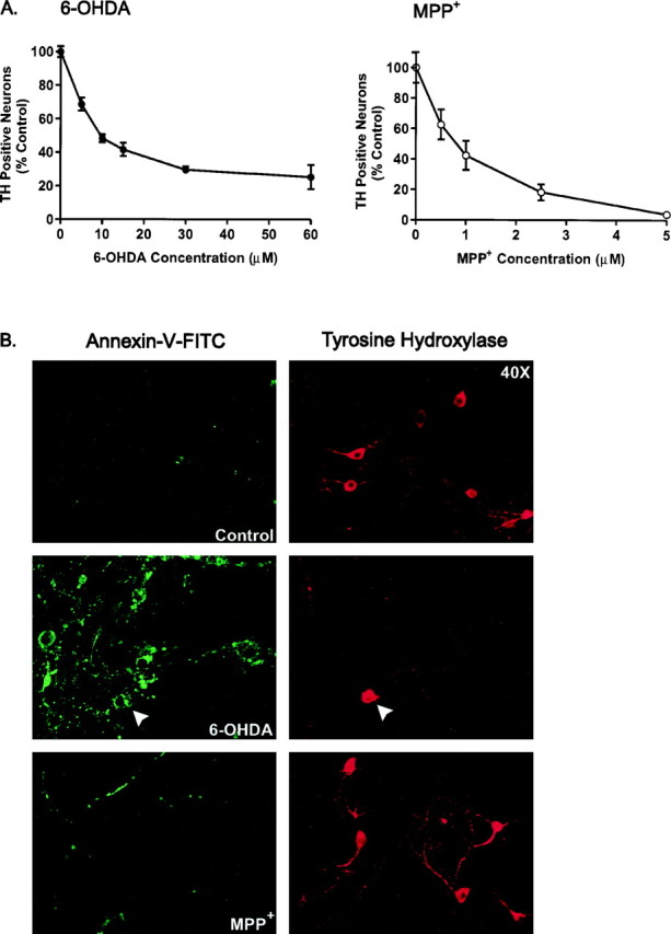
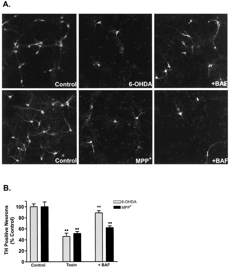
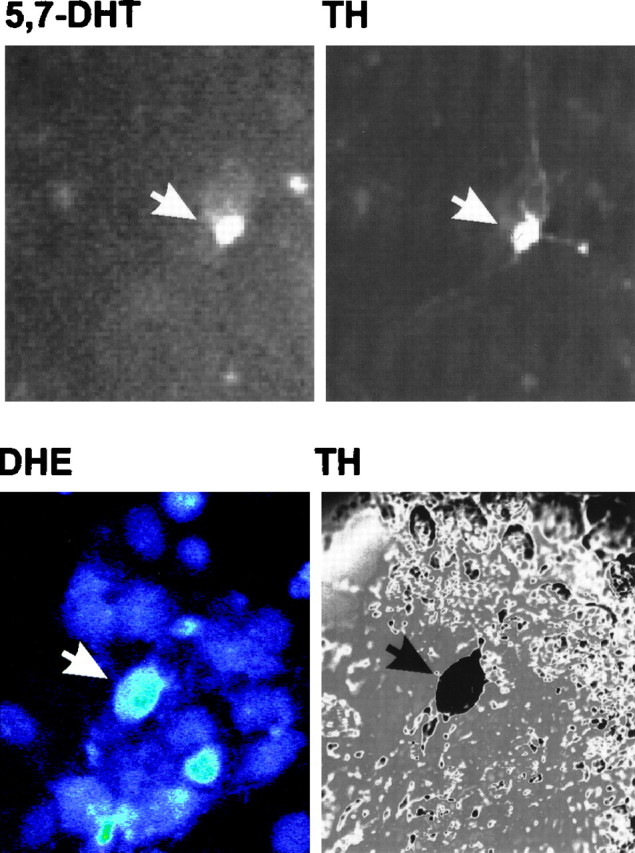
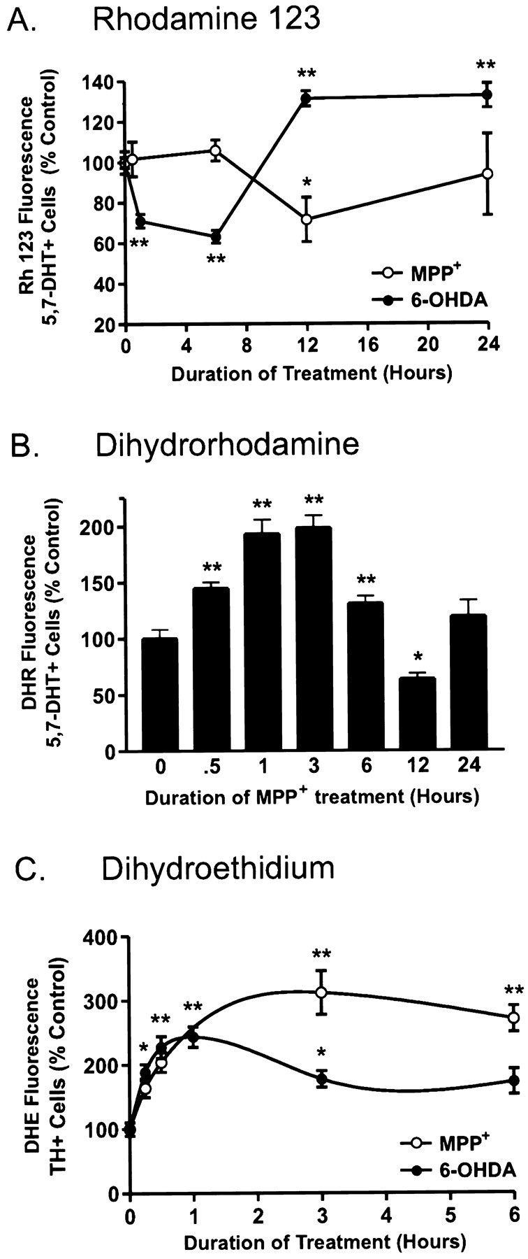
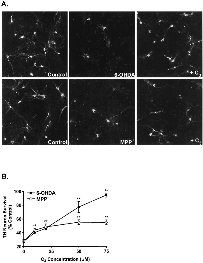
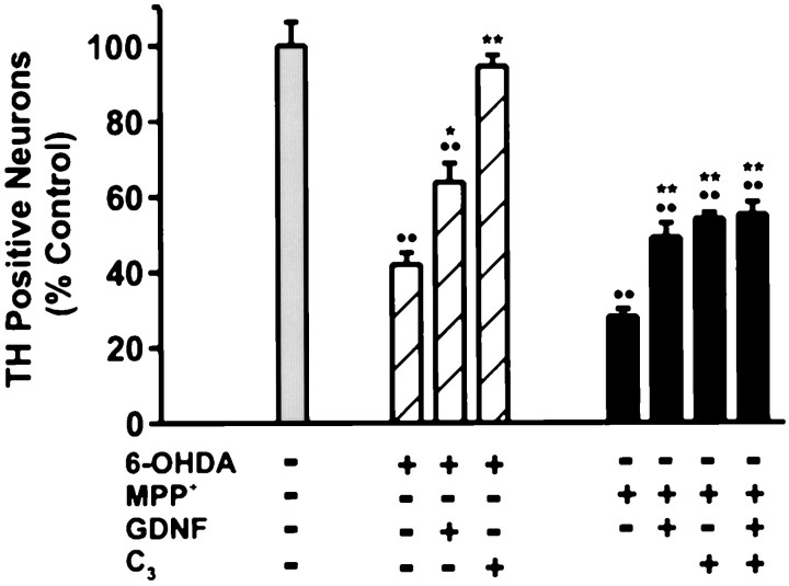
Oxidative stress is thought to contribute to dopaminergic cell death in Parkinson's disease (PD). The neurotoxin 6-hydroxydopamine (6-OHDA), which is easily oxidized to reactive oxygen species (ROS), appears to induce neuronal death by a free radical-mediated mechanism, whereas the involvement of free radicals in N-methyl-4-phenylpyridinium (MPP+) toxicity is less clear. Using free radical-sensitive fluorophores and vital dyes with post hoc identification of tyrosine hydroxylase-positive neurons, we monitored markers of apoptosis and the production of ROS in dopaminergic neurons treated with either 6-OHDA or MPP+. Annexin-V staining suggested that 6-OHDA but not MPP+-mediated cell death was apoptotic. In accordance with this assignment, the general caspase inhibitor Boc-(Asp)-fluoromethylketone only blocked 6-OHDA neurotoxicity. Both toxins exhibited an early, sustained rise in ROS, although only 6-OHDA induced a collapse in mitochondrial membrane potential temporally related to the increase in ROS. Recently, derivatives of buckminsterfullerene (C60) molecules have been shown to act as potent antioxidants in several models of oxidative stress (Dugan et al., 1997). Significant, dose-dependent levels of protection were also seen in these in vitro models of PD using the C3 carboxyfullerene derivative. Specifically, C3 was fully protective in the 6-OHDA paradigm, whereas it only partially rescued dopaminergic neurons from MPP+-induced cell death. In either model, it was more effective than glial-derived neurotrophic factor. These data suggest that cell death in response to 6-OHDA and MPP+ may progress through different mechanisms, which can be partially or entirely saved by carboxyfullerenes.


Figures

Fig. 1.
Fig. 1.
Plasma membrane alterations induced by 6-OHDA and MPP+. A, Cultures were treated with various concentrations of 6-OHDA or MPP+ for 24 and 48 hr, respectively, and processed for TH immunoreactivity, and the number of TH-positive neurons was counted. Data are normalized to control cultures and denote the mean ± SEM of representative determinations made in three separate cultures. Bars with <2% SEM are buried within the symbols. Cells exposed to 6-OHDA or MPP+ exhibited a half-lethal dose of 15 μm and 1 μm, respectively.B, Representative confocal micrographs of mesencephalic cultures stained for TH and annexin-V-FITC visualized at 40× magnification by confocal microscopy. Cultures were treated with 15 μm 6-OHDA or 1 μm MPP+for 6 hr, rinsed, and incubated with annexin-V-FITC for 10 min at room temperature. After imaging, plates were fixed and processed for TH immunoreactivity using a CY3-coupled secondary antibody. Random micrographic fields of annexin-V-stained neurons were relocated by their position on a microwell grid, and the corresponding image of TH immunoreactive neurons was taken.
Fig. 2.
Fig. 2.
The caspase inhibitor BAF rescues cultured dopaminergic neurons from 6-OHDA but not MPP+-induced cell death. A, Representative fluorescent micrographs of mesencephalic cultures stained for TH and visualized using a CY3-conjugated secondary antibody. Cells were exposed to 15 μm 6-OHDA or 1 μm MPP+ in the presence or absence of 50 μm BAF and stained after 24 and 48 hr, respectively.B, TH cell counts of cultures treated with either toxin and BAF. Values are normalized to the number of TH-positive neurons in vehicle-treated cultures and denote the mean ± SEM made of three separate cultures. **p < 0.001 compared with toxin-treated cultures; ••p < 0.001 in relation to vehicle-treated control (ANOVA with post hoc Student’st test).
Fig. 3.
Fig. 3.
Identification of dopaminergic neurons using 5,7-DHT prelabeling or post hoc TH staining. Top panels, Mesencephalic cultures were incubated with 5,7-DHT for 30 min at 37°C, rinsed, and imaged by fluorescence microscopy with a computer-controlled camera. Images were taken at 40× magnification. Cultures were subsequently fixed and stained for TH using a CY3-coupled secondary antibody. Field relocation shows colocalization of TH with 5,7-DHT (white arrows). This methodology was used to assay changes in DHR and Rh 123 fluorescence in dopaminergic cells.Bottom panels, To measure DHE fluorescence in dopaminergic neurons, cultures were stained for TH via a color reaction after incubation with DHE. Bottom left panel, Confocal image of cells exposed to 6-OHDA for 30 min and incubated with DHE for 15 min (60×). On the right is the corresponding differential interference contrast image of TH-stained neurons in a relocated field.
Fig. 4.
Fig. 4.
6-OHDA and MPP+differentially affect mitochondrial membrane potential (ΨΔm) and induce ROS formation.A, Time course of ΨΔm in 5,7-DHT-labeled dopaminergic neurons treated with 15 μm 6-OHDA (•) or 1 μm MPP+ (○). After drug treatments, cells were loaded with 0.3 μm Rh 123 for 20 min and assayed using a laser scanning confocal microscope. Values correspond to average pixel intensity normalized to baseline fluorescence values from vehicle-treated cells. B, Time-dependent changes in ROS production in dopaminergic neurons treated with 1 μmMPP+. After treatment with 1 μmMPP+ for the indicated time period, cells were incubated with 15 μm DHR for 20 min, rinsed, and imaged by confocal microscopy. Values correspond to fluorescence intensity and are normalized to vehicle-treated labeled dopaminergic neurons.C, Time-dependent induction of ROS formation in dopaminergic neurons treated with 15 μm 6-OHDA or 1 μm MPP+. After treatment with either drug for 0, 0.25, 0.5, 1, 3, and 6 hr, cells were loaded with 10 μg/ml DHE for 15 min at 37°C, fixed, stained for TH, and assayed by confocal microscopy. Values represent normalized DHE fluorescence from TH-immunoreactive neurons. Data are mean ± SEM of determinations made in three separate cultures. *p < 0.01; **p< 0.001, compared with values for vehicle-treated cultures (ANOVA withpost hoc Student’s t test).
Fig. 5.
Fig. 5.
The C3 fullerene isomer protects cultured dopaminergic neurons from 6-OHDA-induced toxicity but only partially attenuates MPP+-mediated cell death.A, Representative fluorescent micrographs of TH-positive neurons treated with either 15 μm 6-OHDA or 1 μm MPP+ in the presence or absence of 75 μm C3. B, Dose-dependent effects of C3 on dopaminergic neurons treated with 15 μm 6-OHDA (•) or 1 μmMPP+ (○). The number of surviving dopaminergic neurons was determined 24 hr (6-OHDA) or 48 hr (MPP+) later by TH immunocytochemistry and normalized to the number of TH neurons in vehicle-treated plates. Data are mean ± SEM from determinations made from three cultures. *p < 0.01; **p < 0.001 compared with values from control plates (ANOVA with post hoc Student’st test).
Fig. 6.
Fig. 6.
Effects of C3 and/or GDNF in rescuing dopaminergic neurons from neurotoxin-induced cell death. Primary mesencephalic cultures were pretreated with 1 ng/ml rhGDNF for 3 hr before the addition of either 15 μm 6-OHDA (hatched bars) or 1 μmMPP+ (black bars). The fullerene isomer C3 (75 μm) was tested for its ability to enhance the protective effect of GDNF. Dopaminergic cell viability was assessed 24 hr (6-OHDA) and 48 hr (MPP+) later by TH immunocytochemistry. Data denote the mean ± SEM.Asterisks indicate statistically significant difference between each respective drug condition and GDNF or GDNF with C3. *p < 0.01; **p < 0.001;••p < 0.001 shows difference between vehicle-treated control and all conditions (ANOVA with post hoc Student’s t test).

Similar articles

Cited by

References

    1. Bindokas VP, Jordan J, Lee CC, Miller RJ. Superoxide production in rat hippocampal neurons: selective imaging with hydroethidine. J Neurosci. 1996;16:1324–1336. - PMC - PubMed
    1. Cardozo DL. Midbrain dopaminergic neurons from postnatal rat in long-term primary culture. Neuroscience. 1993;56:409–421. - PubMed
    1. Cardozo DL, Bean BP. Voltage-dependent calcium channels in rat midbrain dopamine neurons: modulation by dopamine and GABAB receptors. J Neurophysiol. 1995;74:1137–1148. - PubMed
    1. Cassarino DS, Fall CP, Smith TS, Bennett JP., Jr Pramipexole reduces reactive oxygen species production in vivo and in vitro and inhibits the mitochondrial permeability transition produced by the parkinsonian neurotoxin methylpyridinium ion. J Neurochem. 1998;71:295–301. - PubMed
    1. Cohen G, Heikkila RE. The generation of hydrogen peroxide, superoxide radical, and hydroxyl radical by 6-hydroxydopamine, dialuric acid, and related cytotoxic agents. J Biol Chem. 1974;249:2447–2452. - PubMed

Publication types

MeSH terms

LinkOut - more resources