. 1999 Feb 15;19(4):1307-16.
doi: 10.1523/JNEUROSCI.19-04-01307.1999.

Molecular analysis of the X11-mLin-2/CASK complex in brain

Affiliations

Molecular analysis of the X11-mLin-2/CASK complex in brain

J P Borg et al. J Neurosci. .

Abstract

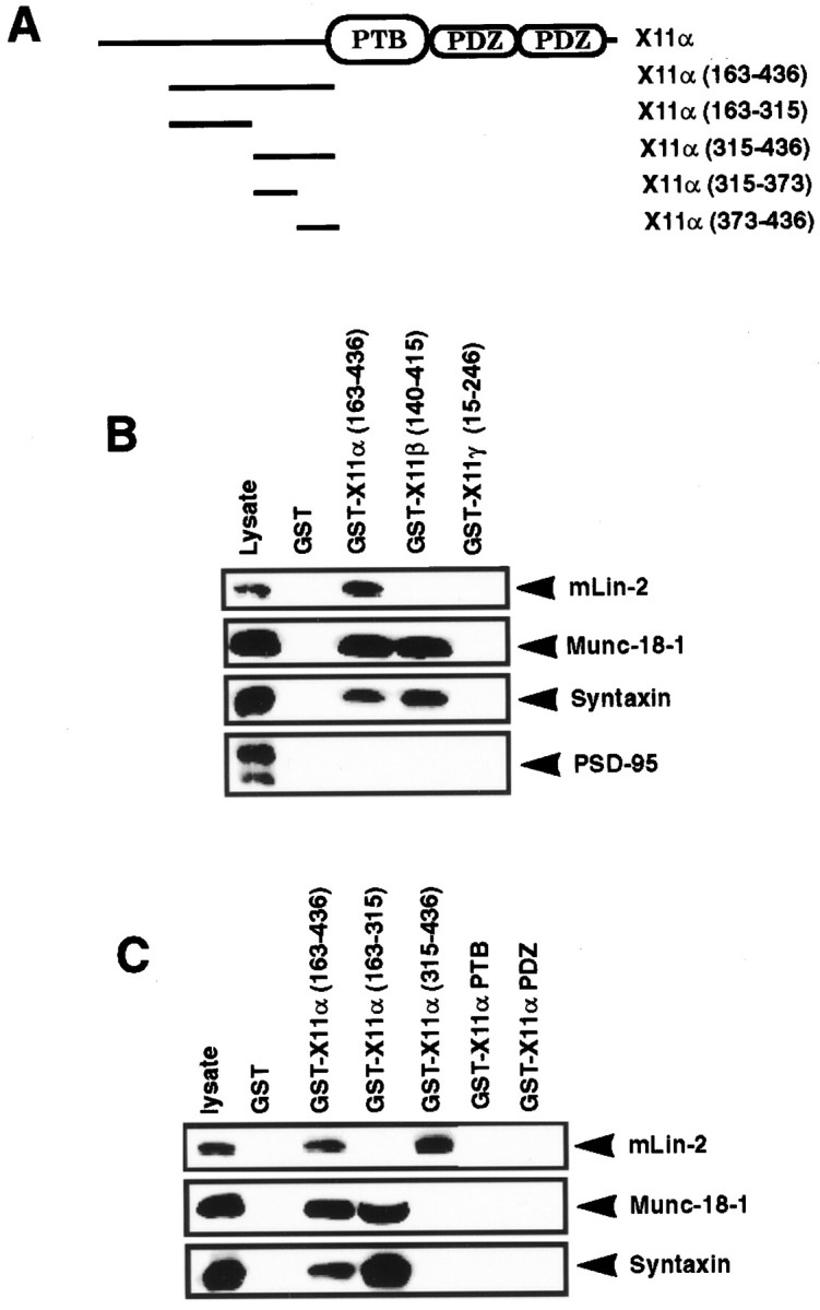
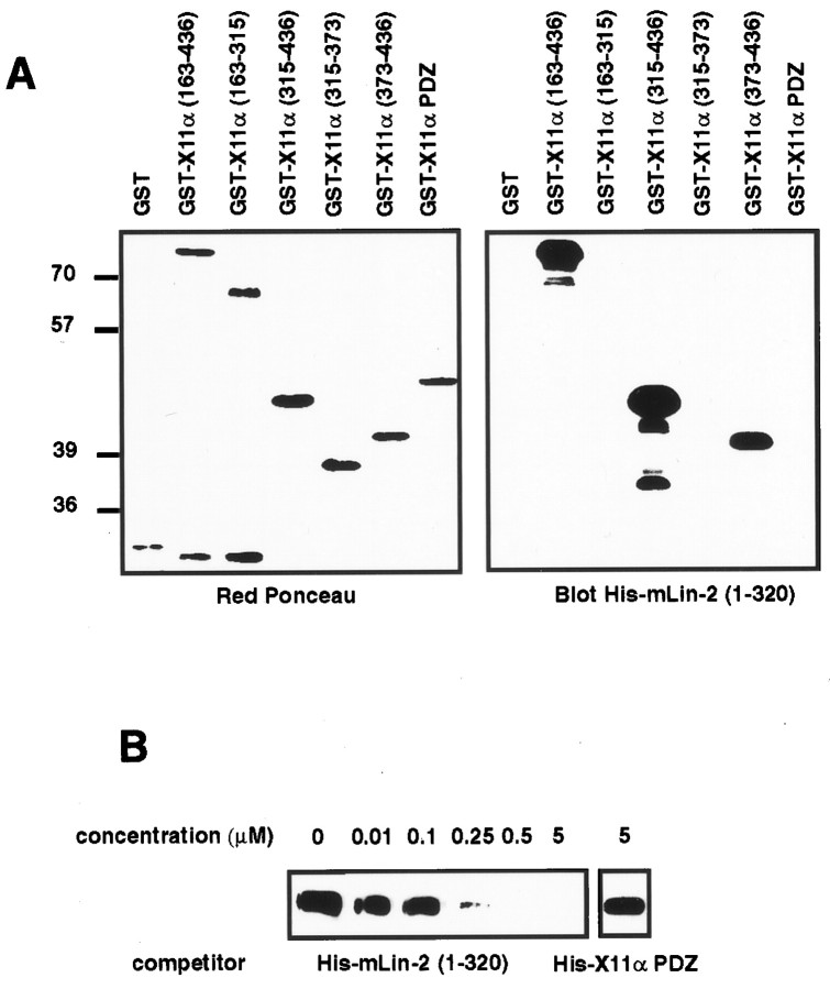
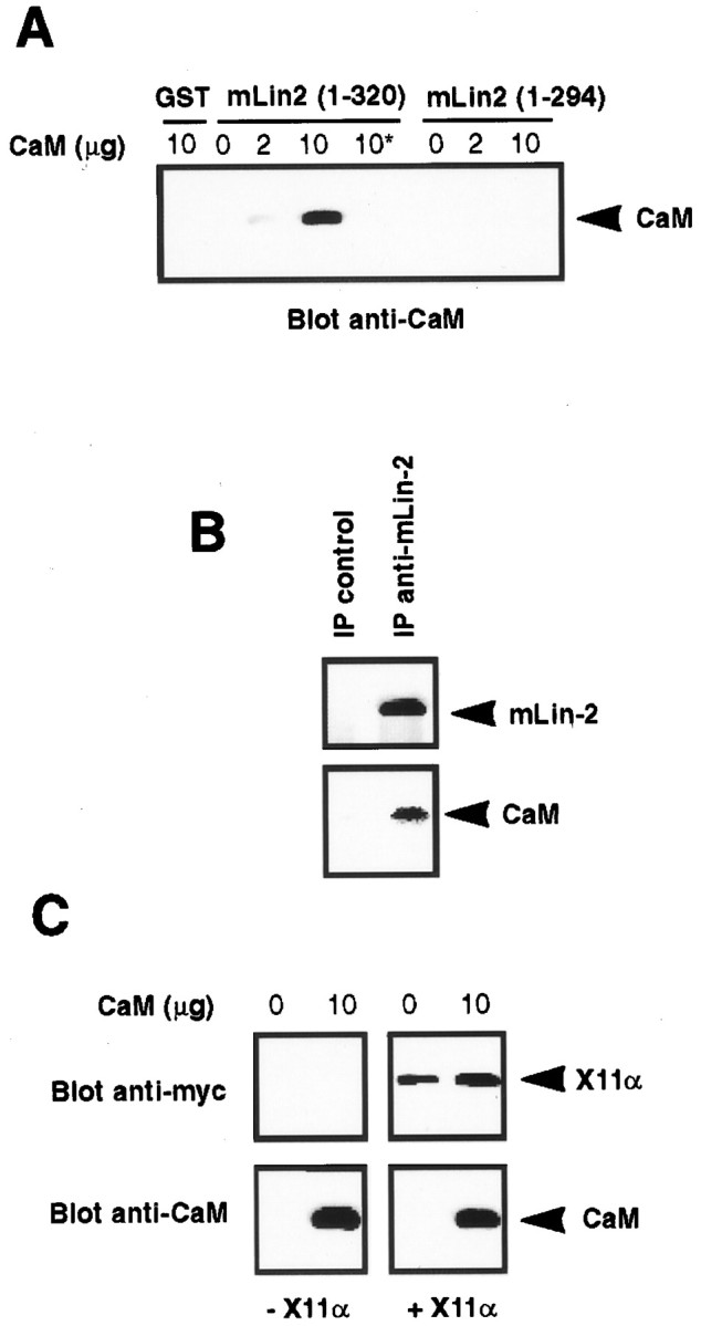
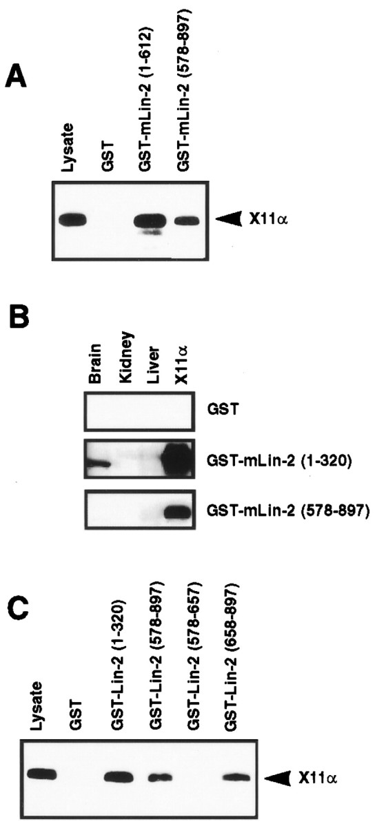
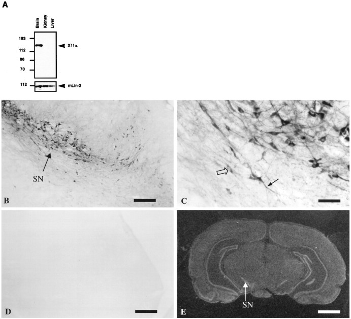
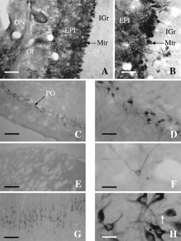
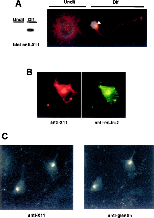
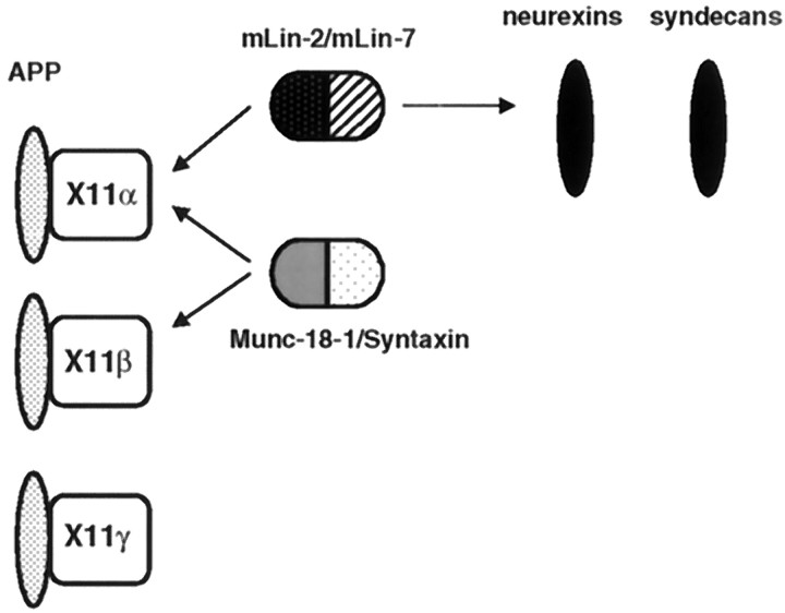
A heterotrimeric complex containing Lin-10/X11alpha, Lin-2/CASK, and Lin-7 is evolutionarily conserved from worms to mammals. In Caenorhabditis elegans, it localizes Let-23, a receptor tyrosine kinase, to the basolateral side of vulval epithelium, a step crucial for proper vulva development. In mammals, the complex may also participate in receptor targeting in neurons. Accordingly, phosphotyrosine binding (PTB) and postsynaptic density-95/Discs large/Zona Occludens-1 domains found in X11alpha and mLin-2/CASK bind to cell-surface proteins, including amyloid precursor protein, neurexins, and syndecans. In this paper, we have further analyzed the X11alpha-mLin-2/CASK association that is mediated by a novel protein-protein interaction. We show that the mLin-2/CASK calmodulin kinase II (CKII) domain directly binds to a 63 amino acids peptide located between the Munc-18-1 binding site and the PTB domain in X11alpha. Ca2+/calmodulin association with mLin-2/CASK does not modify the X11alpha-mLin-2 interaction. A region containing the mLin-2/CASK guanylate kinase domain also interacts with X11alpha but with a lower affinity than the CKII domain. Immunostaining of X11alpha in the brain shows that the protein is expressed in areas shown previously to be positive for mLin-2/CASK staining. Together, our data demonstrate that the X11alpha-mLin-2 complex contacts many partners, creating a macrocomplex suitable for receptor targeting at the neuronal plasma membrane.


Figures

Fig. 1.
Fig. 1.
Delineation of the mLin-2/CASK binding site in X11α protein. A, Schematic representation of the X11α protein. The X11α protein contains a central PTB domain, followed at its C-terminal end by two PDZ domains. N-terminal fragments were produced as GST fusion proteins. B, Proteins extracted from mouse brain were precipitated with GST, GST X11α (region 163–436), GST X11β (region 140–415), or GST X11γ (region 15–246) coupled to glutathione beads. These fusion proteins incorporate peptides from the N terminus of these X11 isoforms. After washing, proteins were separated on 10% SDS-PAGE and transferred to nitrocellulose. The membrane was probed with polyclonal anti-mLin-2/CASK and monoclonal anti-Munc-18-1, anti-Syntaxin, and anti-PSD-95 antibodies. One-tenth of the lysate used for precipitation was run as control (lysate). Detection was performed by chemiluminescence. C, Same as B, with additional X11α fusion proteins. The GST X11α PTB and PDZ proteins comprise the PTB and the two PDZ domains of the protein, respectively.
Fig. 2.
Fig. 2.
The mLin-2/CASK CKII domain directly binds to an X11α (region 373–436) peptide. A, GST fusion proteins described in Figure 1C were subjected to SDS-PAGE and transferred to nitrocellulose. Equivalent amounts of proteins were revealed with Ponceau red stain (left). The membrane was probed with soluble His-mLin-2/CASK (region 1–320) protein, and bound proteins were revealed with anti-T7 antibody, followed by HRP goat anti-mouse and chemiluminescence detection (right).B, The same procedure was performed to detect X11α in 293 cell lysate, except that soluble GST mLin-2/CASK (region 1–320) protein was used as primary reagent and anti-GST antibody–HRP protein A as secondary reagents. Increasing concentrations of soluble His-mLin-2/CASK (region 1–320) were mixed with a fixed concentration of soluble GST mLin-2/CASK (region 1–320) protein to compete for binding to X11α. A soluble His-X11α PDZ containing the two X11α PDZ domains was used as a negative control.
Fig. 3.
Fig. 3.
An integral mLin-2/CASK CKII domain is required for binding to X11α. A, Schematic representation of mLin-2/CASK and GST fusion proteins used in this study.B, Myc-tagged X11α expressed in 293 cells was precipitated by mLin-2/CASK GST coupled to glutathione beads, and bound proteins were revealed by Western blot with anti-myc antibody.C, Same as B, with different mLin-2/CASK constructs.
Fig. 4.
Fig. 4.
Calmodulin binds to mLin-2/CASK but does not affect X11α–mLin-2 interaction. A, GST and GST mLin-2/CASK (regions 1–320 and 1–294) were incubated with 0, 2, or 10 μg of calmodulin in the presence of 0.1 mmCa2+ or 1 mm EGTA (asterisk). After washing, bound calmodulin was resolved on SDS-PAGE, transferred to nitrocellulose, and detected with anti-calmodulin antibody. B, Lysates from untransfected A-172 cells were immunoprecipitated with preimmune or immune anti-mLin-2/CASK antibodies, and bound proteins were resolved on SDS-PAGE and transferred to nitrocellulose. Proteins were successively revealed with anti-mLin-2/CASK (top) and anti-calmodulin (bottom) antibodies. C, GST mLin-2/CASK (region 1–320) fusion protein immobilized on glutathione beads was incubated with Ca2+/calmodulin, and then lysate with (+X11α) or without (−X11α) myc-tagged X11α was added. Bound calmodulin and X11α was then assessed using immunoblotting.
Fig. 5.
Fig. 5.
A region of mLin-2/CASK encompassing the GK domain binds to X11α. A, C, Myc-tagged X11α protein expressed in 293 cells was precipitated with different mLin-2/CASK GST fusion proteins and revealed with anti-myc antibody after Western blot. We have also detected binding to nonmyc-tagged constructs (results not shown). B, Brain, liver, and kidney extracts were run on a gel, and proteins were transferred to nitrocellulose. Myc-tagged X11α protein expressed in 293 cells was used as a positive control (X11α). An overlay assay was performed with soluble GST fusion proteins. Bound GST proteins were revealed using anti-GST antibody–HRP protein A chemiluminescence method.
Fig. 6.
Fig. 6.
X11α is a brain-specific protein.A, Total proteins extracted from mouse brain, kidney, and liver were resolved by 8% SDS-PAGE and transferred to nitrocellulose. Immunoblot with anti-X11α antibody detects X11α only in the brain. mLin-2/CASK is present in all tissues, as observed previously (Hata et al., 1996; Cohen et al., 1998). B, X11α-positive immunostaining in substantia nigra (SN). Scale bar, 300 μm. C, High magnification of the substantia nigra showing neuronal somata, with exclusion of the nucleus (open arrow) and positive dendrites (filled arrow). Scale bar, 50 μm.D, Same as B, but the anti-X11α antibody was preincubated with X11α peptide. E,In situ hybridization of rat brain with theX11α probe. The arrow shows the substantia nigra labeling. Scale bar, 0.2 cm.
Fig. 7.
Fig. 7.
X11α immunostaining in the rat brain.A, Photomicrograph of a coronal section through the main olfactory bulb (MOB) immunostained for X11α. Scale bar, 15 μm. B, High-magnification photomicrograph of the main olfactory bulb showing dense staining of the mitral cell layer. Note the intensely labeled dendrites extending to the glomerular layer. Scale bar, 40 μm. C, Coronal section through the piriform cortex (PO). Scale bar, 230 μm.D, High-magnification photomicrograph of the intensely stained pyramidal cells of layer 2 of the piriform. Scale bar, 40 μm.E, X11α-immunostained section exhibiting scattered cells in the striatum. Scale bar, 230 μm. F, Detail of a X11α-positive neuron in the striatum. Note the punctuate labeling along the axon. Scale bar, 40 μm. G, Coronal section through the cortex stained with X11α antibody. The staining is most prominent in layer V. Scale bar, 100 μm. H, High-magnification photomicrograph at the level of the substantia nigra with cells strongly stained for X11α. Note the lack of nuclear staining and the punctuate labeling. Scale bar, 40 μm.
Fig. 8.
Fig. 8.
Colocalization of X11α and mLin-2/CASK in differentiated NT2 cells. A, Undifferentiated (Undif) or differentiated (Dif) NT2 cells treated with retinoic acid were lysed, and equal amounts of proteins were subjected to Western blot. The membrane was revealed with anti-X11 antibody (left). Cells were also stained with anti-X11 antibody and the nuclear stain 4-6-diamidino-2-phenylindole. White arrowindicates the restricted localization of X11α in a differentiated (Dif) neuron. The cell at the leftrepresents an undifferentiated (Undif) NT2 cell.B, Immunostaining of a differentiated NT2 cell with anti-X11 (left, Cy3-coupled secondary antibody) and anti-mLin-2/CASK (right, FITC-coupled secondary antibody). C, Immunostaining of a differentiated neuron with anti-X11 (left, FITC-coupled secondary antibody) and anti-giantin (right, Cy3-coupled secondary antibody) antibodies.
Fig. 9.
Fig. 9.
The X11 protein family participates in multiple protein complexes in neurons. Schematic representation of the different proteins interacting with X11 proteins. X11α and X11β are highly expressed in brain, whereas X11γ is ubiquitously expressed. APP binds to all three X11 PTB domains, whereas the mLin-2/CASK–mLin-7 complex only interacts with X11α. The Munc-18-1–Syntaxin neuronal complex interacts with the neuronal X11 species. Neurexins and syndecans binds to the mLin-2/CASK PDZ domain.

References

    1. Borg J-P, Margolis B. Function of PTB domains. Curr Top Microbiol Immunol. 1998;228:23–38. - PubMed
    1. Borg J-P, Ooi J, Levy E, Margolis B. The phosphotyrosine interaction domains of X11 and FE65 bind to distinct sites on the YENPTY motif of amyloid precursor protein. Mol Cell Biol. 1996;16:6229–6241. - PMC - PubMed
    1. Borg J-P, Straight SW, Kaech SM, De Taddeo-Borg M, Kroon DE, Karnak D, Turner RS, Kim SK, Margolis B. Identification of an evolutionarily conserved heterotrimeric protein complex involved in protein targeting. J Biol Chem. 1998a;273:31633–31636. - PubMed
    1. Borg J-P, Yang Y, De Taddeo-Borg M, Margolis B, Turner RS. The X11α protein slows cellular amyloid precursor protein processing and reduces Aβ40 and Aβ42 secretion. J Biol Chem. 1998b;273:14761–14766. - PubMed
    1. Butz S, Okamoto M, Sudhof TC. A tripartite protein complex with the potential to couple synaptic vesicle exocytosis to cell adhesion in brain. Cell. 1998;94:773–782. - PubMed

Publication types

LinkOut - more resources