. 1999 Feb 15;19(4):1382-92.
doi: 10.1523/JNEUROSCI.19-04-01382.1999.

BEN/SC1/DM-GRASP expression during neuromuscular development: a cell adhesion molecule regulated by innervation

Affiliations

BEN/SC1/DM-GRASP expression during neuromuscular development: a cell adhesion molecule regulated by innervation

C Fournier-Thibault et al. J Neurosci. .

Abstract

BEN/SC1/DM-GRASP is a cell adhesion molecule belonging to the Ig superfamily that is transiently expressed during avian embryogenesis in a variety of cell types, including the motoneurons of the spinal cord. We have investigated the pattern of BEN expression during neuromuscular development of the chick. We show that both motoneurons and their target myoblasts express BEN during early embryonic development and that the protein becomes restricted at neuromuscular contacts as soon as postsynaptic acetylcholine receptor clusters are observed in muscle fibers. Muscle cells grown in vitro express and maintain BEN expression even when they fuse and give rise to mature myotubes. When embryos are deprived of innervation by neural tube ablation, BEN expression is observed in muscle fibers, whereas, in control, the protein is already restricted at neuromuscular synaptic sites. These results demonstrate that all myogenic cells intrinsically express BEN and maintain the protein in the absence of innervation. Conversely, when neurons are added to myogenic cultures, BEN is rapidly downregulated in muscle cells, demonstrating that innervation controls the restricted pattern of BEN expression seen in innervated muscles. After nerve section in postnatal muscles, BEN protein becomes again widely spread over muscle fibers. When denervated muscles are allowed to be reinnervated, the protein is reexpressed in regenerating motor axons, and reinnervation of synaptic sites leads to the concentration of BEN at neuromuscular junctions. Our results suggest that BEN cell adhesion molecule acts both in the formation of neuromuscular contacts during development and in the events leading to muscle reinnervation.


Figures

Fig. 1.
Fig. 1.
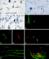
BEN expression during neuromuscular development in chick embryo. A, B, 25 somite stage embryo: labeling of serial transverse sections at the brachial level with BEN probe (A) and 4H5 antibody (B). BEN is detected in motoneurons (m) and dermamyotome (d).C, D, E5 stage: whole-mount in situ hybridization of the forelimb bud with BEN probe (C) and 4H5 antibody staining on a transverse section at the level of the ventral muscle primordia (D). Dorsal and ventral muscle masses express BEN mRNA and protein; motor nerve trunks that begin to invade the bud are also stained with BEN antibody (D,arrow). EH, E7 stage:in situ hybridization with BEN probe on a transverse section at the wing level (E; t, triceps muscle and d, deltoid muscle). Simultaneous staining with 4H5 antibody (F, G) and TRITC-αBGT (F, H) on the same transverse (G, H) or longitudinal (F) sections from triceps muscle. BEN mRNA and protein appear restricted to a subset of fibers within a muscle. Intramuscular nerve terminals express BEN protein, and the first signs of colocalization with AChR clusters are observed (FH, arrows).IK, E18 stage: in situhybridization with BEN probe (I) and simultaneous detection of 4H5 antibody (J) and TRITC-αBGT (K) on the same longitudinal section from PLD wing muscle. Myotubes appear nearly devoid of BEN messenger, whereas BEN protein and AChR clusters perfectly overlap at neuromuscular junctions. Scale bars: C, 400 μm;A, B, E, 100 μm;D, GK, 60 μm;F, 40 μm.
Fig. 2.
Fig. 2.
BEN expression during in vitromyogenesis. AE, In vitromyogenesis without neurons. A, C,D, Muscle cells cultivated for 24 hr: in situ hybridization with BEN probe (A), simultaneous labeling with 4H5 (C), and fast-myosin antibodies (D) in the same culture. Proliferating and postmitotic myoblasts express both BEN mRNA and protein. B, E, Muscle cells cultivated for 6 d: in situ hybridization with BEN probe (B); 4H5 antibody staining (E). Numerous myotubes are differentiated, expressing both BEN mRNA and protein.FI, In vitro myogenesis with neurons. FH, Myoblasts and neurons cocultivated for 3 d: in situ hybridization with BEN probe (F) and simultaneous labeling with 4H5 antibody (G) and TRITC-αBGT (H) in the same culture. BEN mRNA is accumulated in cell bodies of motoneurons (F,thin arrow), whose axons (FH, arrowheads) have invaded myogenic cultures and express BEN protein (G). Conversely, both mRNA (F) and protein (G) are downregulated in muscle fibers (F, H,large arrows) in which clustering of AChR has not yet occurred at this time (H).I, Simultaneous staining with 4H5 antibody and TRITC-αBGT in myotubes and neurons cocultivated for 6 d. BEN-positive axons begin contacting AChR clusters (arrowhead; m, myotube). Scale bars: FH, 125 μm; A, B, 60 μm;E, 45 μm; C, D, 30 μm;I, 10 μm.
Fig. 3.
Fig. 3.
BEN expression in embryonic muscles after neural tube ablation. In situ hybridization with BEN probe on transverse sections from the brachial level of E8 control (A) and neural tube-ablated (B,C) embryos. At this stage, BEN mRNA is nearly repressed in most fibers of axial muscles in control (A,arrows), whereas it is expressed in all fibers of axial muscles from the same level in neural tube-ablated embryos of the same age (B, arrowheads). Aneural muscles in the forelimb also exhibit an homogeneous expression of BEN mRNA in all muscle cells (C; t, triceps). Scale bars:A, B, 200 μm; C, 80 μm.
Fig. 4.
Fig. 4.
Effects of muscle denervation and reinnervation on BEN expression during neuromuscular postnatal development.AC, Longitudinal sections of PLD muscle from a 14-d-old chicken in which the common LD nerve has been severed 10 d before: in situ hybridization with BEN probe (A) and simultaneous staining of a serial section with 4H5 antibody (B) and TRITC-αBGT (C). Denervation induces an accumulation of BEN mRNA and protein at the surface of muscle fibers, which is synchronous of AChR cluster dispersal. DG, Longitudinal sections of PLD muscle from a 20-d-old chicken in which the common LD nerve has been severed 16 d before and allowed to regenerate and reinnervate the muscle: in situhybridization with BEN probe (D) and simultaneous staining of a serial section with 4H5 antibody (E) and TRITC-αBGT (F). Progressive reinnervation of denervated muscle leads to a downregulation of BEN mRNA and a decrease of the protein at the membrane of muscle fibers (E, arrowhead). Conversely, BEN protein is reexpressed in regenerating axons (E,arrow). At this time, clustering of AChRs in reinnervated muscle has not yet occurred (F). Simultaneous staining with 4H5 antibody and TRITC-αBGT of a longitudinal section from a reinnervated muscle in a 25-d-old chicken (G). BEN-positive regenerating nerve terminals progressively contact AChR clusters (G,arrows). H, Biochemical characterization of BEN by immunoblotting with 4H5 antibody. Lane 1, Normal ALD muscle; lane 2, denervated ALD muscle;lane 3, normal PLD muscle; lane 4, denervated PLD muscle; lane 6, purified protein from bursal epithelia; lane 7, purified protein from embryonic spinal cord. Immunoblots evidence a muscle isoform (lanes 1-4) distinct from the neural purified protein (lane 7) but similar to that expressed in the bursal epithelia (lane 6). This isoform is strongly accumulated in denervated muscles (comparelanes 1 and 3 with lanes 2and 4). Scale bars: E,F, 125 μm; A, D, 60 μm; B, C, 45 μm; G, 30 μm.

Similar articles

Cited by

References

    1. Anderson MJ, Cohen MW. Nerve-induced and spontaneous redistribution of acetylcholine receptors on cultured muscle cells. J Physiol (Lond) 1977;268:757–773. - PMC - PubMed
    1. Auda-Boucher G, Jarno V, Fournier-Thibault C, Butler-Browne G, Fontaine-Perus J. Acetylcholine receptor formation in mouse chick chimera. Exp Cell Res. 1997;236:29–42. - PubMed
    1. Bowen MA, Patel DD, Li X, Modrell AR, Malacko AR, Wang WC, Marquardt H, Neubauer M, Pesando JM, Francke U. Cloning, mapping and characterization of activated leucocyte-cell adhesion molecule (ALCAM), a CD6 ligand. J Exp Med. 1995;181:2213–2220. - PMC - PubMed
    1. Bowen MA, Bajorath J, D’Egidio M, Whitney GS, Palmer D, Kobarg J, Starling GC, Siadak AW, Aruffo A. Characterization of mouse ALCAM(CD166): the CD6-binding domain is conserved in different homologs and mediates cross-species binding. Eur J Immunol. 1997;27:1469–1478. - PubMed
    1. Bruses JL, Oka S, Rutishauser U. NCAM-associated polysialic acid on ciliary ganglion neurons is regulated by polysialyltransferase levels and interaction with muscle. J Neurosci. 1995;15:8310–8319. - PMC - PubMed

Publication types

Substances

LinkOut - more resources