. 1999 Feb 15;19(4):1424-36.
doi: 10.1523/JNEUROSCI.19-04-01424.1999.

Selective inhibition of kindling development by intraventricular administration of TrkB receptor body

Affiliations

Selective inhibition of kindling development by intraventricular administration of TrkB receptor body

D K Binder et al. J Neurosci. .

Abstract

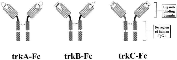
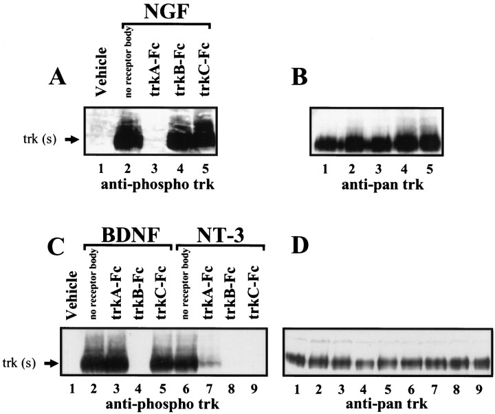
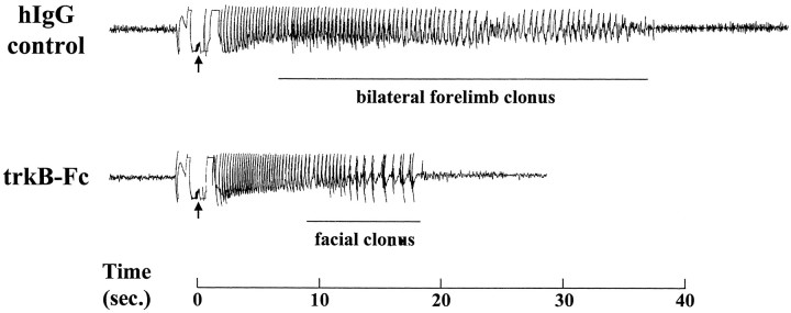
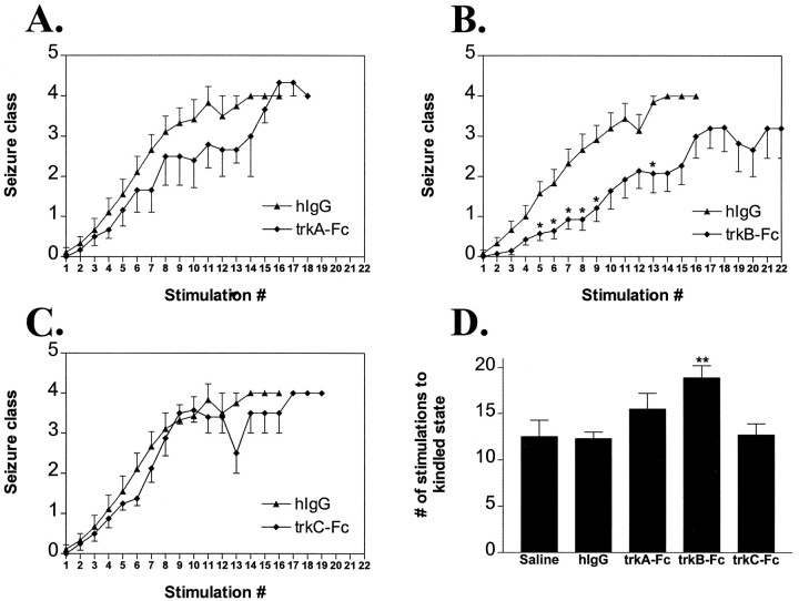
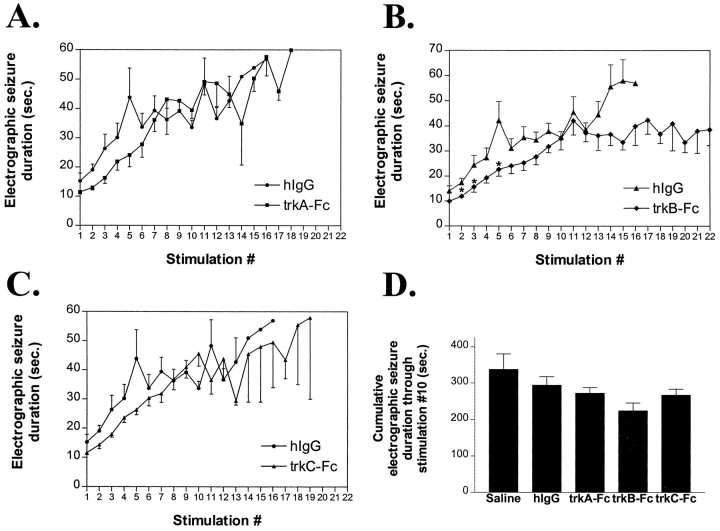
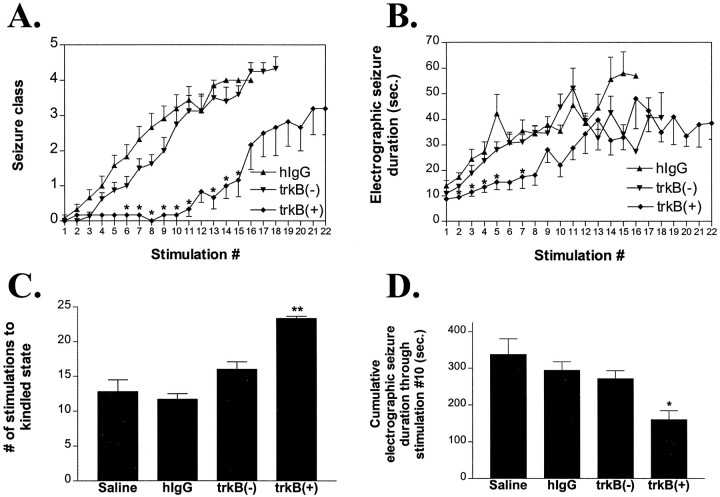
Recent work has shown that neurotrophin gene expression is increased after seizures evoked in the kindling model of epilepsy, but whether neurotrophins regulate kindling development is as yet unclear. In this study, we attempted to block selectively the activation of distinct neurotrophin receptors throughout kindling development in the rat via chronic intracerebroventricular administration of trk receptor bodies. The efficacy and selectivity of the trk receptor bodies were established by inhibition of neurotrophin-induced trk receptor phosphorylation in pheochromocytoma (PC12) cells and primary cultures of cortical neurons. The intracerebroventricular infusion of trkB receptor body (trkB-Fc) inhibited development of kindling in comparison with that seen with saline or human IgG controls, trkA-Fc, or trkC-Fc. These results imply that activation of trkB receptors contributes to the development of kindling, a form of activity-dependent behavioral plasticity in the adult mammalian brain.


Figures

Fig. 1.
Fig. 1.
Structure of trk receptor bodies. The structure is a divalent homodimer containing two trk (trkA,trkB, or trkC) extracellular ligand-binding domains linked via hinge regions to the humanIgG1 Fc region. The putative function is to compete with native trk receptors for endogenous ligand(s) to sequester ligand(s) and prevent trk receptor activation.
Fig. 2.
Fig. 2.
Efficacy and specificity of trk receptor bodies.A, Western blot using anti-phospho trk antibody performed on PC12 cell extracts from vehicle-treated cultures (lane 1) or cultures treated with NGF(lanes 2–5) in the presence or absence of the indicated trk receptor bodies. B, Blot shown in Astripped and reprobed with anti-pan trk antibody that labels all trk proteins regardless of phosphorylation state. C, Western blot performed on cortical cell extracts from vehicle-treated cultures (lane 1) or cultures treated with BDNF(lanes 2–5) or NT-3 (lanes 6–9) in the presence or absence of the indicated trk receptor bodies. D, Blot shown in C stripped and reprobed with the anti-pan trk antibody.
Fig. 3.
Fig. 3.
Sample electroencephalograms from seizures inhIgG versus trkB–Fc groups. The 12th stimulation for each animal is shown. Each arrow marks a stimulation artifact, and the time base is shown at thebottom. Whereas the hIgG-treated animal had a 36 sec seizure discharge with a clonic motor component of 30 sec (class 4 seizure), the trkB–Fc-treated animal had only a 19 sec seizure discharge with facial clonus (class 1 seizure). Such a mild seizure on the 12th stimulation was never seen in the control animals.
Fig. 4.
Fig. 4.
Effects of trk receptor bodies on behavioral seizure development. A–C, Behavioral seizure class (mean ± SEM) as a function of stimulation number forhIgG (n = 12),trkA–Fc (A; n = 9),trkB–Fc (B; n = 14), and trkC–Fc (C; n = 9) groups. Single asterisks in B refer top < 0.05 by nonparametric one-way ANOVA withpost hoc Dunn’s test. D, Number of stimulations to kindled state (three consecutive clonic motor seizures; mean ± SEM) by treatment group. All groups (hIgG, trkA–Fc, trkB–Fc, and trkC–Fc) except saline (n = 8) refer to a dose of 50 μg/d. Double asterisks inD refer to p < 0.01 fortrkB–Fc versus hIgG by one-way ANOVA with post hoc Bonferroni’s test.
Fig. 5.
Fig. 5.
Effects of trk receptor bodies on electrographic seizure duration. A–C, Electrographic seizure duration (in seconds; mean ± SEM) as a function of stimulation number forhIgG, trkA–Fc (A),trkB–Fc (B), andtrkC–Fc (C) groups. Single asterisks in B refer to p < 0.05 by nonparametric one-way ANOVA with post hocDunn’s test. D, Cumulative electrographic seizure duration (mean ± SEM) via kindling stimulation 10 by treatment group. There was no significant difference between groups in cumulative electrographic seizure duration (one-way ANOVA, p> 0.05).
Fig. 6.
Fig. 6.
Immunohistochemistry for anti-hIgG Fcγ. A, Portion of Nissl-stained horizontal section at approximate level of B. Selected structures are labeled. B, Horizontal section from an animal infused with hIgG showing the typical distribution of immunoreactivity (right side, infusion site). In this animal, note the immunoreactivity in the septum bilaterally, the corpus callosum, the right striatum, the righthippocampus, and the fimbria and light immunoreactivity in theleft hippocampus and the fimbria. Arrowsmark the immunoreactivity in pia surrounding the brain.
Fig. 7.
Fig. 7.
Presence of Fc immunoreactivity in the hippocampus correlates with the effect of trkB–Fc on kindling development.A, Right hippocampal section of a trkB–Fc-treated animal with marked inhibition of kindling development (did not reach the kindled state in 22 stimulations). Ependymal immunoreactivity is visible (arrow) lining the right lateral ventricle (asterisk), and significant hippocampal parenchymal immunoreactivity is evident. B, Right hippocampal section of the trkB–Fc-treated animal in which there was the least effect on kindling development (12 stimulations required to reach the kindled state). Ependymal immunoreactivity is visible (arrow) lining the right lateral ventricle (asterisk), but no hippocampal parenchymal immunoreactivity is evident. Scale bar: A,B, 200 μm.
Fig. 8.
Fig. 8.
Anatomic distribution of Fc immunoreactivity versus the effect on kindling development. A–D, Relative hippocampal immunoreactivity (left) or relative striatal immunoreactivity (right) versus the number of stimulations to the kindled state for each animal intrkA–Fc (A),trkB–Fc (B),trkC–Fc (C), andhIgG (D) groups. Spearman rank correlation is significant only for trkB–Fc hippocampal immunoreactivity versus kindling effect (B,left; p < 0.01) and not for trkB–Fc striatal immunoreactivity versus kindling effect (B, right; p > 0.05) or for hippocampal or striatal immunoreactivity versus kindling effect in any other group (all p > 0.05).
Fig. 9.
Fig. 9.
Effect of hippocampal immunoreactivity on kindling parameters within the trkB–Fc group when the original trkB–Fc group (n = 14) is separated into intensely immunoreactive [trkB(+); n = 6] versus poorly immunoreactive [trkB(−); n = 8] animals. A, Behavioral seizure class (mean ± SEM) by stimulation number. Single asterisks refer top < 0.05 versus hIgG by nonparametric one-way ANOVA with post hoc Dunn’s test.B, Electrographic seizure duration (mean ± SEM) by stimulation number. C, Number of stimulations to the kindled state (mean ± SEM) by group. D, Cumulative electrographic seizure duration (mean ± SEM) via stimulation 10 by group. Single and double asterisks inB–D refer to p < 0.05 andp < 0.01, respectively, for trkB(+) versus hIgG by one-way ANOVA with post hoc Bonferroni’s test.

References

    1. Alcantara S, Frisen J, del Rio JA, Soriano E, Barbacid M, Silos-Santiago I. TrkB signalling is required for postnatal survival of CNS neurons and protects hippocampal and motor neurons from axotomy-induced cell death. J Neurosci. 1997;17:3623–3633. - PMC - PubMed
    1. Bengzon J, Soderstrom S, Kokaia Z, Kokaia M, Ernfors P, Persson H, Ebendal T, Lindvall O. Widespread increase of nerve growth factor protein in the rat forebrain after kindling-induced seizures. Brain Res. 1992;587:338–342. - PubMed
    1. Bengzon J, Kokaia Z, Ernfors P, Kokaia M, Leanza G, Nilsson OG, Persson H, Lindvall O. Regulation of neurotrophin and trkA, trkB and trkC tyrosine kinase receptor messenger RNA expression in kindling. Neuroscience. 1993;53:433–446. - PubMed
    1. Blochl A, Thoenen H. Characterization of nerve growth factor (NGF) release from hippocampal neurons: evidence for a constitutive and an unconventional sodium-dependent regulated pathway. Eur J Neurosci. 1995;7:1220–1228. - PubMed
    1. Bugra K, Pollard H, Charton G, Moreau J, Ben-Ari Y, Khrestchatisky M. aFGF, bFGF and flg mRNAs show distinct patterns of induction in the hippocampus following kainate-induced seizures. Eur J Neurosci. 1994;6:58–66. - PubMed

Publication types

Substances

LinkOut - more resources