. 1999 Feb;119(2):385-97.
doi: 10.1104/pp.119.2.385.

Structure, properties, and tissue localization of apoplastic alpha-glucosidase in crucifers

Affiliations

Structure, properties, and tissue localization of apoplastic alpha-glucosidase in crucifers

J D Monroe et al. Plant Physiol. 1999 Feb.

Abstract

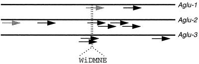
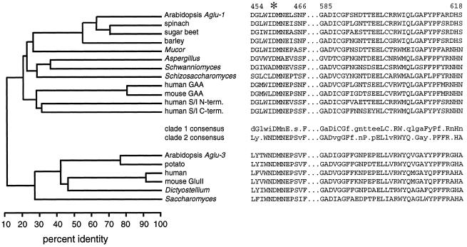
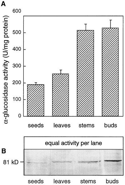
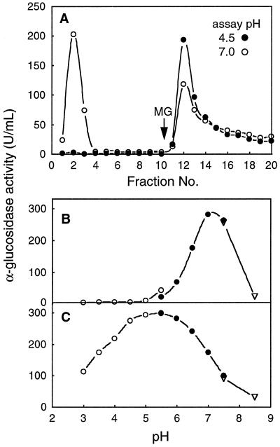
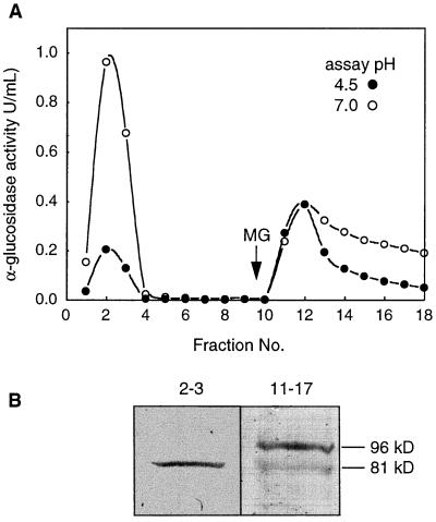
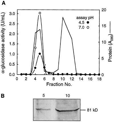
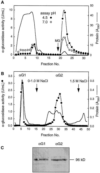
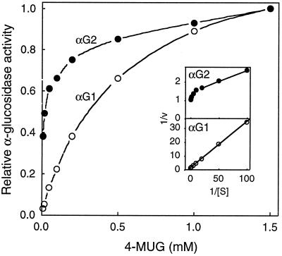
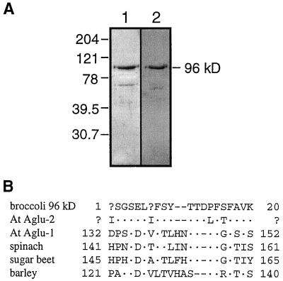
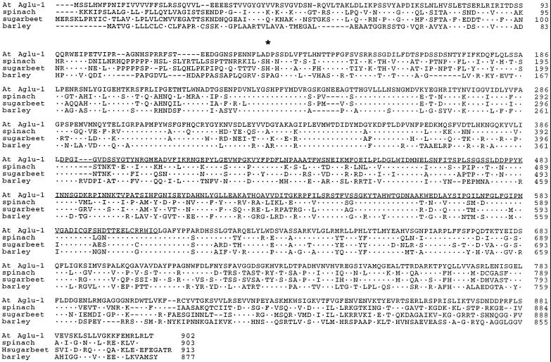
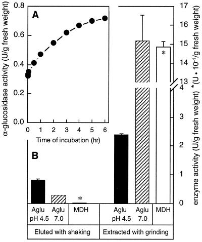
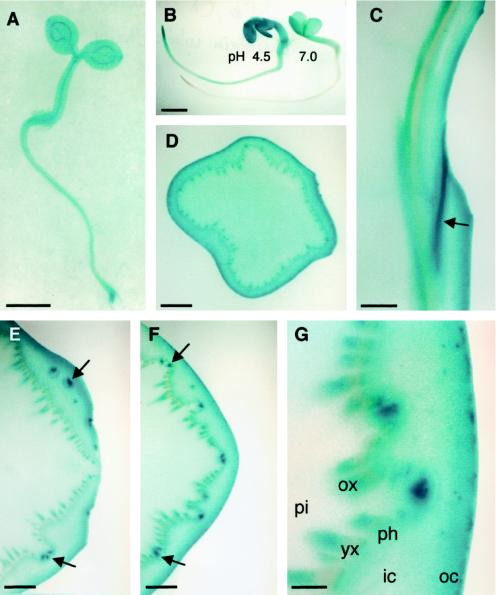
Apoplastic alpha-glucosidases occur widely in plants but their function is unknown because appropriate substrates in the apoplast have not been identified. Arabidopsis contains at least three alpha-glucosidase genes; Aglu-1 and Aglu-3 are sequenced and Aglu-2 is known from six expressed sequence tags. Antibodies raised to a portion of Aglu-1 expressed in Escherichia coli recognize two proteins of 96 and 81 kD, respectively, in vegetative tissues of Arabidopsis, broccoli (Brassica oleracea L.), and mustard (Brassica napus L.). The acidic alpha-glucosidase activity from broccoli flower buds was purified using concanavalin A and ion-exchange chromatography. Two active fractions were resolved and both contained a 96-kD immunoreactive polypeptide. The N-terminal sequence from the 96-kD broccoli alpha-glucosidase indicated that it corresponds to the Arabidopsis Aglu-2 gene and that approximately 15 kD of the predicted N terminus was cleaved. The 81-kD protein was more abundant than the 96-kD protein, but it was not active with 4-methylumbelliferyl-alpha-D-glucopyranoside as the substrate and it did not bind to concanavalin A. In situ activity staining using 5-bromo-4-chloro-3-indolyl-alpha-D-glucopyranoside revealed that the acidic alpha-glucosidase activity is predominantly located in the outer cortex of broccoli stems and in vascular tissue, especially in leaf traces.


Figures

Figure 1
Figure 1
Maps of the relative positions of 11 Arabidopsis α-glucosidase ESTs on the coding sequences of three α-glucosidase genes, Aglu-1, Aglu-2, and Aglu-3. Reading from left to right, ESTs (and accession numbers) from Aglu-1: 38A2T7 (T04333) and 151P15T7 (T76451); Aglu-2: H8A7T7 (W43892), G10B11T7 (N96165), 141B3T7 (T46694), 208 h21T7 (N37141), 169E22T7 (R64965), and 192B2T7 (R90271); Aglu-3: 47E11T7 (T14117), 35C12T7 (T04464), and OBO389 (ATTS5896). The relative position of the conserved active-site peptide WiDMNE is illustrated. ESTs used in this study are illustrated as shaded arrows; length of arrows corresponds roughly to the length of the known EST sequence.
Figure 2
Figure 2
Phylogenetic relationship among Family 31 α-glucosidases and aligned signature peptides indicate that there are two clades. Left, Phylogenetic tree indicating percent identity at the amino acid level using about 60% of each protein. Approximately 250 amino acids from the N termini and 150 amino acids from the C termini of each protein were omitted from this analysis. Right, Sequences of two signature peptide regions along with consensus sequences for each clade. Consensus amino acids are uppercase if perfectly conserved and lowercase if conserved in more than half of the sequences. The sequences (and accession numbers) used in the analysis from clade 1 were: Aglu-1 from Arabidopsis (AF014806), α-glucosidases from spinach (D86624), sugar beet (D89615), barley (U22450), Mucor javanicus (D67034), Aspergillus niger aglA (D45356), Schwanniomyces occidentalis glucoamylase GAM1 (M60207), Schizosaccharomyces pombe C30D11.01C (1723210), lysosomal acid α-glucosidase (GAA) from human (M34424) and mouse (P70699), and the N-terminal and C-terminal halves of the human sucrase/isomaltase (M22616). In clade 2 the sequences (and accession numbers) used were Aglu-3 from Arabidopsis (AB007646), cDNAs from potato (AJ001374) and human (AJ000332), GluII from mouse (U92793), ModA from Dictyostellium discoideum (U72236), and the Saccharomyces cerevisiae open reading frame YBR229c (Z36098).
Figure 3
Figure 3
α-Glucosidase activity and immunoblot of extracts from Arabidopsis tissues probed with anti-Aglu-1. A, α-Glucosidase activity in Arabidopsis tissues. Assays were conducted at pH 4.5 using 4-MUG as the substrate. Bud tissue included some stem tissue, flowers, flower buds, and young siliques. Data represent the means ± sd (n = 3). B, Detection of an 81-kD protein in Arabidopsis tissues with anti-Aglu-1 serum. Equal amounts of α-glucosidase activity (8 units) from the tissues represented in A were separated by SDS-PAGE and probed with anti-Aglu-1 serum. U, Unit.
Figure 4
Figure 4
Separation of neutral and acidic α-glucosidases from an Arabidopsis leaf extract using a ConA-Sepharose column, and the pH profile of each activity peak. A, ConA-Sepharose column. Bound proteins were eluted with 15 mm methyl Glc (MG). α-Glucosidase activity was measured at pH 4.5 (•) and pH 7.0 (○). B, pH profile of pooled fractions 2 and 3 from A. C, pH profile of pooled fractions 12 to 14 from A. For B and C, buffers were sodium acetate (○), Mes (•), or Hepes (▿). U, Unit.
Figure 5
Figure 5
Separation of α-glucosidases from an Arabidopsis stem+bud extract by ConA chromatography and an immunoblot of the pooled active fractions probed with anti-Aglu-1. A, ConA-Sepharose column. Bound proteins were eluted with 15 mm methyl Glc (MG). α-Glucosidase activity was measured at pH 4.5 (•) and pH 7.0 (○). U, Unit. B, Immunoblot of activity peaks separated in A. Nonbinding activity (fractions 2 and 3) and bound activity (fractions 11–17) from A was concentrated 24-, and 215-fold, respectively, and equal volumes of each concentrated fraction were separated by SDS-PAGE and probed with anti-Aglu-1.
Figure 6
Figure 6
Separation of the Arabidopsis α-glucosidase activity from most of the 81-kD polypeptide using Sephadex G-150. A, Sephadex G-150 column. α-Glucosidase activity was assayed at pH 4.5 (•) and pH 7.0 (○). Total protein was monitored at A280 (solid line). U, Unit. B, Immunoblot of fractions 5 and 10 from A. Equal volumes (10 μL) of each fraction were separated by SDS-PAGE and probed with anti-Aglu-1.
Figure 7
Figure 7
Isolation of the major acidic α-glucosidases from broccoli flower buds using ConA- and CM-cellulose chromatography and an immunoblot of pooled active fractions probed with anti-Aglu-1. A, ConA-Sepharose column. Bound proteins were eluted with 15 mm methyl Glc (MG). α-Glucosidase activity was assayed at pH 4.5 (•) and pH 7.0 (○). Total protein was monitored at A280 (solid line) in A and B. B, Separation of fractions 21 to 24 from A on a CM-cellulose column. α-Glucosidase activity was assayed at pH 4.5 (•). C, Immunoblot of activity peaks separated in B. αG1 (fractions 5–6) and αG2 (fractions 28–32) from B were pooled and concentrated. Equal amounts of activity at pH 4.5 (37 units) were separated by SDS-PAGE and probed with anti-Aglu-1. U, Unit.
Figure 8
Figure 8
Effect of 4-MUG on the activity of two broccoli α-glucosidase isoforms separated by CM-cellulose chromatography. αG1 (○) and αG2 (•) from the CM-cellulose column (Fig. 7B) were assayed at pH 4.5. Data were normalized to the level of activity at 1.5 mm 4-MUG. Inset, Lineweaver Burk plots of the same data.
Figure 9
Figure 9
Purified 96-kD broccoli α-glucosidase stained with Coomassie blue or probed with anti-Aglu-1 serum, and the N-terminal sequence from the same protein aligned with other plant α-glucosidase sequences. A, SDS-PAGE and immunoblot of the 96-kD broccoli α-glucosidase. Proteins (4 μg) were separated by SDS-PAGE and either stained with Coomassie blue (lane 1) or probed with anti-Aglu-1 serum (lane 2). The numbers on the left represent molecular mass standards in kilodaltons. B, N-terminal sequence from the broccoli 96-kD α-glucosidase aligned with other plant α-glucosidases. Sequences (and accession numbers) were from Arabidopsis Aglu-1 (AF014806), Arabidopsis Aglu-2 (sequence from the EST H8A7T7), spinach (D86624), sugar beet (D89615), and barley (U22450).
Figure 10
Figure 10
Alignment of the known plant α-glucosidase sequences. Sequences are from Arabidopsis Aglu-1, spinach, sugar beet, and barley. For accession numbers, see legend for Figure 9. Dots represent amino acids that are identical to the Arabidopsis Aglu-1 sequence. The N terminus of the broccoli 96-kD protein is indicated with an asterisk. The 24-kD Aglu-1 polypeptide expressed in E. coli for antibody production is underlined.
Figure 11
Figure 11
Localization of acidic α-glucosidase activity in the apoplastic fraction of mustard seedlings. A, Time course of elution of α-glucosidase activity from intact mustard seedlings. Three-day-old mustard seedlings grown under water were shaken in extraction buffer containing 1 m NaCl for 6 h. α-Glucosidase in the medium was assayed periodically with 4-MUG at pH 4.5. B, Activity of α-glucosidase (Aglu) at either pH 4.5 or 7.0 and malate dehydrogenase (MDH) in the elution buffer from A after 6 h, and in extracts from similar but untreated plants. Data represent the means ± sd (n = 3). U, Unit.
Figure 12
Figure 12
In situ activity of α-glucosidase assayed with α-X-gluc at pH 4.5 for 12 h, except where noted. A, Three-day-old Arabidopsis seedling. B, Three-day-old mustard seedlings stained at pH 4.5 (left) or 7.0 (right) for 3 h. C, Longitudinal section through a broccoli stem. The curved surface on the right side is a leaf abscission zone. Unstained tissue on the left is pith. Strong staining is seen in the leaf trace indicated by the arrow. D, Cross-section of a broccoli stem approximately 3 cm from the apex. E, Cross-section of a broccoli stem through a leaf abscission zone. Arrows point to strong staining in leaf traces at or above the point where they separate from the vascular cylinder. A leaf abscission zone is on the right side of the image. F, Same as E but approximately 4 mm basal. Arrows point to the same leaf traces as in E, but both leaf traces are below the point of separation from the vascular cylinder. G, Finer detail of F. pi, Pith; ox, older xylem; yx, young xylem; ph, phloem; ic, inner cortex; oc, outer cortex. Note the increased staining toward the outer cortex and in the older xylem and in the leaf traces. Bars = 1 mm (A, B, and D); 2 mm (C, E, and F); or 0.5 mm G.

Similar articles

Cited by

References

    1. Arendt CW, Ostergaard HL. Identification of the CD45-associated 116-kDa and 80-kDa proteins as the alpha- and beta-subunits of alpha-glucosidase II. J Biol Chem. 1997;272:13117–13125. - PubMed
    1. Beckerman JL, Ebbole DJ. MPG1, a gene encoding a fungal hydrophobin of Magnaporthe grisea, is involved in surface recognition. Mol Plant Microbe Interact. 1996;9:450–456. - PubMed
    1. Beers EP, Duke SH, Henson CA. Partial characterization and subcellular localization of three α-glucosidase isoforms in pea (Pisum sativumL.) seedlings. Plant Physiol. 1990;94:738–744. - PMC - PubMed
    1. Chantret I, Lacasa M, Chevalier G, Ruf J, Islam I, Mantei N, Edwards Y, Swallow D, Rousset M. Sequence of the complete cDNA and the 5′ structure of the human sucrase-isomaltase gene: possible homology with a yeast glucoamylase. Biochem J. 1992;285:915–923. - PMC - PubMed
    1. Chiba S, Inomata S, Matsui H, Shimomura T. Purification and properties of an α-glucosidase (glucoamylase) in sugar beet seed. Agric Biol Chem. 1978;42:241–245.

Publication types

MeSH terms

Substances

LinkOut - more resources