. 1999 Feb;119(2):435-44.
doi: 10.1104/pp.119.2.435.

Change in apoplastic aluminum during the initial growth response to aluminum by roots of a tolerant maize variety

Affiliations

Change in apoplastic aluminum during the initial growth response to aluminum by roots of a tolerant maize variety

MD Vazquez et al. Plant Physiol. 1999 Feb.

Abstract

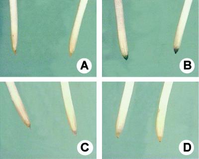
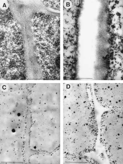
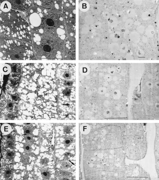
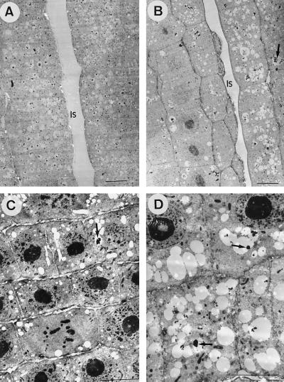
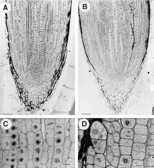
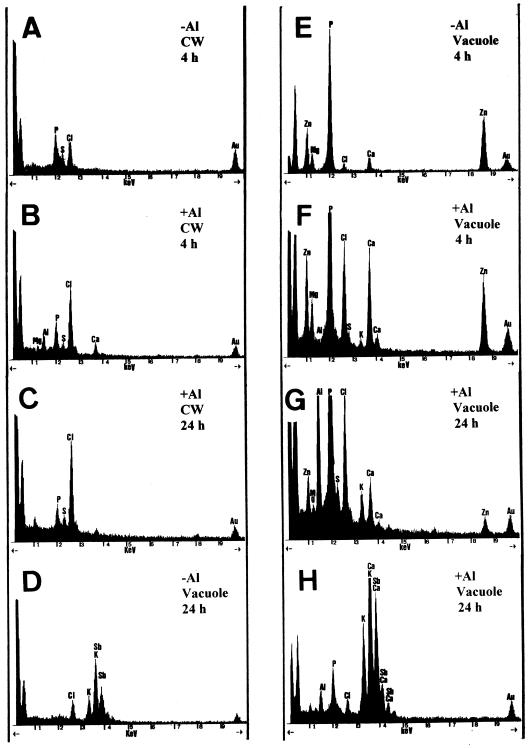
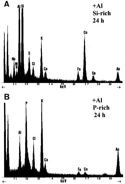
Root elongation, hematoxylin staining, and changes in the ultrastructure of root-tip cells of an Al-tolerant maize variety (Zea mays L. C 525 M) exposed to nutrient solutions with 20 &mgr;M Al (2.1 &mgr;M Al3+ activity) for 0, 4, and 24 h were investigated in relation to the subcellular distribution of Al using scanning transmission electron microscopy and energy-dispersive x-ray microanalysis on samples fixed by different methods. Inhibition of root-elongation rates, hematoxylin staining, cell wall thickening, and disturbance of the distribution of pyroantimoniate-stainable cations, mainly Ca, was observed only after 4 and not after 24 h of exposure to Al. The occurrence of these transient, toxic Al effects on root elongation and in cell walls was accompanied by the presence of solid Al-P deposits in the walls. Whereas no Al was detectable in cell walls after 24 h, an increase of vacuolar Al was observed after 4 h of exposure. After 24 h, a higher amount of electron-dense deposits containing Al and P or Si was observed in the vacuoles. These results indicate that in this tropical maize variety, tolerance mechanisms that cause a change in apoplastic Al must be active. Our data support the hypothesis that in Al-tolerant plants, Al can rapidly cross the plasma membrane; these data clearly contradict the former conclusions that Al mainly accumulates in the apoplast and enters the symplast only after severe cell damage has occurred.


Figures

Figure 1
Figure 1
Seminal roots from maize plants stained with hematoxylin. A and C, Control (0 μm Al) plants after 4 and 24 h, respectively. B and D, Plants treated with 20 μm Al containing nutrient solution for 4 and 24 h, respectively.
Figure 2
Figure 2
TEM images from longitudinal sections of root tips of maize plants exposed for 4 h to control (0 μm Al) (A and C) or 20 μm Al (B and D) in nutrient solution. A and B, Non-osmified, glutaraldehyde-fixed samples. C and D, PA-stained samples. Note thickening of cell walls (B) and higher amount of PA precipitates at the internal site of cell walls (D) in samples from Al-treated plants. Scale bars represent 0.5 μm in A and B, and 1.0 μm in C and D.
Figure 3
Figure 3
TEM images from longitudinal sections of root tips of maize plants exposed for 0 (A and B) or 4 h (C–F) to control (0 μm Al) (A–D) or 20 μm Al (E and F) containing nutrient solution. A, C, and E, Glutaraldehyde-fixed samples. B, D, and F, PA-stained samples. Note the abundance of electron-translucent vacuoles with only some peripheric electron-dense deposits (C and E, arrows) in conventionally fixed samples and the abundance of electron-dense precipitates in the central part of vacuoles from PA-stained samples (B, D, and F). All scale bars represent 10 μm, except B, where bar is 1 μm.
Figure 4
Figure 4
TEM images from longitudinal sections of root tips of maize plants exposed for 24 h to control (0 μm Al) (A and C) or 20 μm Al in nutrient solution (B and D). A and B, PA-stained samples. C and D, Non-osmified, glutaraldehyde-fixed samples. Note the abundance of electron-dense vacuolar deposits (arrows) in conventionally fixed samples from plants exposed to Al (D) in comparison with the scarce presence of deposits in controls (C). All scale bars represent 10 μm. Is, Intercellular space.
Figure 5
Figure 5
Light-microscopy images from longitudinal sections (PA stained) of root tips of maize plants exposed for 24 h to control (0 μm Al) (A and C) or 20 μm Al in nutrient solution (B and D). Controls (A and C) show well-organized cortical cell lines with almost horizontal cell-division planes. Al-exposed plants (B and D) exhibit irregular cell-division planes in internal cortical cells. A and B, Scale bars represent 100 μm; C and D, scale bars represent 10 μm.
Figure 6
Figure 6
Representative EDXMA spectra from electron-dense deposits in cell walls (A–C) and vacuoles (D–H) of root-tip (0–1.5 mm) cells after 4-h (A, B, E, and F) and 24-h treatments (C, D, G, and H). A, D, and E are from control plants and B, C, F, G, and H are from Al-treated plants. All spectra are from glutaraldehyde-fixed samples, except D and H, which are from PA-stained samples. All spectra are printed at 1000 counts. Au is from the grid.
Figure 7
Figure 7
Representative EDXMA spectra from electron-dense deposits in the vacuoles of root-tip cortex cells (3 mm) of plants exposed to Al for 24 h. A, Al-containing deposit with high Si content. B, Al-containing deposit with P, but without Si. All samples were prepared by freeze-substitution. All spectra are printed at 1000 counts; Fe and Co are instrument contaminants; and Au is from the grid.

References

    1. Barceló J, Guevara P, Poschenrieder CH. Silicon amelioration of aluminium toxicity in teosinte (Zea mays L. ssp. Mexicana) Plant Soil. 1993;54:249–255.
    1. Calba H, Jaillard B. Effect of aluminium on ion uptake and H+ release by maize. New Phytol. 1997;137:607–616.
    1. Corrales I, Poschenrieder CH, Barceló J. Influence of silicon pretreatment on aluminium toxicity in maize roots. Plant Soil. 1997;190:203–209.
    1. Delhaize E, Craig S, Beaton CD, Bennet RJ, Jagadish VC, Randall PJ. Aluminum tolerance in wheat (Triticum aestivum L.). I. Uptake and distribution of aluminum in root apices. Plant Physiol. 1993;103:685–693. - PMC - PubMed
    1. Demarty M, Morvan C, Thellier M. Calcium and the cell wall. Plant Cell Environ. 1984;7:449–456.

LinkOut - more resources