Comparative Study
. 1999 Feb;119(2):635-44.
doi: 10.1104/pp.119.2.635.

Molecular cloning and expression analysis of the mitochondrial pyruvate dehydrogenase from maize

Affiliations
Comparative Study

Molecular cloning and expression analysis of the mitochondrial pyruvate dehydrogenase from maize

J J Thelen et al. Plant Physiol. 1999 Feb.

Abstract

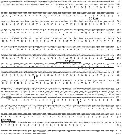
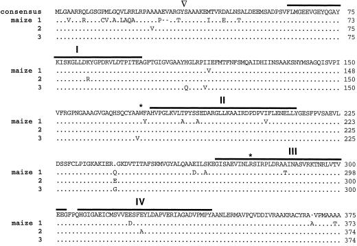
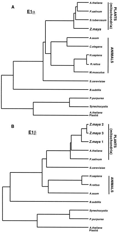
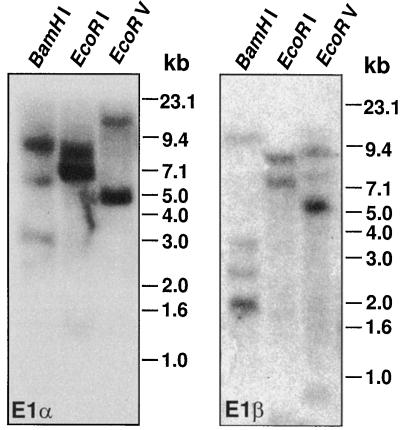
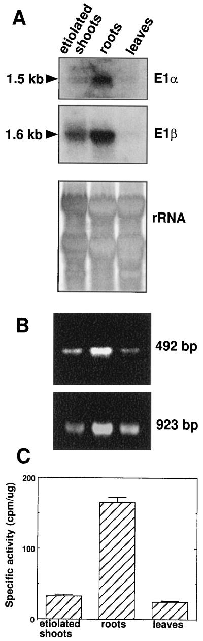
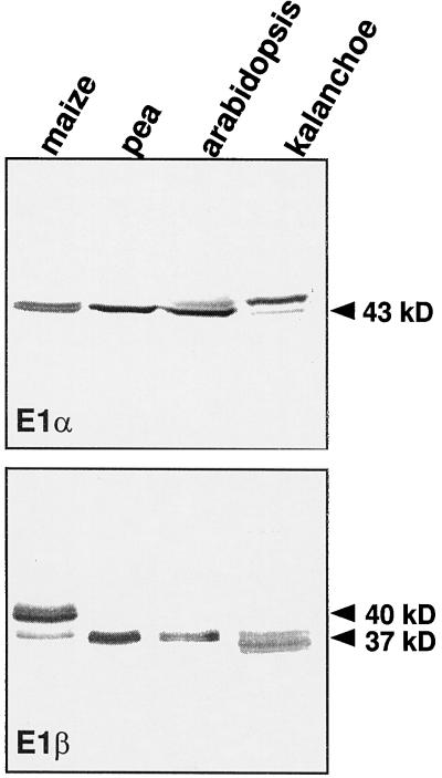
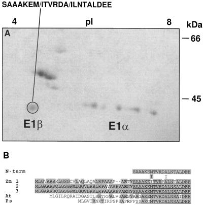
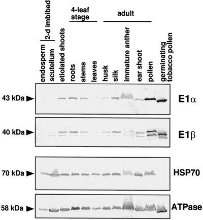
Four cDNAs, one encoding an alpha-subunit and three encoding beta-subunits of the mitochondrial pyruvate dehydrogenase, were isolated from maize (Zea mays L.) libraries. The deduced amino acid sequences of both alpha- and beta-subunits are approximately 80% identical with Arabidopsis and pea (Pisum sativum L.) homologs. The mature N terminus was determined for the beta-subunit by microsequencing the protein purified from etiolated maize shoot mitochondria and was resolved by two-dimensional gel electrophoresis. This single isoelectric species comprised multiple isoforms. Both alpha- and beta-subunits are encoded by multigene families in maize, as determined by Southern-blot analyses. RNA transcripts for both alpha- and beta-subunits were more abundant in roots than in young leaves or etiolated shoots. Pyruvate dehydrogenase activity was also higher in roots (5-fold) compared with etiolated shoots and leaves. Both subunits were present at similar levels in all tissues examined, indicating coordinated gene regulation. The protein levels were highest in heterotrophic organs and in pollen, which contained about 2-fold more protein than any other organ examined. The relative abundance of these proteins in nonphotosynthetic tissues may reflect a high cellular content of mitochondria, a high level of respiratory activity, or an extra plastidial requirement for acetate.


Figures

Figure 2
Figure 2
Nucleotide and deduced amino acid sequence for maize E1α cDNA. Amino acids are denoted by the single-letter abbreviations. The stop codon is indicated by a period. Underlined region indicates the putative TPP-binding domain. The targeting peptide-processing site, indicated by the triangle, is putative and based on characteristics of cleavage sites (von Heijne et al., 1989). The putative intron splice site is shaded and the polyadenylation signal is underlined. Oligonucleotides used as primers for RT-PCR in Figure 6B are overlined. Numbers to the right indicate base pairs or amino acid number. Asterisks and • denote residues referred to in the text.
Figure 1
Figure 1
Amino acid sequence comparison for maize E1β isoforms. Consensus sequence is noted at the top. Dots indicate identity. Gaps, indicated by dashes, were inserted to maximize homology. Overlines indicate the four conserved domains as first pointed out by Wexler et al. (1991). Asterisks denote residues referred to in text. The targeting peptide-processing site is indicated by the inverted triangle. GeneWorks (IntelliGenetics) software was used to perform the alignment algorithm.
Figure 3
Figure 3
Dendrogram analysis of E1α (A) and β (B) subunits. Clustal alignments were performed using the GeneWorks software package (IntelliGenetics). The length of the horizontal lines indicates inverse degree of relatedness. Accession numbers to the sequences are: Porphyra purpurea (U38804); Solanum tuberosum (Z26949); Synechocystis sp. (D90915); Arabidopsis (U21214, U09137); Mus musculus (M76727); Arabidopsis plastid (U80185, U80186); Caenorhabditis elegans (Z47812); P. sativum (U51918, U56697); Homo sapiens (L13318, D90086); Rattus rattus (Z12158, P49432); Saccharomyces cerevisiae (P16387, M98476); Ascaris suum (M76554, M38017); B. subtilis (M31542).
Figure 4
Figure 4
Genomic Southern analysis of maize E1α (left) and E1β (right). Genomic DNA isolated from maize leaves was digested with the indicated restriction enzymes. Approximately 30 μg of DNA was fractionated by electrophoresis on an agarose gel, transferred to a Nytran membrane, and probed with random-prime-labeled DNA. Marker sizes are indicated to the right in kilobases.
Figure 5
Figure 5
RNA, RT-PCR, and activity analysis of PDH from maize tissues. A, Approximately 30 μg of total RNA isolated from dark-grown seedlings, roots, or light-adapted leaves was fractionated on an agarose formaldehyde gel, transferred to a Nytran membrane, and probed with the entire E1α or E1β cDNA. RNA marker sizes are indicated. As a gel-loading control, rRNA is included for comparison in the bottom panel. B, Oligonucleotides specific for the E1α cDNA were used for RT-PCR analysis with maize RNA isolated from dark-grown seedlings, roots, or light-adapted leaves as the template. The top panel displays the product obtained with primers DDR212 and DDR229. The product in the bottom panel was obtained with primers DDR206 and DDR229 (Fig. 2). C, In vivo PDH specific activity was determined from maize organs (dark-grown seedlings, roots, or light-adapted leaves) using a radioisotopic assay. Values are the means of five independent reactions. Error bars indicate sd.
Figure 6
Figure 6
Immunoblot analysis of mitochondria purified from various plants. Approximately 10 μg of purified mitochondrial protein was loaded in each lane. Maize mitochondria were obtained from etiolated shoots, whereas pea, Arabidopsis, and K. daigremontianum mitochondria were isolated from light-grown leaves. Molecular masses of polypeptides are indicated.
Figure 7
Figure 7
N-terminal microsequencing of PDH subunits. A, Coomassie-blue-stained two-dimensional gel electrophoresis of highly purified maize mitochondrial PDC from etiolated shoots. The pI is indicated at the top and the size in kilodaltons is indicated to the right. The circled polypeptide was microsequenced from a replica-blot transferred to a PVDF membrane. At cycles 7 and 12 two residues were obtained (indicated by the slash). B, Comparison of the deduced amino acid sequence for the plant E1β subunits. Zm, Z. mays; At, Arabidopsis; Ps, P. sativum. Shading indicates amino acid identity. Gaps denoted by dashes were inserted to maximize homology. The N termini of the mature maize and pea polypeptides are underlined. Conserved Arg residues involved with peptide processing are indicated in bold type.
Figure 8
Figure 8
Immunoblot analyses showing the expression of PDH subunits from various organs. Maize kernels were allowed to soak for 2 d prior to isolating the endosperm and scutellum. Etiolated shoots were grown for 5 d in complete darkness. The remaining samples were obtained from light-grown plants, grown in either a growth chamber (four-leaf stage) or a greenhouse (adult). As a control, the blots were reprobed with antibodies specific to other mitochondrial proteins, HSP70 and the β-subunit to ATP synthase. Approximately 25 μg of a total protein preparation was loaded per lane. Molecular masses of polypeptides are indicated on the left (in kilodaltons).

References

    1. Ali MS, Shenoy BC, Eswaran D, Andersson LA, Roche TE, Patel MS. Identification of the tryptophan residue in the thiamin pyrophosphate binding site of mammalian pyruvate dehydrogenase. J Biol Chem. 1995;270:4570–4574. - PubMed
    1. Brown J. A catalogue of splice junction and putative branch point sequences from plant introns. Nucleic Acids Res. 1986;14:9549–9559. - PMC - PubMed
    1. Budde RJA, Randall DD. Pea leaf mitochondrial pyruvate dehydrogenase complex is inactivated in vivo in a light-dependent manner. Proc Natl Acad Sci USA. 1990;87:673–676. - PMC - PubMed
    1. Camp PJ, Randall DD. Purification and characterization of the pea chloroplast pyruvate dehydrogenase complex. Plant Physiol. 1985;77:571–577. - PMC - PubMed
    1. Eswaran D, Ali MS, Shenoy BC, Korotchkina LG, Roche TE, Patel MS. Arginine-239 in the beta subunit is at or near the active site of bovine pyruvate dehydrogenase. Biochim Biophys Acta. 1995;1252:203–208. - PubMed

Publication types

MeSH terms

Associated data

LinkOut - more resources