. 1999 Feb 8;144(3):519-32.
doi: 10.1083/jcb.144.3.519.

delta-catenin, an adhesive junction-associated protein which promotes cell scattering

Affiliations

delta-catenin, an adhesive junction-associated protein which promotes cell scattering

Q Lu et al. J Cell Biol. .

Abstract

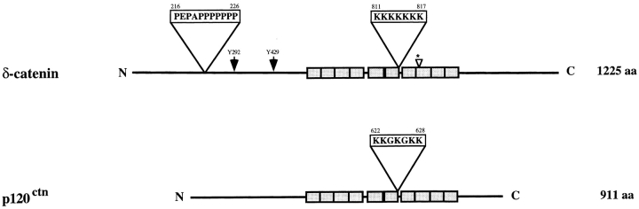
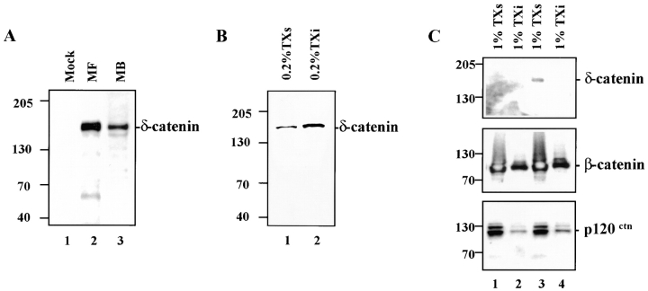
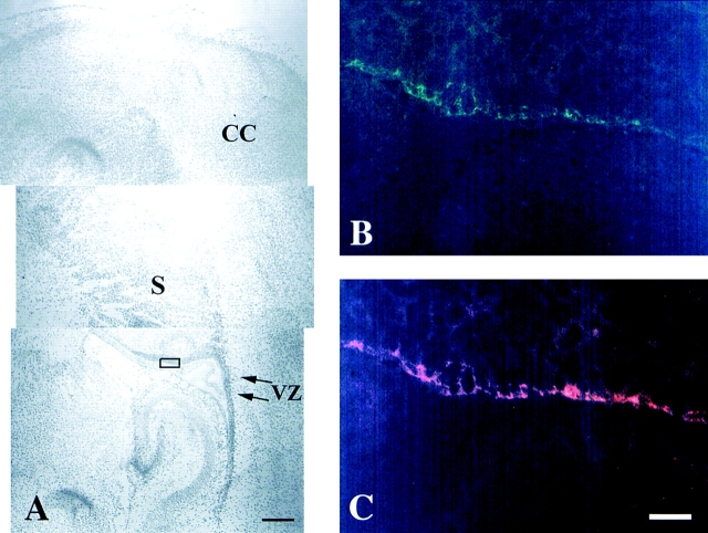
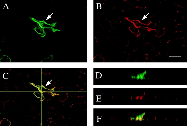
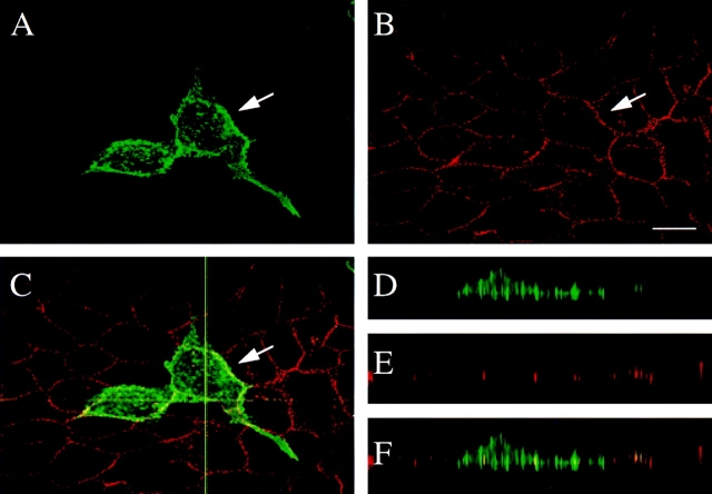
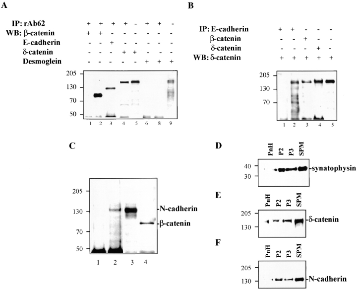
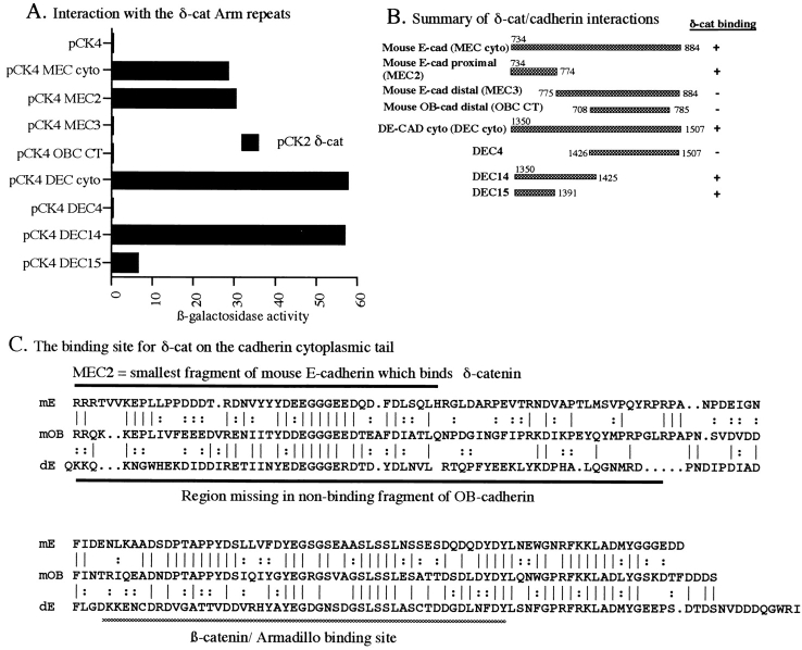
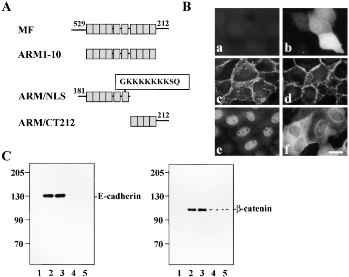
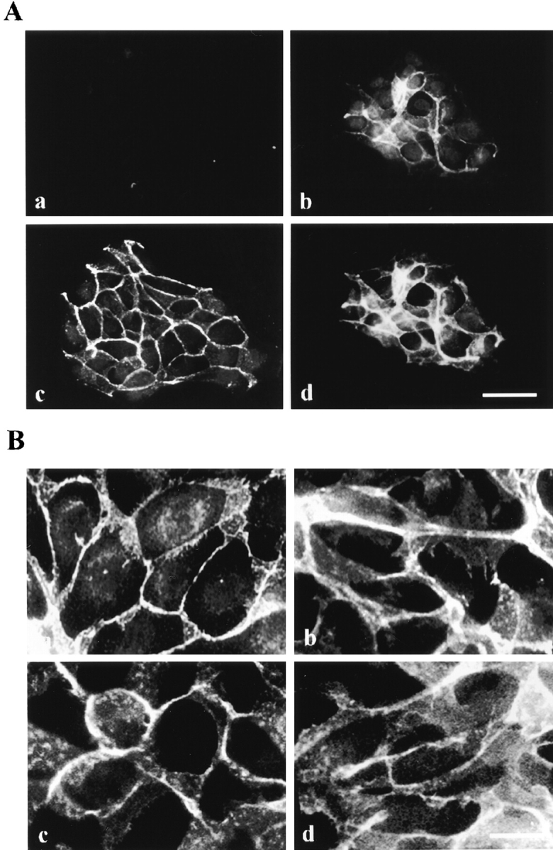
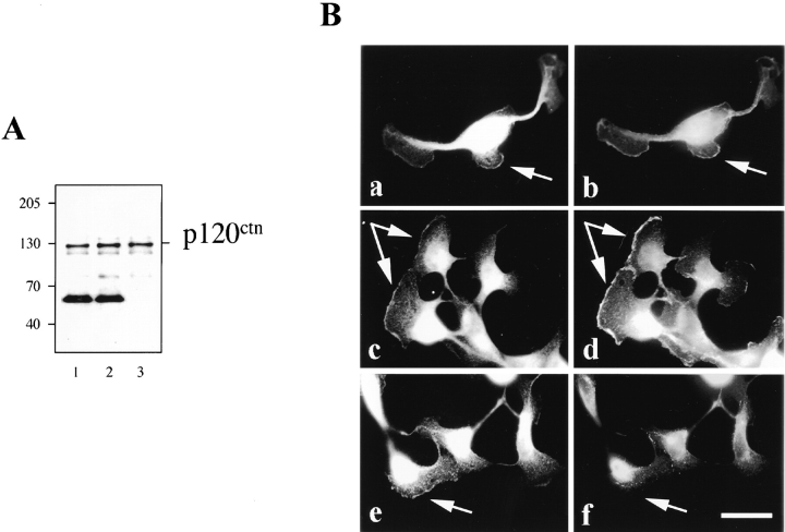
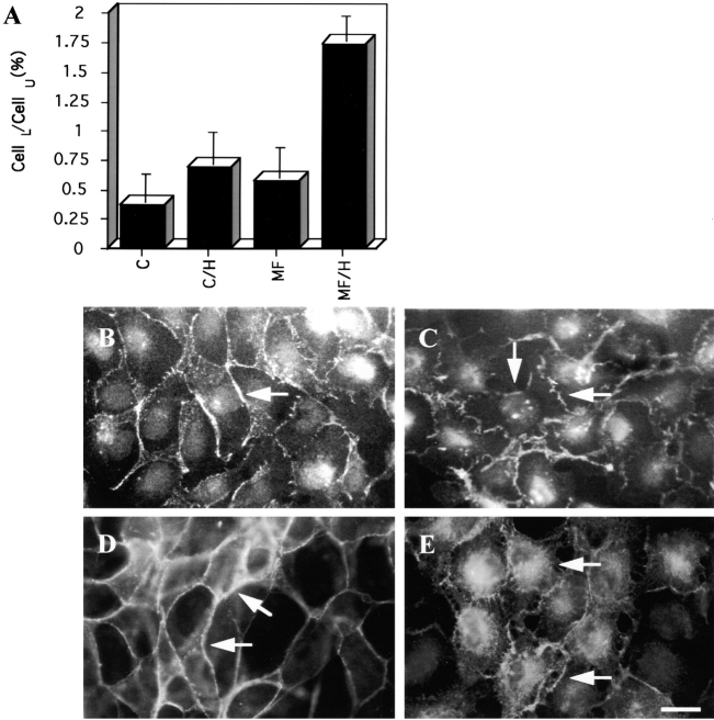
The classical adherens junction that holds epithelial cells together consists of a protein complex in which members of the cadherin family linked to various catenins are the principal components. delta-catenin is a mammalian brain protein in the Armadillo repeat superfamily with sequence similarity to the adherens junction protein p120(ctn). We found that delta-catenin can be immunoprecipitated as a complex with other components of the adherens junction, including cadherin and beta-catenin, from transfected cells and brain. The interaction with cadherin involves direct contact within the highly conserved juxtamembrane region of the COOH terminus, where p120(ctn) also binds. In developing mouse brain, staining with delta-catenin antibodies is prominent towards the apical boundary of the neuroepithelial cells in the ventricular zone. When transfected into Madin-Darby canine kidney (MDCK) epithelial cells delta-catenin colocalized with cadherin, p120(ctn), and beta-catenin. The Arm domain alone was sufficient for achieving localization and coimmunoprecipitation with cadherin. The ectopic expression of delta-catenin in MDCK cells altered their morphology, induced the elaboration of lamellipodia, interfered with monolayer formation, and increased scattering in response to hepatocyte growth factor treatment. We propose that delta-catenin can regulate adhesion molecules to implement the organization of large cellular arrays necessary for tissue morphogenesis.


Figures

Figure 1
Figure 1
Schematic representation of the domain structure of full-length human δ-catenin compared with p120ctn. Amino acids 216–226 contain a proline rich motif that is absent in p120ctn. For δ-catenin, the amino acids 532–1013 are boxed and correspond to the 10 Armadillo repeats. Two Abl tyrosine phosphorylation consensus sites (Y292 and Y429) are indicated by arrows. The small arrowhead with an asterisk indicates a 25–amino acid insertion site that was observed in murine δ-catenin but not in human clones. Amino acids 811–817 represent a lysine rich motif that is a potential nuclear localization signal (NLS) sequence. p120ctn has a similar, albeit somewhat weaker, potential NLS at amino acids 622–628. A partial human δ-catenin cDNA sequence was previously published (Zhou et al., 1997) and the full-length sequence is now updated (GenBank accession number U96136).
Figure 2
Figure 2
Expression of δ-catenin cDNA in transfected MDCK cells and endogenous δ-catenin in developing mouse brains. (A) Immunoblot showing δ-catenin expression in MDCK cells and developing mouse brains. (1) Mock-transfected MDCK cells; (2) MF (MDCK cells transfected with full-length δ-catenin cDNA); (3) MB (mouse brain lysate). (B) Immunoblot of transfected MDCK cells showing δ-catenin in the 0.2% Triton X-100 soluble (TXs, lane 1) and insoluble (Txi, lane 2) fractions. (C) Immunoblots of mock (1 and 2) and δ-catenin transfected (3 and 4) MDCK cells extracted in 1.0% Triton X-100. Soluble and insoluble fractions were blotted with the indicated antibodies. In all panels the molecular weight standard is indicated at the left.
Figure 3
Figure 3
Double immunofluorescence microscopy showing the colocalization of δ-catenin with β-catenin in neocortical neuroepithelia of postnatal day 2 mouse brain. (A) Low magnification view in which the rectangle shows the location of the immunofluorescent images in B and C. CC, cerebral cortex. VZ, ventricular zone. S, striatum. Bar, 0.3 mm. (B) Frozen sagittal section stained by rAb62. (C) Same section immunostained by mouse monoclonal anti–β-catenin. Bar, 20 μm.
Figure 4
Figure 4
Confocal immunofluorescence microscopy of MDCK cells transiently transfected with δ-catenin cDNA. The cells were double labeled with (A) δ-catenin antibodies and with (B) E-cadherin antibody. The arrow points to the transfected cell. (C) Merged fluorescent image showing colocalization of δ-catenin and E-cadherin. The horizontal line indicates where the XZ plane was selected for D–F. (D–F) Respective XZ vertical sections of A–C. Bar, 15 μm.
Figure 5
Figure 5
Confocal immunofluorescence microscopy of MDCK cells transiently transfected with δ-catenin. The cells were double labeled with (A) δ-catenin antibodies and with (B) desmoplakin antibody. The arrow points to the transfected cell. (C) Merged fluorescent image showing minimal colocalization of δ-catenin and desmoplakin. The horizontal line indicates where the XZ plane was selected for D–F. (D–F) Respective XZ vertical sections of A–C. Bar, 15 μm.
Figure 6
Figure 6
Association of δ-catenin with adhesive junction proteins in brain and in MDCK cells stably expressing δ-catenin. (A) δ-catenin coimmunoprecipitates E-cadherin and β-catenin, but not desmoglein. (1 and 6) Mock-transfected MDCK cells. (2–5, 8, and 9) MF cells. (B) Reverse immunoprecipitation showing δ-catenin coprecipitated with E-cadherin and β-catenin. (1) Mock-transfected MDCK cells. (2–5) MF cells. (C) δ-catenin coimmunoprecipitates N-cadherin and β-catenin in brains. (1) N-cadherin immunoblot of brain fractions immunoprecipitated using nonimmune rabbit IgG. (2) N-cadherin immunoblot of brain fractions immunoprecipitated using rAb62. (3) N-cadherin immunoblot of brain lysate. (4) β-catenin immunoblot of brain fractions immunoprecipitated using rAb62. (D–F) Cofractionation of brain δ-catenin with N-cadherin and synaptophysin. PnH, postnuclear homogenates. P2, heavy membranes consisting of myelin, mitochondria, and crude synaptosomes. P3, mostly microsomes. SPM, synaptic plasma membranes. (D) Immunoblot showing brain fractionation profile of synaptophysin. (E) Immunoblot showing brain fractionation profile of δ-catenin. (F) Immunoblot showing brain fractionation profile of N-cadherin. Molecular weight markers are indicated at the left of each panel.
Figure 7
Figure 7
δ-catenin interacts directly with the juxtamembrane region of cadherins in the yeast two-hybrid system. (A) Interaction of δ-catenin with various cadherin fragments. The Arm repeat region of human δ-catenin in the pCK2 “bait” vector was tested against pCK4 vector alone or with pCK4 fusions encoding the full-length murine E-cadherin cytoplasmic domain (pCK4 ME-CAD cyto), two complementary fragments of murine E-cadherin carrying either the membrane-proximal region (pCK4 MEC2) or the distal region with the β-catenin binding site (pCK4 MEC3), a COOH-terminal fragment of OB-cadherin not containing the juxtamembrane region (pCK4 OB-CAD CT), the entire cytoplasmic domain of Drosophila E-cadherin (pCK4 DEC), and smaller fragments of Drosophila E-cadherin as shown in B (pCK4 DEC 4, 14, and 15). (B) Schematic summary of the cadherin fragments and their interaction with δ-catenin. (C) Sequence alignment of the cytoplasmic tails of mouse E-cadherin, mouse OB-cadherin, and Drosophila DE-cadherin. Above the sequences are shown the smallest fragment of mouse E-cadherin which bound δ-catenin in the yeast two-hybrid assay, while below are diagrammed the amino acids missing from the OB-cadherin clone which does not bind to δ-catenin. The β-catenin/Armadillo binding site is also indicated.
Figure 8
Figure 8
Deletion analysis of δ-catenin localization and interaction with adhesive junction proteins. (A) Schematic drawings show the design of deletion mutants. MF, full-length δ-catenin. ARM1-10, δ-catenin sequence containing only the Arm repeats. ARM/NLS, δ-catenin containing partial NH2 terminus including six Arm repeats. Within Arm repeat 6, the boxed sequence indicates a putative nuclear localization signal. ARM/CT212: δ-catenin containing the complete COOH terminus but with only four Arm repeats from the COOH-terminal end. (B) Localization of mutant δ-catenin in MDCK cells. (a) Mock transfection. (b) GFP reporter. (c) Full-length δ-catenin. (d) Armadillo domain alone. (e) ARM/NLS. (f) ARM/CT212. Bar, 10 μm. (C) Coimmunoprecipitation of δ-catenin deletion mutants from transfected MDCK cells. Fractions were immunoprecipitated with GFP antibody and labeled with either E-cadherin or β-catenin antibody. (1) Mock-transfected cells. (2) MF. (3) ARM1-10. (4) ARM/ CT212. (5) ARM/NLS. Molecular weight markers are indicated at the left of each panel.
Figure 9
Figure 9
δ-catenin transfection altered MDCK cell morphology. (A) Double immunofluorescent labeling showing colocalization of δ-catenin with E-cadherin in MDCK cells stably expressing δ-catenin cDNA. (a and b) Anti–δ-catenin immunofluorescent microscopy. (c and d) Monoclonal anti–E-cadherin immunofluorescent microscopy. (a and c) Mock-transfected MDCK cells. (b and d) MDCK cells stably expressing δ-catenin cDNA. Note in b and d multilayers of MDCK cells can be observed while in a and c the monolayer is intact. Bar, 20 μm. (B) Double immunofluorescent microscopy showing the localization of adherens junction–associated proteins β-catenin and p120ctn in mock (a and c) and δ-catenin–transfected MDCK cells (b and d). (a and b) Anti– β-catenin. (c and d) Anti-p120ctn. Note p120ctn localization to cell–cell contact in mock- and δ-catenin–transfected MDCK cells. Bar, 5 μM.
Figure 11
Figure 11
Ectopic expression of δ-catenin does not lead to displacement of p120ctn from adhesive junction proteins. (A) Immunoblot showing p120ctn coimmunoprecipitation with E-cadherin in mock (1), and MF cells (2). (3) MDCK cell lysate. (B) Double immunofluorescent microscopy showing redistribution of junctional proteins in δ-catenin–transfected MDCK cells after HGF stimulation. (a, c, and e) Anti–δ-catenin. (b) Anti– β-catenin. (d) Anti-p120ctn. (f) Anti-desmoplakin. Note the colocalization of δ-catenin with β-catenin and p120ctn, but not desmoplakin, at the leading edges of cells treated with HGF. Arrows point to the lamellipodia formation. Bar, 5 μm.
Figure 10
Figure 10
Ectopic expression of δ-catenin promotes HGF-stimulated cell spreading and scattering. (A) Two-chamber system showing the enhanced migration of δ-catenin–expressing cells. CellL/CellU is the ratio of cells which migrated from the upper chamber to the lower chamber. C, control MDCK cells. C/H, control cells treated with HGF. MF, δ-catenin–transfected MDCK cells. MF/H, δ-catenin–transfected MDCK cells treated with HGF. (B and C) Monoclonal anti–E-cadherin immunofluorescence after overnight treatment with HGF. (B) Mock-transfected MDCK cells. (C) MDCK cells stably expressing δ-catenin. In B, cell–cell contact is still largely intact while in C cell–cell contact points appear disrupted (see arrows). (D and E) Anti–δ-catenin immunofluorescent microscopy showing the effect of HGF on δ-catenin distribution. (D) δ-catenin–expressing cells before HGF treatment. (E) δ-catenin–expressing cells after HGF treatment sometimes remain in clusters but show disruptions at points of cell–cell contact (see arrow) and show a redistribution of δ-catenin to the intracellular compartment. Bar, 5 μm.

References

    1. Aberle H, Butz S, Stappert J, Weissig H, Kemler R, Hoschuetzky H. Assembly of the cadherin-catenin complex in vitro with recombinant proteins. J Cell Sci. 1994;107:3655–3663. - PubMed
    1. Aberle H, Schwartz H, Kemler R. Cadherin-catenin complex: protein interactions and their implications for cadherin function. J Cell Biochem. 1996;61:514–523. - PubMed
    1. Balkovetz DF, Pollack AL, Mostov KE. Hepatocyte growth factor alters the polarity of Madin-Darby canine kidney cell monolayers. J Biol Chem. 1997;272:3471–3477. - PubMed
    1. Barth AI, Nathke IS, Nelson WJ. Cadherins, catenins and APC protein: interplay between cytoskeletal complexes and signaling pathways. Curr Opin Cell Biol. 1997a;9:683–690. - PubMed
    1. Barth AI, Pollack AL, Altschuler Y, Mostov KE, Nelson WJ. NH2-terminal deletion of beta-catenin results in stable colocalization of mutant beta-catenin with adenomatous polyposis coli protein and altered MDCK cell adhesion. J Cell Biol. 1997b;136:693–706. - PMC - PubMed

Publication types

MeSH terms

Associated data