. 1999 Feb 8;144(3):533-47.
doi: 10.1083/jcb.144.3.533.

DE-Cadherin is required for intercellular motility during Drosophila oogenesis

Affiliations

DE-Cadherin is required for intercellular motility during Drosophila oogenesis

P Niewiadomska et al. J Cell Biol. .

Abstract

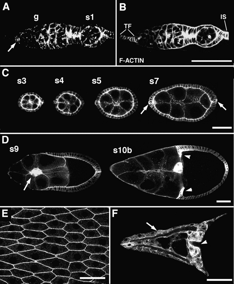
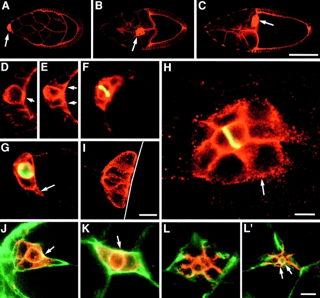
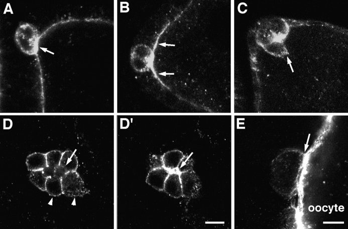
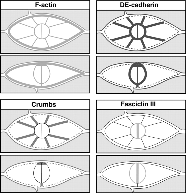
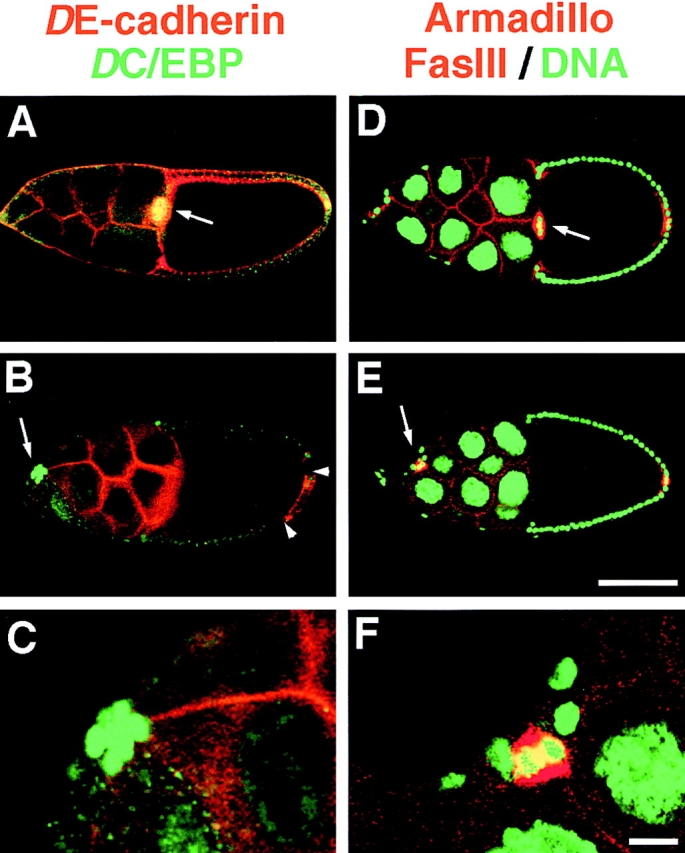
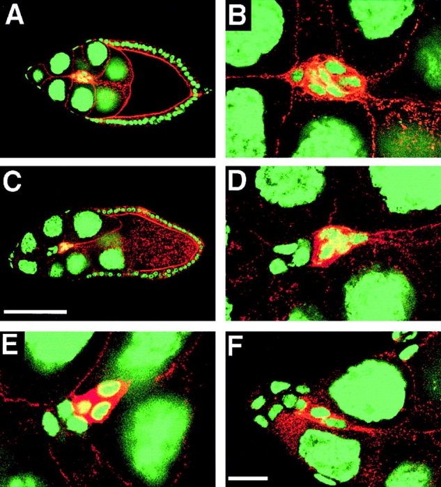
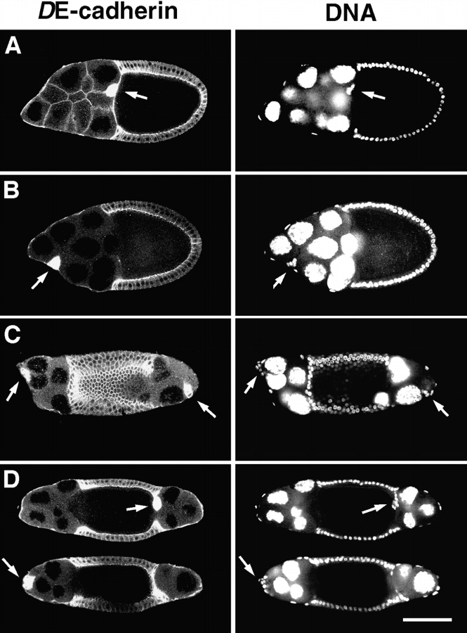
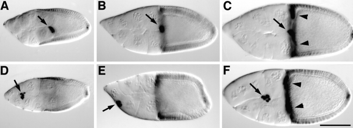
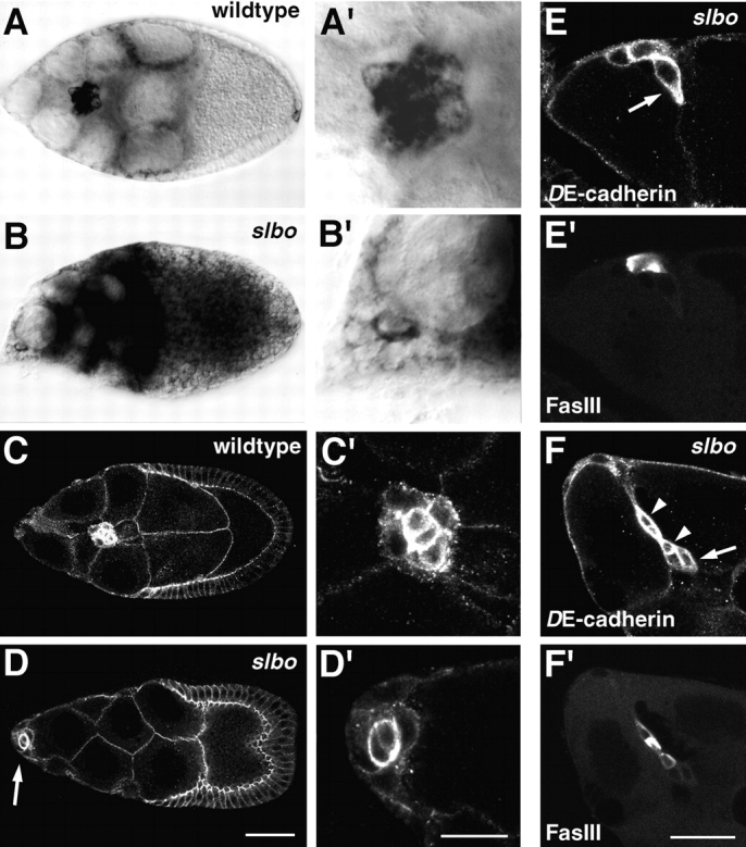
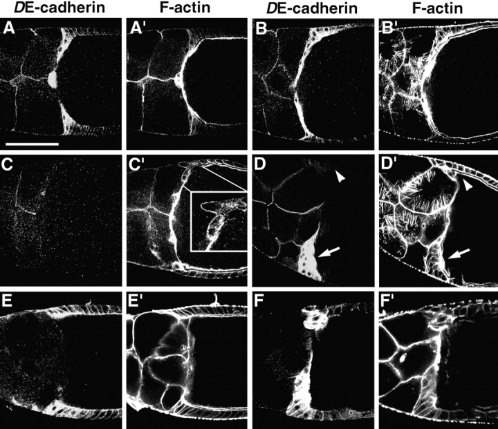
Cadherins are involved in a variety of morphogenetic movements during animal development. However, it has been difficult to pinpoint the precise function of cadherins in morphogenetic processes due to the multifunctional nature of cadherin requirement. The data presented here indicate that homophilic adhesion promoted by Drosophila E-cadherin (DE-cadherin) mediates two cell migration events during Drosophila oogenesis. In Drosophila follicles, two groups of follicle cells, the border cells and the centripetal cells migrate on the surface of germline cells. We show that the border cells migrate as an epithelial patch in which two centrally located cells retain epithelial polarity and peripheral cells are partially depolarized. Both follicle cells and germline cells express DE-cadherin, and border cells and centripetal cells strongly upregulate the expression of DE-cadherin shortly before and during their migration. Removing DE-cadherin from either the follicle cells or the germline cells blocks migration of border cells and centripetal cells on the surface of germline cells. The function of DE-cadherin in border cells appears to be specific for migration as the formation of the border cell cluster and the adhesion between border cells are not disrupted in the absence of DE-cadherin. The speed of migration depends on the level of DE-cadherin expression, as border cells migrate more slowly when DE-cadherin activity is reduced. Finally, we show that the upregulation of DE-cadherin expression in border cells depends on the activity of the Drosophila C/EBP transcription factor that is essential for border cell migration.


Figures

Figure 1
Figure 1
Expression of DE-cadherin during wild-type oogenesis. (A and B) A germarium, double-stained for DE-cadherin (A) and F-actin (B). DE-cadherin expression is seen throughout the germarium (g), in germline cells and somatic cells except for the terminal filament. A previously unidentified group of 6–7 somatic cells close to the base of the terminal filament is strongly stained (arrow). Low levels of DE-cadherin are detected in the anterior region of the germarium where germline stem cells and early cystoblasts are located. Posterior to this region strong DE-cadherin expression is seen in the germline cells and all somatic cells. The DE-cadherin expression in the germarium was described in more detail elsewhere (Godt and Tepass, 1998). (C) Throughout oogenesis DE-cadherin is expressed in the follicular epithelium and the germline cells of follicles. Anterior and posterior polar cells (arrows) show a higher level of expression than the remaining follicle cells during stages 4–7. (D) The highest amounts of DE-cadherin are seen in two migrating follicle cell populations, the border cells (arrow) and the centripetal cells (arrowheads). (E) shows a top view of the DE-cadherin expression pattern in the follicular epithelium at stage 14. (F) shows DE-cadherin distribution in the dorsal appendages (arrow) and in the part of the follicular epithelium that covers the anterior side of the oocyte including the micropyle (arrowhead). This part of the follicular epithelium is composed of the border cells and the centripetal cells. Anterior is to the left in all panels. s, stage; TF, terminal filament; IS, interfollicular stalk. Bars: (A and B) 50 μm; (C, D, and F) 50 μm; (E) 15 μm.
Figure 2
Figure 2
Expression of DE-cadherin in border cells of wild-type ovaries. (A–C) Arrows point to border cells. (A) At early stage 9 border cells have accumulated high levels of DE-cadherin before migration is initiated. (B) Border cells expressing elevated levels of DE-cadherin migrate on a straight path through the center of the follicle towards the oocyte during stage 9. (C) After border cells have reached the oocyte that fills the posterior half of a follicle at stage 10a, they maintain high levels of DE-cadherin expression for some time and move slightly dorsally. (D–I) show high resolution images of DE-cadherin (red) expression in border cells. (F–H) show follicles that are counterstained for Fasciclin III (green) that serves as a marker for the two polar cells. Fasciclin III accumulates at the contact surface between polar cells. (D) Stage 6. Anterior polar cells contain more DE-cadherin than neighboring follicle cells. Polar cells are constricted apically (arrow) and have a rounded shape. DE-cadherin is concentrated at the zonula adherens. (E) Stage 8. Follicle cells adjacent to polar cells have upregulated DE-cadherin expression (arrows). (F) Early stage 9. Same follicle as in A. DE-cadherin distribution in follicle cells adjacent to polar cells (the rosette cells) has depolarized. (G) Early stage 9. Migration is initiated by a rosette cell penetrating between nurse cells (arrow). (H) Late stage 9. Same follicle as in B. During migration polar cells have a central position and are surrounded by rosette cells. Highest concentration of DE-cadherin is seen at the contact surfaces between rosette cells and polar cells and between rosette cells. Lower amounts of DE-cadherin are seen at the interface of rosette cells and nurse cells in a punctate pattern (arrow). (I) Stage 10a. Border cells have established contact to the surface of the oocyte (white line). The polar cells are centrally located and their constricted apical surface contacts the oocyte (see also Peifer et al., 1993). (J–L′) show distribution of DE-cadherin (red) and F-actin (green) in border cells at stage 9. F-actin is concentrated in the periphery of the border cell cluster. (J) Border cells at the onset of migration and (K) during mid migration. Note the round centrally located polar cells with constricted apical cell surfaces that are rich in DE-cadherin and F-actin. (L) and (L′) show two confocal sections of the same cluster. The arrows in L′ point to the constricted apical surfaces of the polar cells. Anterior is to the left in all panels. Bars: (A–C) 50 μm; (D–G, J–L′) 10 μm; (H and I) 10 μm.
Figure 3
Figure 3
Expression of Crumbs in border cells of wild-type ovaries. (A) Crumbs is found at the apical cell surface in all follicle cells. At early stage 8 anterior polar cells have upregulated Crumbs expression (arrow). Crumbs protein is also seen in the cytoplasm of the polar cells. (B) Follicle cells next to the polar cells upregulate Crumbs during stage 8. (C) At early stage 9 when a rosette cell initiates migration (arrow), Crumbs has a nonpolarized distribution in rosette cells. D and D′ show two confocal sections of the same cluster during mid migration. In polar cells Crumbs is concentrated at the apical cell surface (arrow in D′) and not found at the lateral cell surface that contacts the rosette cells (arrow in D). In rosette cells Crumbs accumulates at the contact sides between neighboring rosette cells and is found in a punctate pattern at the interface of rosette cells and nurse cells (arrowheads). (E) At stage 10 when the border cells are in contact with the oocyte Crumbs distribution in rosette cells is again restricted to the apical surface. Bars: (A–D′) 10 μm; (E) 10 μm.
Figure 4
Figure 4
Schematic summary of marker distribution in migrating border cells. For each marker a top view (upper panels) and a side view (lower panels) is shown (see text for further explanations).
Figure 5
Figure 5
DE-cadherin expression in border cells is required for their migration between the nurse cells. (A–C) Double staining of stage 10 follicles with DE-cadherin (red) and the nuclear border cell marker DC/EBP (green). Arrows point to border cell clusters. (A) In a wild-type follicle the border cell cluster that expresses DE-cadherin and DC/EBP has reached the oocyte. (B) shows a shg mutant follicle cell clone that encompasses most of the follicle cells, except for a small patch of DE-cadherin positive cells at the posterior pole (arrowheads). The germline cells express DE-cadherin. A shg mutant border cell cluster expressing DC/EBP has formed that has not moved between the nurse cells towards the oocyte. The cluster is located between nurse cells and the follicular epithelium close to the anterior tip of the follicle. (C) Close-up of the shg mutant border cell cluster shown in B. (D–F) Triple staining of stage 10 follicles for Armadillo (red) that is expressed in the same pattern as DE-cadherin, for the polar cell marker Fasciclin-III (FasIII; also red), and the nuclear marker Picogreen (green). Arrows point to border cell clusters. (D) In the wild-type follicle the border cell cluster has reached the oocyte. (E) shows a shg mutant follicle cell clone derived from shg mutant follicle stem cells that comprises all follicle cells, including the border cells, as indicated by the absence of Armadillo. Red staining in anterior and posterior polar cells is due to expression of Fasciclin III. The anterior polar cells are part of the border cell cluster that has not migrated to the oocyte but remained attached to follicle cells close to the anterior pole of the follicle. (F) Closeup of the shg mutant border cell cluster shown in (E). Bars: (A, B, D, and E) 100 μm; (C and F) 10 μm.
Figure 6
Figure 6
Migration of shg mutant mosaic border cell clusters. Stage 9 follicles were stained with anti-DE-cadherin (red) and the nuclear marker Picogreen (green). (A) A wild-type follicle showing a migrating border cell cluster. (B) Closeup of the border cell cluster shown in A. All border cells express DE-cadherin. (C) shg mutant follicle cell clone covering part of the follicular epithelium. (E) Closeup of the mosaic border cell cluster shown in C. The migrating border cell cluster contains DE-cadherin positive cells at the front, and DE-cadherin negative cells trailing behind. D shows another example of a mosaic border cell cluster with the DE-cadherin expressing border cells moving ahead. F shows a shg mutant mosaic border cell cluster in which the DE-cadherin positive cells have started migrating between the nurse cells and the shg mutant cells are still located at the anterior end of the follicle. Anterior is to the left in all panels. Bars: (A and C) 100 μm; (B, D, E, and F) 20 μm.
Figure 7
Figure 7
Disruption of border cell migration in shg mutant germ- line clones. Stage 10 follicles are double stained with DE-cadherin (left panels) and the nuclear marker Picogreen (right panels). Arrows point to border cell clusters. (A) Wild-type follicle with the border cell cluster attached to the oocyte. (B) shg mutant germline clone with a normally located oocyte. The border cell cluster has not invaded between the nurse cells but remained directly beneath the follicular epithelium close to the anterior pole. (C) shg mutant germline clone with a centrally located oocyte. Two border cell clusters have formed, one at each pole of the follicle, which have not migrated towards the oocyte. (D) Two optical sections of the same shg mutant germline clone that has a centrally located oocyte. From the two border cell clusters one has moved and reached the oocyte (upper section). Note that the border cell cluster is in contact with the follicular epithelium. The other one has not moved away from its original polar position (lower section). Anterior is to the left in all panels. Bar, 100 μm.
Figure 8
Figure 8
Reduced motility of border cells in a weak shg mutant. X-gal staining of follicles at late stage 9 (A and D), at stage 10a (B and E) and stage 10b (C and F) reveals lacZ expression of the shgP34-1 P-element insertion in border cells (arrows) and centripetal cells (arrowheads). (A–C) shgP34-1/CyO follicles show normal border cell migration. The border cell cluster is attached to the oocyte in all three follicles. (D–F) shgP34-1/shgR6 mutant follicles. D and F show border cell clusters that have migrated between nurse cells but have not reached the oocyte. E shows a border cell cluster that has not penetrated between nurse cells but remained in contact with the follicular epithelium. Anterior is to the left in all panels. Bar, 100 μm.
Figure 9
Figure 9
Upregulation of shg expression in border cells depends on slbo. A′–D′ are close-ups of the border cell clusters shown in A–D. (A and A′) shg RNA expression in a wild-type follicle, and (B and B′) a homozygous slbo1 mutant follicle at stage 9 was detected by in situ hybridization using a digoxygenin-labeled shg cDNA probe. The migrating border cell cluster in the wild-type follicle shows high concentration of shg transcript in all border cells. In the slbo mutant follicle the border cell cluster remained at the anterior tip. shg expression in the border cells has not been upregulated, and is much lower than in wild-type. The two polar cells show a higher level of shg expression than the rosette cells (B′). (C and D′) Protein expression as revealed by anti-DE-cadherin staining in a wild-type follicle (C and C′) and a homozygous slbo1 mutant follicle (D and D′) at stage 9. The slbo mutant border cell cluster expresses a much lower level of DE-cadherin than the wild-type cluster. The two polar cells express higher levels of DE-cadherin than the surrounding rosette cells in both genotypes. (E–F′) Double staining of slbo1 mutant follicles with anti-DE-cadherin (E and F) and the polar cell specific marker Fasciclin III (E′ and F′). Migratory activity of border cells (arrows) in stage 10b slbo1 mutant follicles is accompanied by upregulation of DE-cadherin expression. Arrowheads in F point to polar cells. Anterior is to the left in all panels. Bars: (A–D) 50 μm; (A′–D′) 20 μm; (E–F′) 20 μm.
Figure 10
Figure 10
DE-cadherin is required for centripetal cell movement. Follicles are double stained for DE-cadherin (A–F) and F-actin (A′–F′). (A and A′) In an early stage 10b wild-type follicle centripetal cells strongly elongate apical-basally, develop protrusive ends that penetrate between oocyte and nurse cells, and migrate along the surface of the oocyte towards the border cells. Strongest concentration of DE-cadherin is seen in the leading edges of the centripetal cells. (B and B′) At late stage 10b, when cytoplasmic actin fibers have formed in the nurse cells, the invaded centripetal cells form a thin layer covering the anterior surface of the oocyte in a wild-type follicle. (C and C′) Mid-stage 10b follicle with a shgR69 mutant follicle cell clone that comprises all follicle cells. Some shg mutant centripetal cells have segregated from the follicular epithelium but remain in the periphery of the follicle. These cells have a rounded appearance and lack protrusive ends in contrast to wild-type centripetal cells (see inset). (D and D′) The late stage 10b follicle has a shg mutant mosaic follicular epithelium. DE-cadherin negative centripetal cells (arrowhead) show no invasive behavior in contrast to the DE-cadherin positive centripetal cells (arrow) that are elongated and migrated between oocyte and nurse cells. However, the migrating DE-cadherin positive centripetal cells form a thicker layer than in wild-type follicles. (E and E′) Stage 10b follicle with a shg mutant germline. Centripetal cells that show an elevated level of DE-cadherin expression did not move between germline cells. (F and F′) Stage 10b follicle with a shg mutant germline. Centripetal cells moved between oocyte and nurse cells forming clumps of cells with abnormal morphology. Anterior is to the left in all panels. Bar, 100 μm.

References

    1. Bellen HJ, O'Kane CJ, Wilson C, Grossniklaus U, Pearson RK, Gehring WJ. P-element-mediated enhancer detection: a versatile method to study development in Drosophila. . Genes Dev. 1989;3:1288–1300. - PubMed
    1. Bement WM, Forscher P, Mooseker MS. A novel cytoskeletal structure involved in purse string wound closure and cell polarity maintenance. J Cell Biol. 1993;121:565–578. - PMC - PubMed
    1. Birchmeier W. E-cadherin as a tumor (invasion) suppressor gene. BioEssays. 1995;17:97–99. - PubMed
    1. Bixby JL, Zhang R. Purified N-cadherin is a potent substrate for the rapid induction of neurite outgrowth. J Cell Biol. 1990;110:1253–1260. - PMC - PubMed
    1. Braga VM, Machesky LM, Hall A, Hotchin NA. The small GTPases Rho and Rac are required for the establishment of cadherin-dependent cell–cell contacts. J Cell Biol. 1997;137:1421–1431. - PMC - PubMed

Publication types