. 1999 Mar;73(3):1809-17.
doi: 10.1128/JVI.73.3.1809-1817.1999.

Functional interactions of the HHCC domain of moloney murine leukemia virus integrase revealed by nonoverlapping complementation and zinc-dependent dimerization

Affiliations

Functional interactions of the HHCC domain of moloney murine leukemia virus integrase revealed by nonoverlapping complementation and zinc-dependent dimerization

F Yang et al. J Virol. 1999 Mar.

Abstract

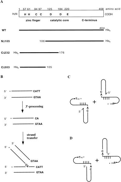
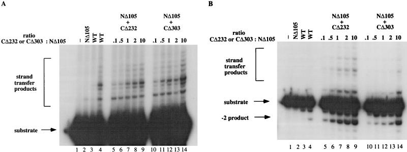
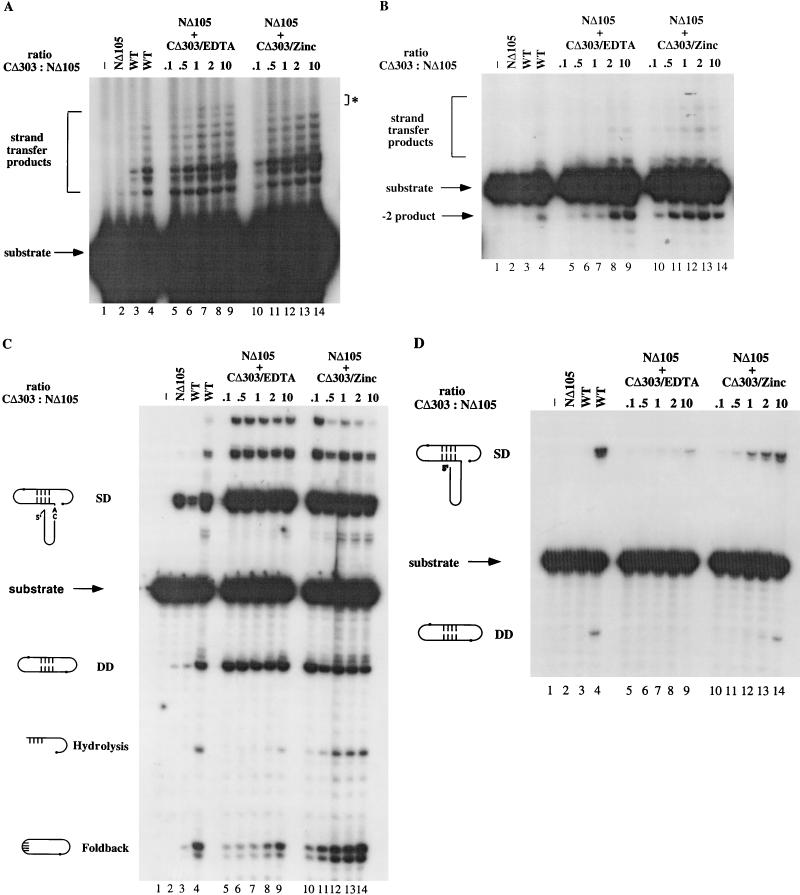
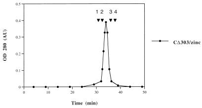
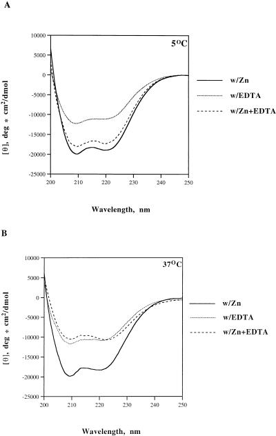
The retroviral integrase (IN) is required for the integration of viral DNA into the host genome. The N terminus of IN contains an HHCC zinc finger-like motif, which is conserved among all retroviruses. To study the function of the HHCC domain of Moloney murine leukemia virus IN, the first N-terminal 105 residues were expressed independently. This HHCC domain protein is found to complement a completely nonoverlapping construct lacking the HHCC domain for strand transfer, 3' processing and coordinated disintegration reactions, revealing trans interactions among IN domains. The HHCC domain protein binds zinc at a 1:1 ratio and changes its conformation upon binding to zinc. The presence of zinc within the HHCC domain stimulates selective integration processes. Zinc promotes the dimerization of the HHCC domain and protects it from N-ethylmaleimide modification. These studies dissect and define the requirement for the HHCC domain, the exact function of which remains unknown.


Figures

FIG. 1
FIG. 1
Illustration of WT M-MuLV IN, mutant INs, and assays used in this study. (A) Schematic representation of WT M-MuLV IN and IN mutants used in this study. The name of each mutant is indicated to the left of each protein. (B) DNA substrates and assays for 3′ processing and strand transfer reactions. (C) DNA substrates and assay for coordinated disintegration reactions with crossbone substrates with 5′-ss overhang in the LTR (tailed crossbone substrates). (D) DNA substrates and assay for coordinated disintegration reaction with substrates without 5′-ss overhang in the LTR (untailed crossbone substrates). The asterisks indicate the ends labeled.
FIG. 2
FIG. 2
Complementation of NΔ105 by nonoverlapping HHCC domain protein. (A) Complementation of NΔ105 by CΔ232/CΔ303 for strand transfer reaction. Lane 1, control buffer incubation; lane 2, 10 pmol of NΔ105; lanes 3 and 4, 10 and 40 pmol of WT IN, respectively; lanes 5 to 9, 10 pmol of NΔ105 plus CΔ232; the ratio of CΔ232 to NΔ105 is indicated at the top of the panel; lanes 10 to 14, 10 pmol of NΔ105 plus CΔ303; the ratio of CΔ303 to NΔ105 is indicated at the top of the panel. (B) Complementation of NΔ105 by CΔ232/CΔ303 for 3′ processing reaction. The lanes are the same as in panel A. Both CΔ232 and CΔ303 were renatured in the presence of EDTA; the reaction buffer contained 10 mM MnCl2.
FIG. 3
FIG. 3
Zinc stimulates CΔ303’s ability to complement NΔ105. (A) Complementation of NΔ105 by CΔ303/EDTA or CΔ303/zinc for strand transfer reaction. Lane 1, control buffer incubation; lane 2, 10 pmol of NΔ105; lanes 3 and 4, 10 and 40 pmol of WT IN, respectively; lanes 5 to 9, 10 pmol of NΔ105 plus CΔ303/EDTA; lanes 10 to 14, 10 pmol of NΔ105 plus CΔ303/zinc. Ratios of CΔ303/zinc or CΔ303/EDTA to NΔ105 are indicated at the top of each panel. Lanes in panels B to D are the same as in panel A. ∗, difference in strand transfer products. (B) Complementation of NΔ105 by CΔ303/EDTA or CΔ303/zinc for 3′ processing reaction. (C) Complementation of NΔ105 by CΔ303/EDTA or CΔ303/zinc for coordinated disintegration reaction with tailed crossbone substrates. (D) Complementation of NΔ105 by CΔ303/EDTA or CΔ303/zinc for coordinated disintegration reaction with untailed crossbone substrate. CΔ303 was renatured in zinc or EDTA as indicated at the top of each panel; all reaction buffers contained MnCl2 as indicated in Materials and Methods. SD, single disintegration product; DD, double disintegration product.
FIG. 4
FIG. 4
Zinc binding protects the HHCC domain from NEM modification. (A) CΔ303/zinc remains active for complementation after NEM treatment. A disintegration assay with oligonucleotide 7440 as substrate was used to test the susceptibility of IN mutants to NEM modification. Lane 1, control buffer incubation; lane 2, WT IN; lane 3, NΔ105; lane 4, complementation of NΔ105 and CΔ303/zinc; lane 5, complementation of NΔ105 and CΔ303/EDTA; lane 6, NEM-modified WT IN; lane 7, NEM-modified NΔ105; lane 8, complementation of NΔ105 and NEM-modified CΔ303/zinc; lane 9, complementation of NΔ105 and NEM-modified CΔ303/EDTA. All NEM-treated proteins are underlined. (B) Only CΔ303/EDTA can be labeled by fluorescent maleimide. CΔ303/zinc and CΔ303/EDTA were modified with N(1-pyrene) maleimide as described in Materials and Methods, and the protein bands were analyzed for fluorescence (lanes 1 and 2) and by Coomassie blue staining (lanes 3 to 5). Lanes 1 and 3, CΔ303/zinc; lanes 2 and 4, CΔ303/EDTA; lane 5, protein molecular mass markers.
FIG. 5
FIG. 5
Fast protein liquid chromatography elution profile of the HHCC domain protein CΔ303/zinc. CΔ303/zinc was chromatographed on a Superose-12 column. The protein standard markers are as follows: 1, bovine serum albumin, 67 kDa; 2, ovalbumin, 43 kDa; 3, chymotrypsinogen A, 25 kDa; 4, RNase A, 13.7 kDa. The elution time of the protein standards is indicated on the panel. OD 280, optical density at 280 nm; AU, absorbance units.
FIG. 6
FIG. 6
CD spectra of the HHCC domain proteins. CD spectra of CΔ303/zinc, CΔ303/EDTA, and CΔ303/zinc plus 5 mM EDTA at 5°C (A) and 37°C (B) are shown.

References

    1. Berg J M, Shi Y. The galvanization of biology: a growing appreciation for the roles of zinc. Science. 1996;271:1081–1085. - PubMed
    1. Berg J M. Zinc finger domains: hypotheses and current knowledge. Annu Rev Biophys Biophys Chem. 1990;19:405–421. - PubMed
    1. Bradford M M. A rapid and sensitive method for the quantitation of microgram quantities of protein utilizing the principle of protein-dye binding. Anal Biochem. 1976;72:248–254. - PubMed
    1. Brahms S, Brahms J. Determination of protein secondary structure in solution by vacuum ultraviolet circular dichroism. J Mol Biol. 1980;138:149–178. - PubMed
    1. Brown P O. Integration. In: Coffin J M, Hughes S H, Varmus H E, editors. Retroviruses. Plainview, N.Y: Cold Spring Harbor Laboratory Press; 1997. pp. 161–203. - PubMed

Publication types

LinkOut - more resources