. 1999 Mar;73(3):1846-52.
doi: 10.1128/JVI.73.3.1846-1852.1999.

vig-1, a new fish gene induced by the rhabdovirus glycoprotein, has a virus-induced homologue in humans and shares conserved motifs with the MoaA family

Affiliations

vig-1, a new fish gene induced by the rhabdovirus glycoprotein, has a virus-induced homologue in humans and shares conserved motifs with the MoaA family

P Boudinot et al. J Virol. 1999 Mar.

Abstract

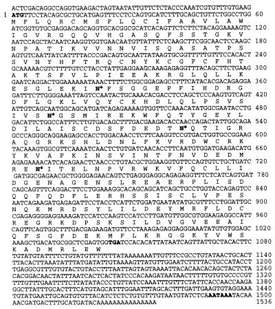
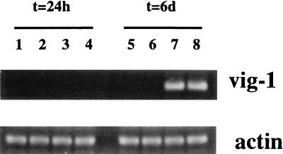
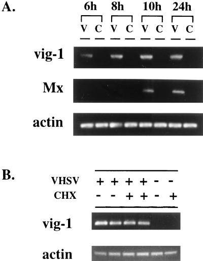
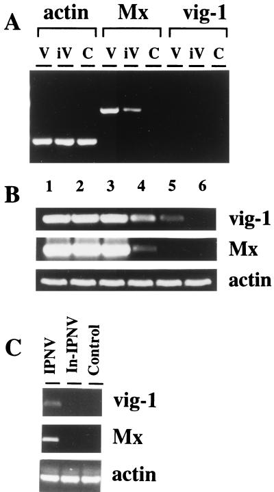
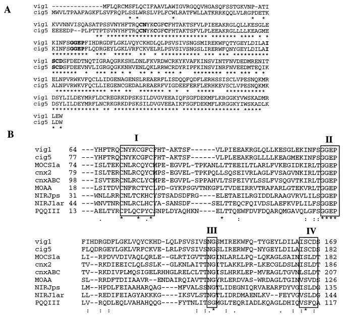
We used mRNA differential display methodology to analyze the shift of transcription profile induced by the fish rhabdovirus, viral hemorrhagic septicemia virus (VHSV), in rainbow trout leukocytes. We identified and characterized a new gene which is directly induced by VHSV. This VHSV-induced gene (vig-1) encodes a 348-amino-acid protein. vig-1 is highly expressed during the experimental disease in lymphoid organs of the infected fish. Intramuscular injection of a plasmid vector expressing the viral glycoprotein results in vig-1 expression, showing that the external virus protein is sufficient for the induction. vig-1 expression is also obtained by a rainbow trout interferon-like factor, indicating that vig-1 can be induced through different pathways. Moreover, vig-1 is homologous to a recently described human cytomegalovirus-induced gene. Accordingly, vig-1 activation may represent a new virus-induced activation pathway highly conserved in vertebrates. The deduced amino acid sequence of vig-1 is significantly related to sequences required for the biosynthesis of metal cofactors. This suggests that the function of vig-1 may be involved in the nonspecific virus-induced synthesis of enzymatic cofactors of the nitric oxide pathway.


Figures

FIG. 1
FIG. 1
VHSV-induced expression of vig-1. (A) Semiquantitative PCR assay on cDNA from rainbow trout head kidney cells cultured as described with VHSV (VHSV), with BPL-inactivated virus (BPL-VHSV) or without virus (Control). The samples were normalized on the basis of actin expression. (B) Northern blot analysis of vig-1 expression in rainbow trout head kidney cells treated as described in panel A. The same blots were hybridized with an actin probe as a control for the total amounts of RNA loaded in the gels.
FIG. 2
FIG. 2
Sequence of vig-1 cDNA. Rainbow trout vig-1 cDNA nucleotidic and deduced amino acid sequences. The initiation codon, the termination codon, and the polyadenylation signal are in bold. Potential glycosylation sites are indicated by a bold-faced N*.
FIG. 3
FIG. 3
VIG-1 has no signal peptide. (A) The VIG-1 Kyte-Doolittle hydropathic profile shows a hydrophobic stretch at the N terminus of the protein (amino acids 6 to 23, arrow). (B) In vitro transcription-translation and microsome assays were performed with a plasmid containing vig-1 cDNA. The same experiments were performed with a plasmid containing gVHSV cDNA in order to control the efficiency of microsome assay. When the extract was supplemented with dog pancreatic microsomes, a mobility shift was detected for gVHSV but not for VIG-1 (lanes 3 and 7). This shift can be abolished by endoglycosidase H treatment (lane 8), which has no effect on the VIG-1 product (lane 4).
FIG. 4
FIG. 4
vig-1 induction during the experimental disease. vig-1 mRNA expression was assessed by RT-PCR in the head kidney during the course of VHSV infection. vig-1 transcripts were searched for 24 h (lanes 3 and 4) or 6 days (lanes 7 and 8) after trout were infected with VHSV. Trout injected with phosphate-buffered saline were used as controls at 24 h (lanes 1 and 2) or at 6 days (lanes 5 and 6). Total amounts of cDNA were controlled by the amplification of actin mRNA from the same samples.
FIG. 5
FIG. 5
vig-1 induction after genetic immunization against the glycoprotein of the VHSV. vig-1 mRNAs were detected in the muscle at the site of injection (lanes 3 and 4) and in the head kidney (lanes 7 and 8) by RT-PCR, 7 days after genetic immunization with pCDNA1_GVHSV. Trout injected with pCDNA1 were used as controls (lanes 1 and 2 for muscle and 5 and 6 for head kidney). Total amounts of mRNA were controlled by the amplification of actin mRNA from the same samples.
FIG. 6
FIG. 6
The induction of vig-1 is directly mediated by viral particles. (A) Kinetics of vig-1 induction in rainbow trout head kidney cells cultured in the presence of VHSV (V) or without virus (C) at 20°C. vig-1 and Mx mRNAs were searched for by a RT-PCR assay at 6, 8, 10, and 24 h. Samples were normalized on the basis of the actin expression. (B) Effect of CHX on vig-1 mRNA induction. vig-1 mRNAs were detected by RT-PCR assay from cells cultured 7 h with VHSV, with or without CHX. Cells cultured without virus in the presence or absence of CHX were used as controls, and samples were normalized on the basis of actin expression.
FIG. 7
FIG. 7
Induction of vig-1 in response to interferon. Actin, Mx, and vig-1 mRNAs were assessed by a RT-PCR assay in different experiments. (A) RTG-2 cells incubated with live VHSV (V) or BPL-inactivated virus (iV) or without virus (C). (B) Head kidney cells were stimulated by serial dilutions of conditioned medium displaying a trout interferon-like activity (lanes: 1, 1/2; 2, 1/10; 3, 1/100; 4, 1/1,000; 5, 1/10,000). Cells cultured with unconditioned medium were processed similarly as a control (lane 6). (C) Head kidney cells cultured with live IPNV (IPNV) or inactivated IPNV (In-IPNV) or without virus (Control).
FIG. 8
FIG. 8
Multiple alignments of the predicted amino acid sequence of VIG-1 with related proteins. (A) VIG-1 is aligned with the sequence deduced from human cig-5 gene (accession no. AF026941). (B) The following proteins are aligned with VIG-1 and CIG-5: MoaA proteins from human (MOCS1a, accession no. AF034374), Arabidopsis thaliana (cnx2, accession no. Z48047), Aspergillus nidulans (cnxABC, accession no. AF027213), and Escherichia coli (MoaA, accession no. AE000181), NIRJ proteins from Pseudomonas aeruginosa (NIRJps, accession no. D84475) and Archaeoglobus fulgidus (NIRJ1ar, accession no. AE001026), and coenzyme pyrrolo-quinoline-quinone biosynthesis protein III from Acinetobacter calcoaceticus (PQQIII, accession no. X06452). Grey shaded boxes represent conserved motifs, and conserved residues are indicated by an asterisk. The three cysteines in the first box (I) constitute a potential iron-sulfur metal binding site.

References

    1. Ankel H, Capobianchi M R, Castilletti C, Dianzani F. Interferon induction by HIV glycoprotein 120: role of the V3 loop. Virology. 1994;205:34–43. - PubMed
    1. Basurco B, Benmansour A. Distant strains of the fish rhabdovirus VHSV maintain a sixth functional cistron which codes for a nonstructural protein of unknown function. Virology. 1995;212:741–745. - PubMed
    1. Bengten E, Stromberg S, Pilstrom L. Immunoglobulin VH regions in Atlantic cod (Gadus morhua L.): their diversity and relationship to VH families from other species. Dev Comp Immunol. 1994;18:109–122. - PubMed
    1. Benmansour A, Paubert G, Bernard J, de Kinkelin P. The polymerase-associated protein (M1) and the matrix protein (M2) from a virulent and an avirulent strain of viral hemorrhagic septicemia virus (VHSV), a fish rhabdovirus. Virology. 1994;198:602–612. - PubMed
    1. Bernard J, Lecocq X F, Rossius M, Thiry M E, de Kinkelin P. Cloning and sequencing the messenger RNA of the N gene of viral haemorrhagic septicaemia virus. J Gen Virol. 1990;71:1669–1674. - PubMed

Publication types

Associated data

LinkOut - more resources