. 1999 Mar;73(3):1918-30.
doi: 10.1128/JVI.73.3.1918-1930.1999.

The chromatin structure of the long control region of human papillomavirus type 16 represses viral oncoprotein expression

Affiliations

The chromatin structure of the long control region of human papillomavirus type 16 represses viral oncoprotein expression

W Stünkel et al. J Virol. 1999 Mar.

Abstract

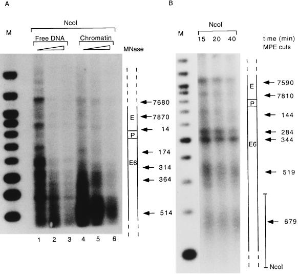
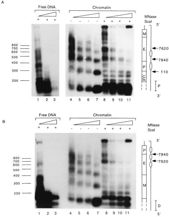
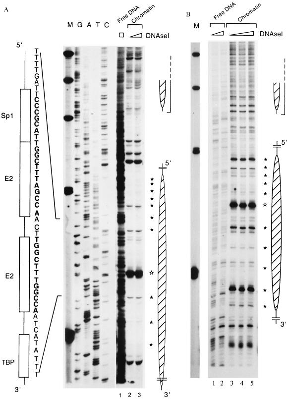
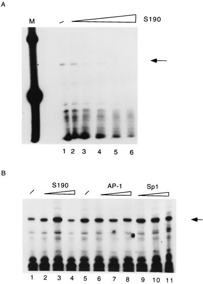
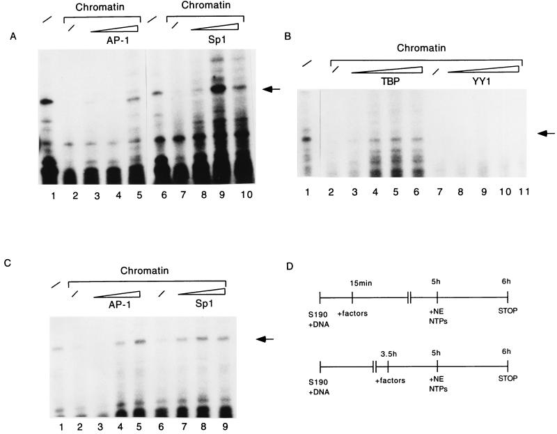
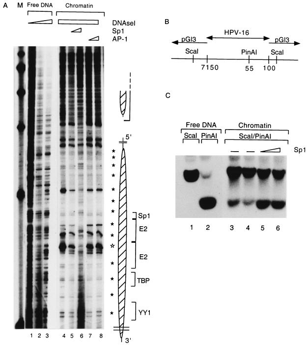
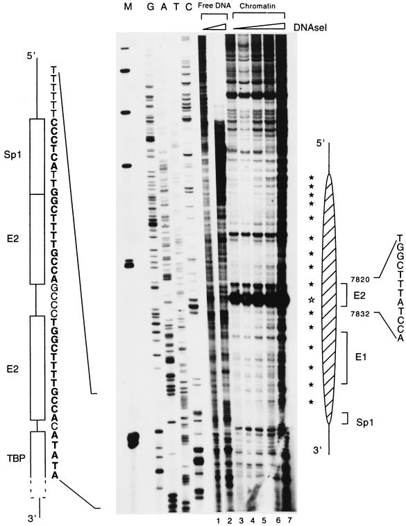
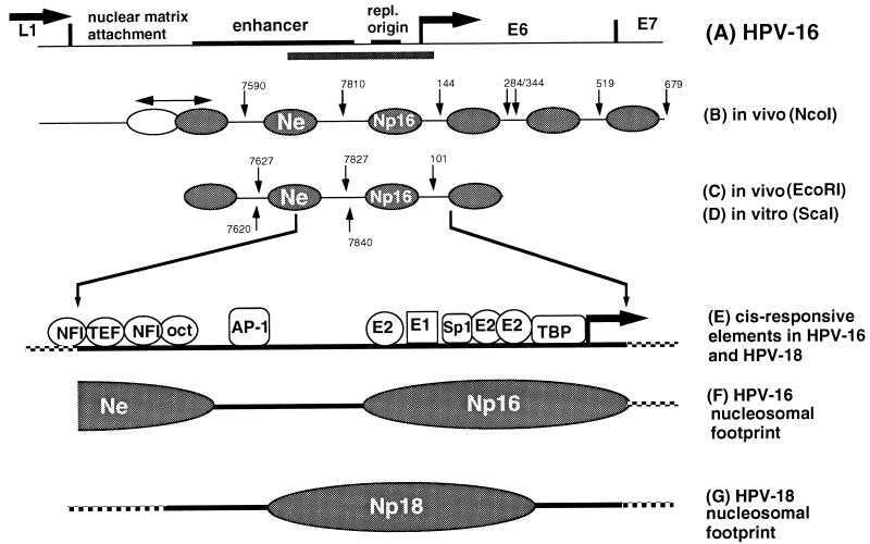
The long control region (LCR) of human papillomavirus type 16 (HPV-16) has a size of 850 bp (about 12% of the viral genome) and regulates transcription and replication of the viral DNA. The 5' segment of the LCR contains transcription termination signals and a nuclear matrix attachment region, the central segment contains an epithelial cell-specific enhancer, and the 3' segment contains the replication origin and the E6 promoter. Here we report observations on the chromatin organization of this part of the HPV-16 genome. Treatment of the nuclei of CaSki cells, a cell line with 500 intrachromosomal copies of HPV-16, with methidiumpropyl-EDTA-Fe(II) reveals nucleosomes in specific positions on the LCR and the E6 and E7 genes. One of these nucleosomes, which we termed Ne, overlaps with the center of the viral enhancer, while a second nucleosome, Np16, overlaps with the replication origin and the E6 promoter. The two nucleosomes become positioned on exactly the same segments after in vitro assembly of chromatin on the cloned HPV-16 LCR. Primer extension mapping of DNase I-cleaved chromatin revealed Np16 to be positioned centrally over E6 promoter elements, extending into the replication origin. Ne covers the center of the enhancer but leaves an AP-1 site, one of the strongest cis-responsive elements of the enhancer, unprotected. Np16, or a combination of Np16 and Ne, represses the activity of the E6 promoter during in vitro transcription of HPV-16 chromatin. Repression is relieved by addition of Sp1 and AP-1 transcription factors. Sp1 alters the structure of Np16 in vitro, while no changes can be observed during the binding of AP-1. HPV-18, which has a similar arrangement of cis-responsive elements despite its evolutionary divergence from HPV-16, shows specific assembly in vitro of a nucleosome, Np18, over the E1 binding site and E6 promoter elements but positioned about 90 bp 5' of the position of Np16 on the homologous HPV-16 sequences. The chromatin organization of the HPV-16 and HPV-18 genomes suggests important regulatory roles of nucleosomes during the viral life cycle.


Figures

FIG. 1
FIG. 1
The LCR and the E6 gene of the HPV-16 genome in CaSki cells are nucleosomally organized. Nuclei were treated with increasing amounts of micrococcal nuclease (MNase) for 5 min (A) or of MPE for 10 to 40 min (B), and total genomic DNA was purified, restricted with NcoI separated by agarose gel electrophoresis, blotted onto nylon membranes, and processed with a radioactive probe close to the NcoI site, as indicated on the right of panel B. Arrows mark fragments with increased accessibility to micrococcal nuclease or MPE. Their sizes were estimated by comparison with a 100-bp size marker (lanes M). A scheme on the right of each panel shows the position of each cleavage site within the LCR of HPV-16.
FIG. 2
FIG. 2
Nucleosomes assembled in vitro on the LCR and the E6 gene of HPV-16 DNA are found in positions similar to those detected in CaSki cells. pHPV-16-Luc DNA was assembled into chromatin with Drosophila S190 extract and treated with 1, 2, 4, and 5 U of micrococcal nuclease (MNase) (lanes 4 to 7 and 8 to 11 of each panel). The nuclease-treated DNA was purified and fractionated on agarose gels without further treatment by restriction endonucleases (lanes 4 to 7 of each panel) or after digestion with ScaI (lanes 8 to 11). The DNA was blotted, and the blots were processed either with a proximal probe (P) (A) or with a distal probe (D) (B). As a control, lanes 1 to 3 of each panel show the digestion patterns of micrococcal nuclease-treated and ScaI-restricted free DNAs. The numbers on the left indicate the sizes of the fragments in base pairs. A schematic presentation on the right of each panel identifies within the HPV-16 LCR the MAR (M), the enhancer (E), the E6 promoter (P), the genomic positions of micrococcal nuclease-sensitive sites (arrows), and the predicted positions of nucleosomes (ovals).
FIG. 3
FIG. 3
A specifically positioned nucleosome covers the promoter of HPV-16. (A) The HPV-16 LCR cloned in pHPV-16-Luc was assembled into chromatin with Drosophila S190 extracts and treated with increasing amounts of DNase I, and the resulting fragments were purified and assayed by primer extension. Lanes 2 and 3, footprints originating from two nucleosomes that overlap with the promoter and the enhancer (large and small oval shapes on the right). Weak 10-bp-spaced bands (filled stars) indicate DNase I accessibility due to the rotational phasing of the nucleosomes, and a strong hypersensitive site (open star) suggests the center of the dyad symmetry of the nucleosome. As controls, lane 1 shows DNase I treatment of free HPV-16 LCR DNA and the left side of the panel indicates a sequencing ladder. Lane M, size marker with bands at 500, 400, 300, 200, and 100 bp. Symbols and nucleotides on the left identify the four cis-responsive elements of the E6 promoter, namely, binding sites for Sp1, the viral factor E2, and TBP. (B) Footprint obtained in a similar experiment and permitting similar interpretations. It highlights the 10-bp periodicity but does not permit clear mapping of the extent of nucleosomal protection.
FIG. 4
FIG. 4
Nucleosomal assembly leads to transcriptional repression of the HPV-16 promoter. (A) Analyses of in vitro transcription from free pHPV-16-Luc DNA (lane 1) or the same plasmid assembled into nucleosomes by increasing amounts of Drosophila S190 extract (lanes 2 to 6). Lane M, 100-bp ladder serving as size standard. The specific transcript initiated at the HPV-16 E6 promoter is indicated by an arrow on the right. (B) The Drosophila S190 extract does not contain nonhistone components that might inhibit in vitro transcription, as shown by the addition of 20, 50, and 70 μl of S190 extract (lanes 2 to 4, respectively) at the beginning of an in vitro transcription reaction with nucleosome-free DNA (lane 1). As a further control, we ensured that the factors Sp1 and AP-1 are not limiting in the basic transcriptional system. Lanes 6 to 11 confirm that an excess of AP-1 and Sp1 does not superstimulate transcription of nonchromatin templates.
FIG. 5
FIG. 5
Relief of nucleosomal transcriptional repression by trans-acting factors. (A) AP-1 (lanes 3 to 5) and Sp1 (lanes 8 to 10), added 15 min after initiation of chromatin assembly on pHPV-16-Luc, activate in vitro transcription from the E6 promoter. Specific transcripts were detected by primer extension (arrow). The same transcript is generated from free DNA in the absence of chromatin and an excess of any additional factor (lanes 1 and 6) but is repressed by chromatin alone (lanes 2 and 7). (B) Under the same conditions as used for panel A, TBP (lanes 3 to 6) marginally induces a nucleosomally organized E6 LCR, while YY1 (lanes 8 to 11) fails to do so. Lane 1, transcription of nucleosome-free DNA; lanes 2 and 7, repression of transcription by nucleosomes. To detect the weak signals of the transcriptional induction by TBP, this blot was exposed five times longer than the blot shown in panel A. YY1 has a strong binding site within the promoter sequence covered by the nucleosome, which does not lead to transcriptional repression in transfection experiments, while additional YY1 sites remote from the promoter and possibly protected by the upstream nucleosome may negatively interfere with transcription independent from the chromatin state of the HPV-16 LCR (50). (C) AP-1 (lanes 3 to 5) and Sp1 (lanes 7 to 9) can activate in vitro transcription from the E6 promoter even when added at a late stage of chromatin assembly. The conditions of this experiment resembled those for panel A, but the transcription factors were added 3.5 h after initiation of chromatin assembly. Lane 1, transcription from naked DNA, lanes 2 and 6, transcription from chromatin in the absence of additional factors. (D) Schematic diagram of the experiments in panels A and C. NTP, nucleoside triphosphate.
FIG. 6
FIG. 6
Sp1 can rearrange the structure of the E6 promoter-bound nucleosome. (A) pHPV-16-Luc DNA was assembled into chromatin, subjected to DNase I treatment, and processed as described for Fig. 3 to visualize the nucleosomal footprints overlapping with the HPV-16 E6 promoter (lanes 4 to 8) and enhancer (large and small oval shapes, respectively, on the right). Weak 10-bp-spaced bands (filled stars) indicate DNase I accessibility due to the rotational phasing of the nucleosomal core, and a strong hypersensitive site (open star) is most likely the center of the dyad symmetry of the nucleosome. As controls, lanes 1 to 3 show DNase I treatment of free HPV-16 LCR DNA. A nucleosome (Np16) protects four cis-responsive elements of the E6 promoter, namely, binding sites for Sp1, the viral factor E2, TBP, and a YY1 site (brackets on the right of the oval shape). Sp1 (lanes 5 and 6) or AP-1 (lanes 7 and 8) was added 3.5 h after start of the chromatin assembly and in the case of Sp1 resulted in the reemergence of the cleavage pattern of the free DNA and the disappearance of nucleosome-specific DNase I-hypersensitive sites. Lane M, markers. (B) Schematic representation of an HPV-16 LCR clone from position 7150 to 100 in the vector pGI3. The enzyme ScaI cleaves in vector sequences on both sides of the cloned segment; PinAI cleaves at position 55 in the promoter sequences of HPV-16. (C) Southern blot of a ScaI digest (lane 1) and a ScaI-PinAI double digest (lane 2). The ScaI-PinAI fragment is released inefficiently in the presence of chromatin (lanes 3 and 4) but efficiently with increasing Sp1 concentrations (lanes 5 and 6). This blot was processed with a radioactive probe specific for the ScaI-PinAI fragment.
FIG. 7
FIG. 7
A specifically positioned nucleosome covers the replication origin of HPV-18 and extends into E6 promoter sequences. The cloned HPV-18 LCR was assembled into chromatin with Drosophila S190 extracts and treated with increasing amounts of DNase I, and the resulting fragments were assayed by primer extension. Lanes 3 to 7 show, with increasing DNase I treatment, the footprint of a nucleosome overlapping the replication origin (distal E2 and E1 binding site), indicated by a large hatched oval on the right. Weak 10-bp-spaced bands (filled stars) indicate DNase I accessibility due to the rotational phasing of the nucleosomal organization, and a strong hypersensitivity site (open star) suggests the center of the dyad symmetry of the nucleosome. As controls, lanes 1 and 2 show DNase I treatment of free HPV-18 LCR DNA, and the left side shows a sequencing ladder of this sequence. Lane M, size marker with bands at 500, 400, 300, 200, and 100 bp. Symbols and nucleotides on the right identify the third E2 binding site from the E6 promoter, the E1 binding site, and one of the four cis-responsive elements of the E6 promoter, namely, the binding site for Sp1.
FIG. 8
FIG. 8
Nucleosomal organization of the LCR of HPV-16. (A) Genomic segment of HPV-16 with a size of approximately 1.5 kb. The LCR has a size of 850 bp and includes an MAR, the epithelial cell-specific enhancer, the replication origin (repl. origin), and the E6 promoter (arrow) and is flanked by the L1 and E6 genes. The bar below the 3′ side of the enhancer, the replication origin, and the promoter highlights the genomic fragment that has been studied in both HPV-16 and HPV-18 by footprint analysis to map specifically positioned nucleosomes. (B) MPE-hypersensitive sites in HPV-16 chromatin in CaSki cells permit the mapping of the positions of six nucleosomes relative to an NcoI site, which is positioned downstream of this segment. A double-headed arrow above the leftmost nucleosome, which overlaps with the MAR, indicates a region of protection whose size exceeds that for the normal protection by a nucleosome, possibly due to alternative positions of a nucleosome. The nucleosome (Ne) between the MPE cuts at position 7590 and 7810 overlaps with the epithelial cell-specific enhancer, and the third nucleosome (Np), between positions 7810 and 144, overlaps with the replication origin and the E6 promoter. Three additional nucleosomes are positioned within the E6 gene and at the 5′ end of the E7 gene, respectively. (C and D) The positions of four nucleosomes on the HPV-16 LCR in CaSki cells mapped with an upstream probe (hybridizing close to the EcoRI site at position 7456) are in agreement with the data obtianed with the NcoI probe, and at least two of these nucleosomes are positioned identically after assembly of chromatin in vitro (data not shown). (E and F) One of these nucleosomes was footprinted, and its center apparently overlaps with two E2 binding sites at the E6 promoter. The protection of this nucleosome includes the TBP binding site, the Sp1 binding site, and the binding site for the replication factor E1. (G) In HPV-18, which has a similar arrangement of elements of the replication origin and the E6 promoter, a nucleosome (Np18) it positioned about 90 bp further upstream from the E6 promoter, extending to and partially protecting the promoter elements. The HPV-16 DNA, and possibly also the DNA of HPV-18, may position in vitro the Ne nucleosome, which overlaps with the core of the epithelial cell-specific enhancer, but leave the 3′ side of the enhancer with a strong AP-1 site unprotected.

References

    1. Alexiadis V, Varga-Weisz P D, Bonte E, Becker P B, Gruss C. In vitro chromatin remodeling by chromatin accessibility complex (CHRAC) at the SV40 origin of DNA replication. EMBO J. 1998;17:3428–3438. - PMC - PubMed
    1. Archer T K, Cordingley M G, Wolford R G, Hager G L. Transcription factor access is mediated by accurately positioned nucleosomes on the mouse mammary tumor virus promoter. Mol Cell Biol. 1991;11:688–698. - PMC - PubMed
    1. Baker C C, Phelps W C, Lindgren V, Braun M J, Gonda M A, Howley P M. Structural and transcriptional analysis of human papillomavirus type 16 sequences in cervical carcinoma cell lines. J Virol. 1987;61:962–971. - PMC - PubMed
    1. Batson S C, Rimsky S, Sundseth R, Hansen U. Association of nucleosome-free regions and basal transcription factors with in vivo-assembled chromatin templates active in vitro. Nucleic Acids Res. 1993;21:3459–3468. - PMC - PubMed
    1. Bauer W R, Hayes J J, White J H, Wolffe A P. Nucleosome structural changes due to acetylation. J Mol Biol. 1994;236:685–690. - PubMed

LinkOut - more resources