. 1999 Mar;73(3):2045-51.
doi: 10.1128/JVI.73.3.2045-2051.1999.

Conditions for copackaging rous sarcoma virus and murine leukemia virus Gag proteins during retroviral budding

Affiliations

Conditions for copackaging rous sarcoma virus and murine leukemia virus Gag proteins during retroviral budding

R P Bennett et al. J Virol. 1999 Mar.

Abstract

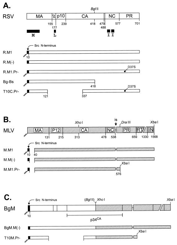
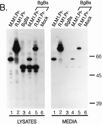
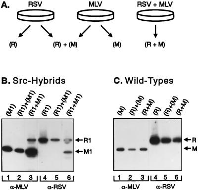
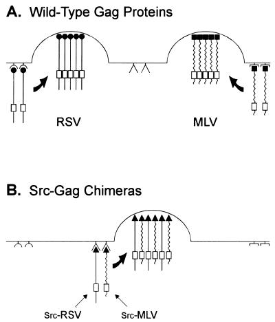
Rous sarcoma virus (RSV) and murine leukemia virus (MLV) are examples of distantly related retroviruses that normally do not encounter one another in nature. Their Gag proteins direct particle assembly at the plasma membrane but possess very little sequence similarity. As expected, coexpression of these two Gag proteins did not result in particles that contain both. However, when the N-terminal membrane-binding domain of each molecule was replaced with that of the Src oncoprotein, which is also targeted to the cytoplasmic face of the plasma membrane, efficient copackaging was observed in genetic complementation and coimmunoprecipitation assays. We hypothesize that the RSV and MLV Gag proteins normally use distinct locations on the plasma membrane for particle assembly but otherwise have assembly domains that are sufficiently similar in function (but not sequence) to allow heterologous interactions when these proteins are redirected to a common membrane location.


Figures

FIG. 1
FIG. 1
Derivatives of the RSV and MLV Gag proteins. RSV sequences are represented by open boxes, and those of MLV are represented by hatched boxes. The restriction sites used to create the chimeras are noted above each sequence. Myristylation is indicated by a squiggly line, and M(−) indicates that the glycine encoded by the second codon is replaced, eliminating the site of myristic acid addition. Numbers below the Gag proteins refer to amino acids counted from the N terminus of each Gag protein and mark positions cleaved by the viral protease. (A) Derivatives of the RSV Gag protein. The wild-type molecule is illustrated at the top, where the positions of domains essential for budding are indicated with black bars (M, L, and I). Gag derivatives in which the first 10 residues are replaced with those of the Src oncoprotein are also depicted. The location of the inactivating amino acid substitution in the RSV protease, D37S, is noted. (B) Derivatives of the MLV Gag protein. The wild-type protein is illustrated at the top, and versions in which the first 39 amino acids are replaced with the first 10 residues of the Src protein are also shown. The site of in-frame suppression (“is”) of the stop codon between the gag and pol genes is indicated. (C) RSV-MLV Gag chimeras. The RSV BglII site was destroyed, while the MLV XhoI site was restored during construction of the chimeric gene.
FIG. 2
FIG. 2
Expression of RSV and MLV Gag derivatives. COS-1 cells in 35-mm dishes were transfected with the indicated DNAs and labeled 48 h later with [3H]leucine for 2.5 h. The RSV and MLV proteins from the cell lysates or media samples were immunoprecipitated with anti-RSV and anti-MLV serum, respectively. The proteins were electrophoresed on an SDS–10% polyacrylamide gel and visualized by fluorography. Positions of the Gag precursors (Pr) for RSV, MLV, and M.M1, along with the capsid (CA) cleavage products, are indicated on the left. The positions of molecular mass standards are indicated (in kilodaltons) on the right.
FIG. 3
FIG. 3
Complementation analysis with RSV and MLV Gag derivatives. COS-1 cells transfected with the indicated DNAs or combinations of DNAs were metabolically labeled with [3H]leucine for 2.5 h. Immunoprecipitations were performed with a mixture of RSV and MLV antisera. The proteins were separated on an SDS–10% polyacrylamide gel and visualized by fluorography. The positions of molecular mass markers are indicated (in kilodaltons) on the right. (A) Analysis of T10C derivatives. These mutants lack the L domain function of RSV and are blocked at a late step in budding, after membrane binding, but retain the I domain sequences involved in interactions among Gag proteins. (B) Analysis of mutant Bg-Bs. This RSV deletion mutant lacks the region of Gag-Gag interaction.
FIG. 3
FIG. 3
Complementation analysis with RSV and MLV Gag derivatives. COS-1 cells transfected with the indicated DNAs or combinations of DNAs were metabolically labeled with [3H]leucine for 2.5 h. Immunoprecipitations were performed with a mixture of RSV and MLV antisera. The proteins were separated on an SDS–10% polyacrylamide gel and visualized by fluorography. The positions of molecular mass markers are indicated (in kilodaltons) on the right. (A) Analysis of T10C derivatives. These mutants lack the L domain function of RSV and are blocked at a late step in budding, after membrane binding, but retain the I domain sequences involved in interactions among Gag proteins. (B) Analysis of mutant Bg-Bs. This RSV deletion mutant lacks the region of Gag-Gag interaction.
FIG. 4
FIG. 4
Coimmunoprecipitation analysis of RSV and MLV Gag proteins. (A) Diagram of the transfection and mixing protocol. COS-1 cells expressing derivatives of the RSV Gag protein, the MLV Gag protein, or both were metabolically labeled for 6 h with [3H]leucine at 48 h after transfection. (B) Analysis of Src-Gag chimeras. The labeled particles were collected by centrifugation through a cushion of 20% sucrose, resuspended in buffer, and divided into two samples of equal volume. In the case of the singly transfected cells, one sample was immunoprecipitated with anti-MLV or anti-RSV serum. The remaining two samples were mixed, split into equal aliquots, and then immunoprecipitated with anti-MLV or anti-RSV serum. In the case of the particles obtained from cotransfected cells, the two aliquots were immunoprecipitated with either anti-MLV or anti-RSV serum. The proteins were electrophoresed on an SDS–10% polyacrylamide gel and visualized by fluorography. The identity of each protein in the gel is indicated on the right. (C) Analysis of wild-type Gag proteins. The experiment was set up exactly as described above except that wild-type constructs were used in place of the Src chimeras.
FIG. 5
FIG. 5
Distinct sites utilized by Gag proteins during particle assembly on the plasma membrane. (A) The wild-type Gag proteins of RSV and MLV have unrelated membrane-binding domains (depicted by solid circles and solid squares, respectively) and do not encounter one another when expressed in the same cell. Molecular interactions between Gag molecules of a single type occur through the I domain located within the NC sequence (open rectangles) and lead to the emergence of particles of a single type. (B) When the membrane-binding domains of each type of Gag molecule are replaced with that of the Src protein (solid triangles), particles containing both species are released from the cell. This model suggests that the membrane-binding domains of RSV Gag, MLV Gag, and the Src protein interact with distinct regions on the inner surface of the plasma membrane. Therefore, Gag proteins must be targeted to the same membrane location for copackaging to occur. The molecular nature of the proposed receptors is unknown.

References

    1. Bennett R P, Nelle T D, Wills J W. Functional chimeras of the Rous sarcoma virus and human immunodeficiency virus Gag proteins. J Virol. 1993;67:6487–6498. - PMC - PubMed
    1. Bowzard J B, Bennett R P, Krishna N K, Ernst S M, Rein A, Wills J W. Importance of basic residues in the nucleocapsid sequence for retrovirus Gag assembly and complementation rescue. J Virol. 1998;72:9034–9044. - PMC - PubMed
    1. Buser C A, Sigal C T, Resh M D, McLaughlin S. Membrane binding of myristylated peptides corresponding to the NH2 terminus of Src. Biochemistry. 1994;33:13093–13101. - PubMed
    1. Craven R C, Bennett R P, Wills J W. Role of the avian retroviral protease in the activation of reverse transcriptase during virion assembly. J Virol. 1991;65:6205–6217. - PMC - PubMed
    1. Craven R C, Parent L J. Dynamic interactions of the Gag polyprotein. Curr Top Microbiol Immunol. 1996;214:65–94. - PubMed

Publication types

MeSH terms

Substances