. 1999 Mar;73(3):2115-25.
doi: 10.1128/JVI.73.3.2115-2125.1999.

CD21-Dependent infection of an epithelial cell line, 293, by Epstein-Barr virus

Affiliations

CD21-Dependent infection of an epithelial cell line, 293, by Epstein-Barr virus

J D Fingeroth et al. J Virol. 1999 Mar.

Abstract

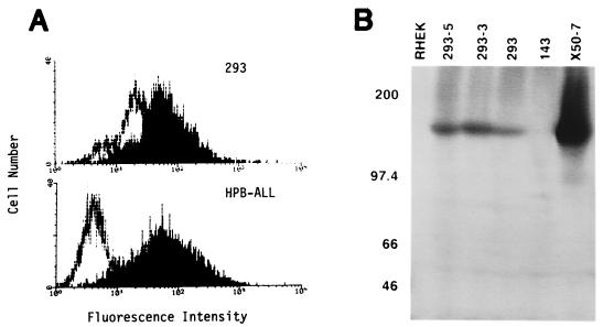
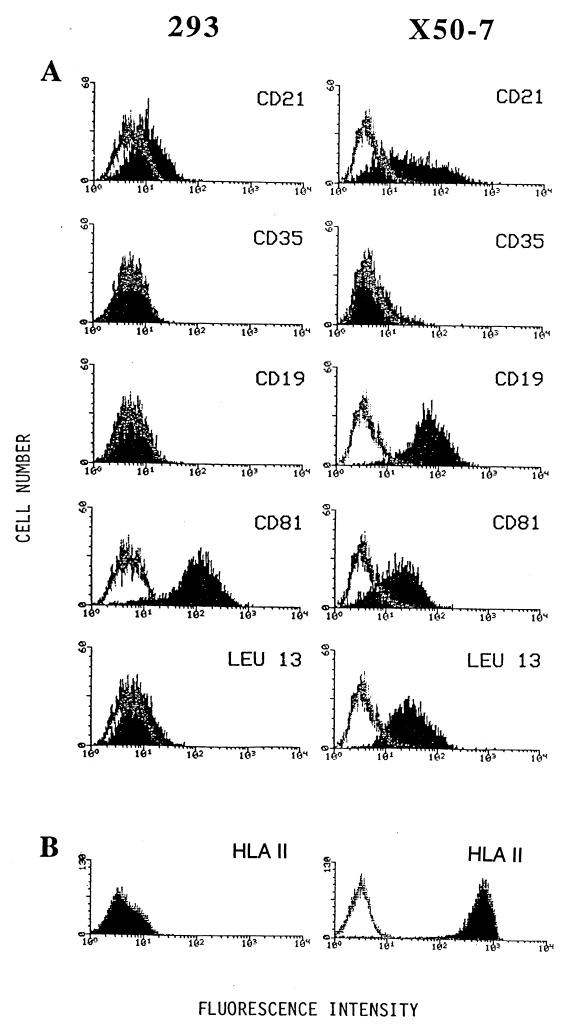
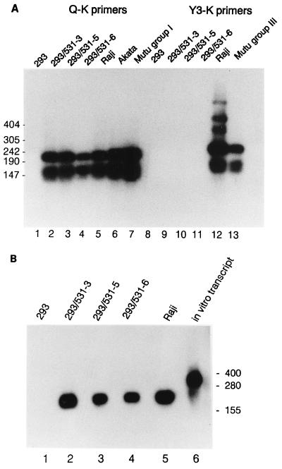
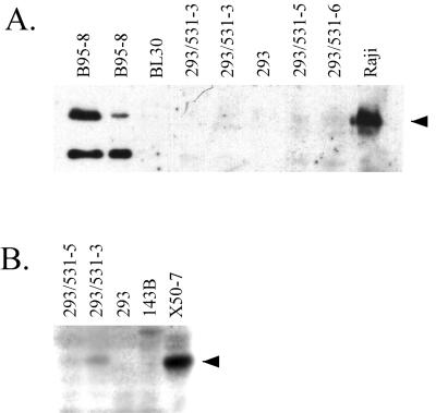
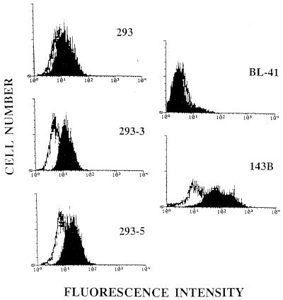
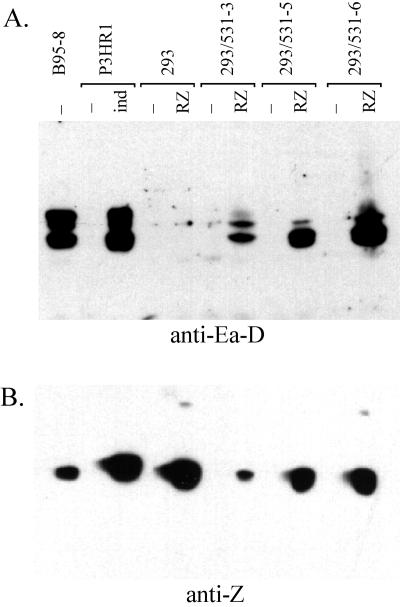
Epstein-Barr virus (EBV) is invariably present in undifferentiated nasopharyngeal carcinomas, is found sporadically in other carcinomas, and replicates in the differentiated layer of the tongue epithelium in lesions of oral hairy leukoplakia. However, it is not clear how frequently or by what mechanism EBV infects epithelial cells normally. Here, we report that a human epithelial cell line, 293, can be stably infected by EBV that has been genetically marked with a selectable gene. We show that 293 cells express a relatively low level of CD21, that binding of fluorescein-labeled EBV to 293 cells can be detected, and that both the binding of virus to cells and infection can be blocked with antibodies specific for CD21. Two proteins known to form complexes with CD21 on the surface of lymphoid cells, CD35 and CD19, could not be detected at the surface of 293 cells. All infected clones of 293 cells exhibited tight latency with a pattern of gene expression similar to that of type II latency, but productive EBV replication and release of infectious virus could be induced inefficiently by forced expression of the lytic transactivators, R and Z. Low levels of mRNA specific for the transforming membrane protein of EBV, LMP-1, as well as for LMP-2, were detected; however, LMP-1 protein was either undetectable or near the limit of detection at less than 5% of the level typical of EBV-transformed B cells. A slight increase in expression of the receptor for epidermal growth factor, which can be induced in epithelial cells by LMP-1, was detected at the cell surface with two EBV-infected 293 cell clones. These results show that low levels of surface CD21 can support infection of an epithelial cell line by EBV. The results also raise the possibility that in a normal infection of epithelial cells by EBV, the LMP-1 protein is not expressed at levels that are high enough to be oncogenic and that there might be differences in the cells of EBV-associated epithelial cancers that have arisen to allow for elevated expression of LMP-1.


Figures

FIG. 1
FIG. 1
Detection of CD21 on 293 cells with a monospecific rabbit antiserum. (A) Detection of CD21 on the cell surface of 293 compared with HPB-ALL by flow cytometry. Cells were stained either with rabbit anti-CD21 (block) or with normal rabbit serum (line). (B) Detection of CD21 protein in extracts of 293 cells by Western analysis. Total soluble protein from Nonidet P-40 lysis of 500,000 cells was analyzed for the cell lines indicated. Two clones of 293 that are stably infected with the EBV recombinant P3-531 are indicated as 293-5 and 293-3. At the left, positions are indicated (in kilodaltons) for standards.
FIG. 2
FIG. 2
CD21-dependent binding of FITC-labeled EBV to 293 cells, as determined by flow cytometry. (A) Binding of FITC-labeled EBV to 293 cells without pretreatment. (B) Before incubating 293 cells with FITC-labeled EBV, surface CD21 was cross-linked with anti-CD21 mouse MAb HB-5 followed by goat F(ab′)2 against mouse IgG. (C) 293 cells were treated identically except that the irrelevant control MAb UPC10 was used for cross-linking.
FIG. 3
FIG. 3
Analysis of surface expression of CD21 and proteins reported to form membrane complexes with CD21 for 293 cells and X50-7 cells. Binding is indicated (block) for mouse MAbs specific to CD21 (HB-5), to CD35/CR1 (543), to CD19 (B-C3), to CD81/TAPA-1 (5A6), and to Leu-13 (anti-Leu-13). Each is shown in comparison to binding by an irrelevant antibody, UPC10 (line).
FIG. 4
FIG. 4
Detection of circular EBV genomes in stably infected 293 cell clones. Cells were lysed in the wells of a 0.8% agarose gel, allowing nonintegrated viral genomes to be electrophoresed into the gel (12). After electrophoresis and transfer of DNA to a nylon membrane, EBV genomes were detected by hybridization to random-prime-labeled BamHI W repeat. “c” and “l” indicate positions of supercoiled and linear (broken) EBV genomes, respectively. Integrated and nicked circular EBV genomes do not migrate through the gel appreciably. Lanes 1 through 5 contained 200,000 293 cells or 293 cell clones infected with P3-531, as indicated. Lanes 6 to 8 contained 10,000, 40,000, and 200,000, Raji cells, with EBV-negative DG75 cells also added to lanes 6 and 7 so that a total of 200,000 cells were loaded into every well.
FIG. 5
FIG. 5
Detection of EBNA1 mRNA and EBER1 RNA in infected 293 cells. (A) Spliced EBNA1 transcripts were detected in 2 μg of total RNA from the indicated cell lines by RT-PCR using primers specific for the 3′K (coding) exon and for one of the upstream exons, either Q or Y3 as indicated, to reveal type I or type III latency promoter usage (46), as previously described (26). Amplification products were detected by Southern analysis using labeled internal exon U as a probe. Positions of DNA size markers are indicated (in base pairs) at the left. (B) EBER1 RNA was detected by Northern analysis of 20 μg of total RNA extracted from each cell line. An in vitro transcript (lane 6) includes the EBER1 sequence and additional RNA from the vector, p386 (54). Positions of RNA size markers are shown (in bases) at the right.
FIG. 6
FIG. 6
Northern analysis of RNA transcripts specific to LMP-1 and LMP-2A/2B genes in infected 293 cells. Fifteen micrograms of total RNA from each cell line was analyzed. The blot was first probed with exon 3 of LMP-1 (A), then stripped and reprobed to detect cellular GAPDH mRNA (C), and finally stripped and reprobed to detect LMP-2A/2B mRNA (B). Probes are described in Materials and Methods. Because 293 cells contain significantly less GADPH mRNA than the B-cell line X50-7 and the pre-B-cell line NALM-6, a photograph of the ethidium bromide-stained gel is shown (D) to demonstrate that similar amounts of rRNA were loaded on the gel. RNA was analyzed from three different sources of 293 cells, designated 293 C, 293 J, and 293 Y. (E) Detection of LMP-1 mRNA by another Northern blot performed in the same manner.
FIG. 7
FIG. 7
LMP-1 protein, if present in infected 293 cells, is present at less than 5% of the level typical of EBVC-immortalized B cells. (A) Western analysis using affinity-purified rabbit antibodies directed against LMP-1, detected by chemiluminescence using a horseradish peroxidase-conjugated secondary antibody. From left to right, extracts from 40,000 B95-8 cells, 10,000 B95-8 cells, and 200,000 cells of each of the remaining cell lines were analyzed. (B) Western analysis using LMP-1-specific MAb S12, detected by 125I-labeled secondary antibody. Extracts from 500,000 cells were analyzed for each cell line.
FIG. 8
FIG. 8
Detection by fluorescence cytometry of EGFR on 293 and on EBV-infected 293 cells. Cells stained with a mouse IgG2a Mab to the human EGFR (block) were compared with an isotype-matched irrelevant control MAb, UPC10 (black line), and analyzed by flow cytometry. BL-41, an EBV-negative Burkitt’s lymphoma line, was used as a negative control; 143B, an osteosarcoma cell line that expresses moderate amounts of EGFR, was used as a positive control.
FIG. 9
FIG. 9
Induction of lytic antigens in infected 293 cells. (A) Detection of EA-D by Western analysis using MAb R3 and a horseradish peroxidase-conjugated secondary antibody. Infected clones of 293 cells were either transfected with pCMV-RZ (17) by the calcium phosphate method (14) with a 20% glycerol shock 5 h later or mock transfected, as indicated. P3HR1 c1 16 cells were induced by diluting a nearly saturated culture into fresh medium containing 3 mM sodium butyrate. Lysates from 200,000 cells of each cell line were analyzed. (B) Detection of EBV Z transactivator by Western analysis using MAb BZ1 as for panel A.

References

    1. Anagnostopoulos I, Hummel M, Kreschel C, Stein H. Morphology, immunophenotype, and distribution of latently and/or productively Epstein-Barr virus-infected cells in acute infectious mononucleosis: implications for the interindividual infection route of Epstein-Barr virus. Blood. 1995;85:744–750. - PubMed
    1. Baichwal V R, Sugden B. Posttranslational processing of an Epstein-Barr virus-encoded membrane protein expressed in cells transformed by Epstein-Barr virus. J Virol. 1987;61:866–875. - PMC - PubMed
    1. Birkenbach M, Tong X, Bradbury L E, Tedder T F, Kieff E. Characterization of an Epstein-Barr virus receptor on human epithelial cells. J Exp Med. 1992;176:1405–1414. - PMC - PubMed
    1. Dawson C W, Rickinson A B, Young L S. Epstein-Barr virus latent membrane protein inhibits human epithelial cell differentiation. Nature. 1990;344:777–780. - PubMed
    1. Delecluse H J, Hilsendegen T, Pich D, Zeidler R, Hammerschmidt W. Propagation and recovery of intact, infectious Epstein-Barr virus from prokaryotic to human cells. Proc Natl Acad Sci USA. 1998;95:8245–8250. - PMC - PubMed

Publication types

LinkOut - more resources