. 1999 Mar;73(3):2126-35.
doi: 10.1128/JVI.73.3.2126-2135.1999.

Human immunodeficiency virus type 1 integrase protein promotes reverse transcription through specific interactions with the nucleoprotein reverse transcription complex

Affiliations

Human immunodeficiency virus type 1 integrase protein promotes reverse transcription through specific interactions with the nucleoprotein reverse transcription complex

X Wu et al. J Virol. 1999 Mar.

Abstract

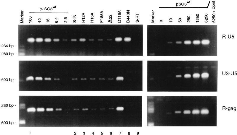
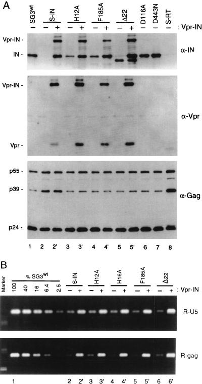
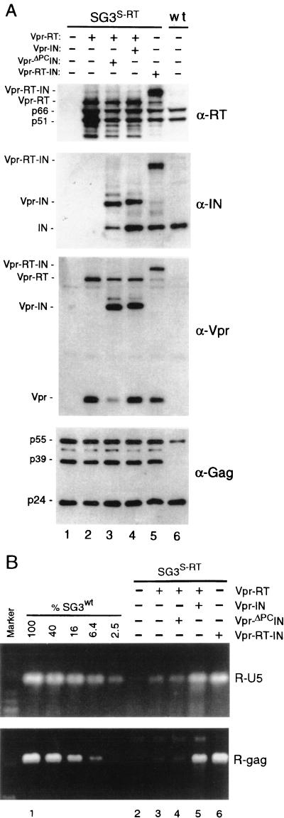
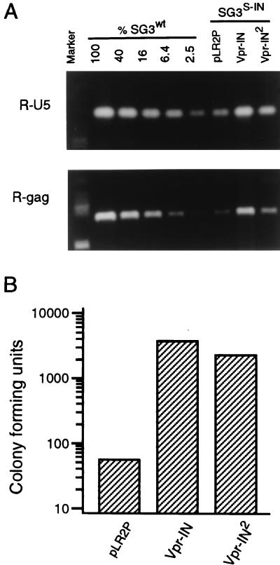
The human immunodeficiency virus type 1 (HIV-1) integrase protein (IN) is essential for integration of the viral DNA into host cell chromosomes. Since IN is expressed and assembled into virions as part of the 160-kDa Gag-Pol precursor polyprotein and catalyzes integration of the provirus in infected cells as a mature 32-kDa protein, mutations in IN are pleiotropic and may affect virus replication at different stages of the virus life cycle in addition to integration. Several different phenotypes have been observed for IN mutant viruses, including defects in virion morphology, protein composition, reverse transcription, nuclear import, and integration. Because the effects of mutations in the IN domain of Gag-Pol can not always be distinguished from those of mutations in the mature IN protein, there remains a significant gap in our understanding of IN function in vivo. To directly analyze the function of the mature IN protein itself, in the context of a replicating virus but independently from that of Gag-Pol, we used an approach developed in our laboratory for incorporating proteins into HIV virions by their expression in trans as fusion partners of either Vpr or Vpx. By providing IN in trans as a Vpr-IN fusion protein, our analysis revealed, for the first time, that the mature IN protein is essential for the efficient initiation of reverse transcription in infected cells and that this function does not require the IN protein to be enzymatically (integration) active. Our findings of a direct physical interaction between IN and reverse transcriptase and the failure of heterologous HIV-2 IN protein to efficiently support reverse transcription indicate that this novel function occurs through specific interactions with other viral components of the reverse transcription initiation complex. Studies involving complementation between integration- and DNA synthesis-defective IN mutants further support this conclusion and reveal that the highly conserved HHCC motif of IN is important for both activities. These findings provide important new insights into IN function and reverse transcription in the context of the nucleoprotein reverse transcription complex within the infected cell. Moreover, they validate a novel approach that obviates the need to mutate Gag-Pol in order to study the role of its individual mature components at the virus replication level.


Figures

FIG. 1
FIG. 1
Mutations in IN can impair reverse transcription. Wild-type (pSG3wt) and mutant (S-IN, H12A, H16A, F185A, Δ22, D116A, S-RT, and D443N) proviral clones were introduced into 293T cells by calcium phosphate DNA transfection methods. Forty-eight hours later, culture supernatants were filtered through 0.45-μm-pore-size filters and analyzed by HIV-1 p24 antigen ELISA (Coulter Inc.). The virus-containing culture supernatants were normalized to 500 ng of p24 antigen (CA), treated with RNase-free DNase H (20 U/ml for 2 h) (Promega Corp.), and placed on cultures of HeLa-CD4 cells at 37°C. After 4 h, the cell monolayers were washed, trypsinized, resuspended in fetal bovine serum, and divided into two aliquots. One aliquot set (which contained 1/10 of the total number of cells) was lysed in phosphate-buffered saline containing 1% Triton X-100 and analyzed by p24 antigen ELISA to quantify intracellular CA protein (Table 1). The other aliquot set was placed back in culture medium at 37°C for an additional 14 h. The cells were then washed, and total DNA was extracted by organic methods. For each DNA extract, 250-pg equivalents (p24 antigen) were analyzed by PCR methods for early (R-U5), intermediate (U3-U5), and late (R-gag) viral DNA products of reverse transcription. The amplified products were resolved on 1.5% agarose gels and stained with ethidium bromide. To assess the relative amount of each of the amplified DNA products, four serial 2.5-fold dilutions of the wild-type (SG3wt) DNA were analyzed in parallel. The undiluted 250-pg sample was arbitrarily set to 100. As standards, 10 to 6,250 copies of the pSG3wt clone were also analyzed by PCR under identical conditions. As a control for the efficiency of DpnI cleavage of potential carryover plasmid DNA, 6,250 copies of pSG3wt DNA were analyzed after digestion with DpnI as described previously (26). The virus origin of the ethidium bromide-stained DNA products was confirmed by Southern blot analysis with a homologous nick-translated probe (data not shown). The ethidium bromide staining intensity of each amplified DNA produce was measured with a Lynx 5000 molecular biology workstation (Applied Imaging, Santa Clara, Calif.). The data shown are from a representative experiment that was repeated three times, each time with independent transfection-derived virus preparations.
FIG. 2
FIG. 2
Analysis of Vpr-IN-complemented virions. Four micrograms of the wild-type and mutant proviral DNA clones was individually transfected (−) into 293T cells and cotransfected (+) with the pLR2P-vprIN expression plasmid. Forty-eight hours later, the culture supernatants were collected, passed through 0.45-μm-pore-size filters, and analyzed for HIV-1 p24 antigen concentration by ELISA. (A) Immunoblot analysis. One-half of the filtered supernatant was centrifuged (125,000 × g for 2 h) over cushions of 20% sucrose. The pellets were lysed and examined by immunoblot analysis with anti-IN (α-IN) (top), anti-Vpr (middle), and anti-Gag (bottom) antibodies as described earlier (57). Vpr-IN-containing H16A virions were identical to the H12A virions (data not shown). (B) The trans-IN protein rescues viral DNA synthesis. Five hundred nanograms of wild-type virus and each of the mutant viruses was used to infect cultures of HeLa-CD4 cells. After 4 h, the cell monolayers were washed, trypsinized, resuspended in fetal bovine serum, and divided into two aliquots. One aliquot set was analyzed by p24 antigen ELISA as described for Fig. 1. The other aliquot set was placed back in culture medium at 37°C. At 18 h postinfection, the cells were washed and total DNA was extracted by organic methods. The extracts were normalized for intracellular CA protein concentration and analyzed by PCR for viral DNA products of reverse transcription as described for Fig. 1. The data are from a representative experiment that was repeated three times, each time with independent virus preparations.
FIG. 3
FIG. 3
The trans-IN protein functions after virus assembly and proteolytic processing. Four micrograms of pSG3S-RT DNA was transfected into 293T cells (−) or cotransfected (+) with the Vpr-RT, Vpr-ΔPCIN, and Vpr-RT-IN expression vectors, respectively. (A) Immunoblot analysis. Transfection-derived virions were concentrated from the culture supernatants by ultracentrifugation (125,000 × g for 2 h) through cushions of 20% sucrose. The pellets were lysed and examined by immunoblot analysis with anti-RT (α-RT), anti-IN, anti-Vpr, and anti-Gag antibodies as indicated. (B) The trans-IN protein is required for viral DNA synthesis. Five hundred nanograms of the transfection-derived viruses was used to infect cultures of HeLa-CD4 cells. DNA products of reverse transcription were prepared and analyzed exactly as described above. The data are from a representative experiment that was repeated three times.
FIG. 4
FIG. 4
Complementation between different IN mutants. (A) Enzymatically defective trans-IN protein supports reverse transcription. Four micrograms of the S-IN, H12A, H16A, F185A, and Δ22 IN mutant proviral clones was transfected alone and separately cotransfected into 293T cells with 2 μg of the Vpr-IND116A or Vpr-IN expression vector. Forty-eight hours later, supernatant virions were prepared and used to infect HeLa-CD4 cells exactly as described in the legend to Fig. 1. The infected cells were washed 18 h later, and total DNA was extracted and treated with DpnI endonuclease. The late R-gag DNA product of reverse transcription was PCR amplified and analyzed as described above. The data are from a representative experiment that was repeated two times. (B) Complementation of proviral DNA integration. The D116A IN mutant was inserted into the SG3 hygromycin-resistant clone, generating Hy-SG3D116A. The Hy-SG3D116A mutant virus produces wild-type levels of viral DNA yet is integration defective. Four micrograms of Hy-SG3D116A was transfected with 2 μg of the control vector (pLR2P) and individually cotransfected with 2 μg of the Vpr-IN, Vpr-INH12A, Vpr-INH16A, Vpr-INF185A, and Vpr-INΔ22 IN mutant expression vectors, respectively. Since the env region of Hy-SG3D116A contains the hygromycin resistance marker, the virions were pseudotyped by including the pCMV-VSV-G env vector in the transfection reactions. Forty-eight hours after transfection, the culture supernatants were filtered through 0.45-μm-pore-size filters and analyzed for HIV-1 p24 antigen concentration by ELISA. Twenty-five nanograms (p24 antigen) of each pseudotyped virus stock was used to infect cultures of HeLa cells. The infected cells were maintained in hygromycin selection medium for 12 days and then stained to identify resistant colonies. These results were highly reproducible in three independent experiments. The data shown are from a single representative experiment.
FIG. 5
FIG. 5
Analysis of heterologous IN. (A) HIV-2 IN protein (IN2) does not efficiently support HIV-1 reverse transcription. Four micrograms of pSG3S-IN was cotransfected into 293T cells with 2 μg of the Vpr-IN, Vpr-IN2, and pLR2P (vector only) expression vectors, respectively. Four micrograms of pSG3wt was also transfected as a control. Forty-eight hours later, supernatant virions were prepared and used to infect HeLa-CD4 cells exactly as described in the legend to Fig. 1. The infected cells were washed 18 h later, and total DNA was extracted and treated with DpnI endonuclease. Early (R-U5) and late (R-gag) viral DNA products of reverse transcription were amplified by PCR and analyzed as described above. The data are from a representative experiment that was repeated three times, each time with independent virus preparations. (B) Complementation of proviral DNA integration. To directly compare the ability of the heterologous trans-IN2 protein to support integration of the provirus with that of the homologous IN protein, the hygromycin-resistant, integration-defective Hy-SG3 INAA35A clone was used for analysis. Hy-SG3 INAA35A contains a mutation in each of the three residues that comprise the catalytic center of the IN protein (D64A, D116A, and E152A) and efficiently synthesizes viral DNA after entry. Four micrograms of Hy-SG3AA35A was cotransfected with 2 μg of the Vpr-IN, and Vpr-IN2 expression plasmids, respectively. The virions were pseudotyped by including the pCMV-VSV-G env vector in the transfection reactions. Forty-eight hours after transfection, the culture supernatants were filtered through 0.45-μm-pore-size filters and analyzed for HIV-1 p24 antigen concentration by ELISA. Twenty-five nanograms (p24 antigen) of each of the pseudotyped virus stocks was used to infect cultures of HeLa cells. The infected cells were maintained in hygromycin selection medium for 12 days and then stained to identify resistant colonies as described earlier (35). These results were highly reproducible in three independent experiments. The data shown are from a single representative experiment.
FIG. 6
FIG. 6
Interaction between recombinant HIV-1 RT and IN proteins. (A) Coomassie blue-stained gel showing the G-bead-bound GST and GST-IN proteins used to pull-down the RT heterodimer. Equal quantities of G-beads bound to no protein (lane 1), GST (lane 2), or GST-IN (lane 3) were incubated with crude bacterial lysates in HND buffer containing RT heterodimer. Following extensive washing, the bound proteins were analyzed by SDS-PAGE. (B) Immunoblot analysis showing RT-IN interaction. A duplicate gel run in parallel to that shown in panel A was transferred to nitrocellulose and probed with the 5B2B2 anti-RT monoclonal antibody. The positions of p66 and p51 polypeptides are indicated. (C) The RT-IN interaction is resistant to micrococcal nuclease digestion. The experiment was similar to that described above except that the HND buffer contained 50 mM Tris-HCl (pH 8.0) and 1 mM CaCl2, and prior to addition of the bacterial lysates, the samples were preincubated with 100 U of micrococcal nuclease at 37°C for 10 min, and the nuclease was inactivated with EGTA.

References

    1. Aiken C. Pseudotyping human immunodeficiency virus type 1 (HIV-1) by the glycoprotein of vesicular stomatitis virus targets HIV-1 entry to an endocytic pathway and suppresses both the requirement for Nef and the sensitivity to cyclosporin A. J Virol. 1997;71:5871–5877. - PMC - PubMed
    1. Aiken C, Trono D. Nef stimulates human immunodeficiency virus type 1 proviral DNA synthesis. J Virol. 1995;69:5048–5056. - PMC - PubMed
    1. Ansari-Lari M A, Gibbs R A. Expression of human immunodeficiency virus type 1 reverse transcriptase in trans during virion release and after infection. J Virol. 1996;70:3870–3875. - PMC - PubMed
    1. Braaten D, Franke E K, Luban J. Cyclophilin A is required for an early step in the virus life cycle of human immunodeficiency virus type 1 before the initiation of reverse transcription. J Virol. 1996;70:3551–3560. - PMC - PubMed
    1. Brown P. Integration. In: Coffin J M, Hughes S H, Varmus H E, editors. Retroviruses. Cold Spring Harbor, N.Y: Cold Spring Harbor Laboratory; 1997. pp. 161–204. - PubMed

Publication types

LinkOut - more resources