. 1999 Mar;73(3):2434-41.
doi: 10.1128/JVI.73.3.2434-2441.1999.

Moloney murine leukemia virus-induced preleukemic thymic atrophy and enhanced thymocyte apoptosis correlate with disease pathogenicity

Affiliations

Moloney murine leukemia virus-induced preleukemic thymic atrophy and enhanced thymocyte apoptosis correlate with disease pathogenicity

C Bonzon et al. J Virol. 1999 Mar.

Abstract

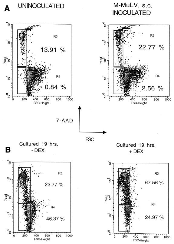
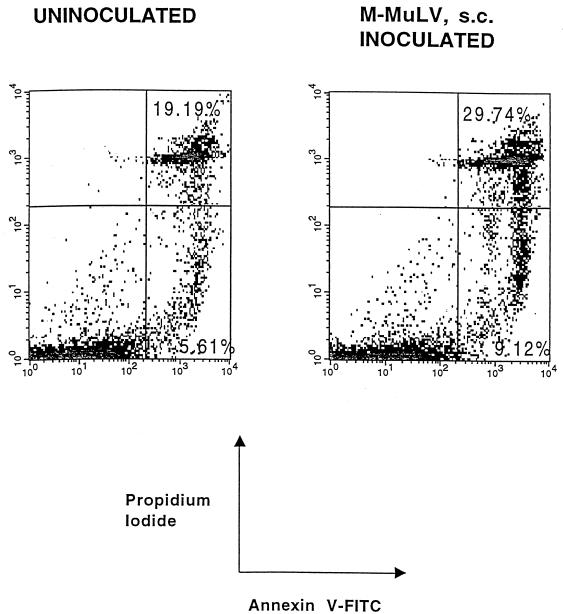
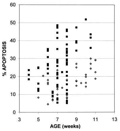
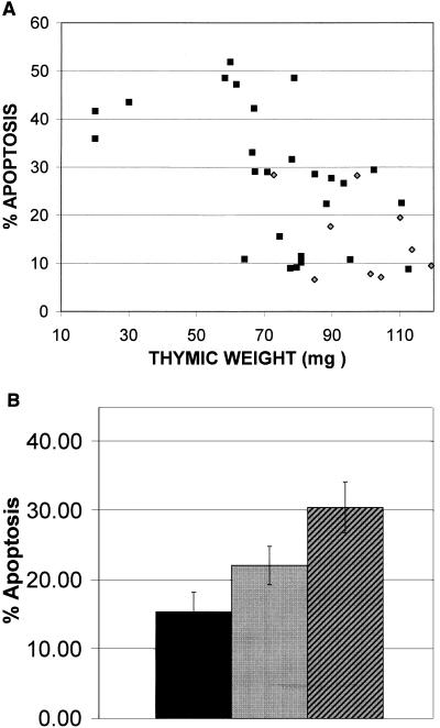
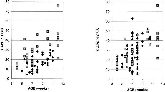
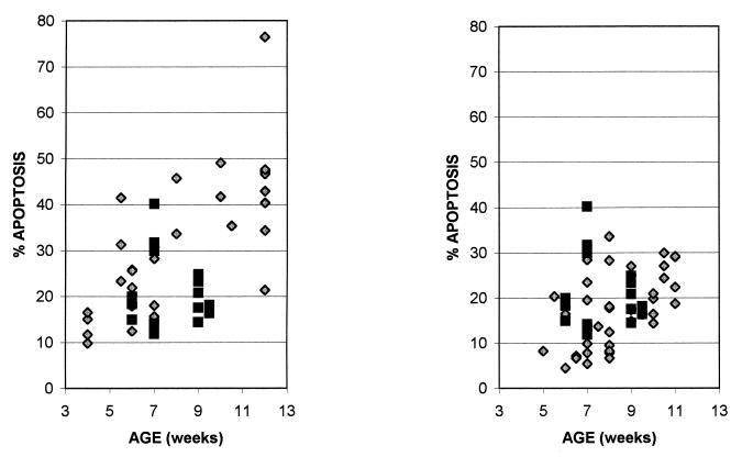
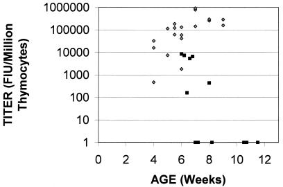
Moloney murine leukemia virus (M-MuLV) is a replication-competent, simple retrovirus that induces T-cell lymphoma with a mean latency of 3 to 4 months. During the preleukemic period (4 to 10 weeks postinoculation) a marked decrease in thymic size is apparent for M-MuLV-inoculated mice in comparison to age-matched uninoculated mice. We were interested in studying whether the thymic regression was due to an increased rate of thymocyte apoptosis in the thymi of M-MuLV-inoculated mice. Neonatal NIH/Swiss mice were inoculated subcutaneously (s.c.) with wild-type M-MuLV (approximately 10(5) XC PFU). Mice were sacrificed at 4 to 11 weeks postinoculation. Thymic single-cell suspensions were prepared and tested for apoptosis by two-parameter flow cytometry. Indications of apoptosis included changes in cell size and staining with 7-aminoactinomycin D or annexin V. The levels of thymocyte apoptosis were significantly higher in M-MuLV-inoculated mice than in uninoculated control animals, and the levels of apoptosis were correlated with thymic atrophy. To test the relevance of enhanced thymocyte apoptosis to leukemogenesis, mice were inoculated with the Mo+PyF101 enhancer variant of M-MuLV. When inoculated intraperitoneally, a route that results in wild-type M-MuLV leukemogenesis, mice displayed levels of enhanced thymocyte apoptosis comparable to those seen with wild-type M-MuLV. However, in mice inoculated s.c., a route that results in attenuated leukemogenesis, significantly lower levels of apoptosis were observed. This supported a role for higher levels of thymocyte apoptosis in M-MuLV leukemogenesis. To examine the possible role of mink cell focus-forming (MCF) recombinant virus in raising levels of thymocyte apoptosis, MCF-specific focal immunofluorescence assays were performed on thymocytes from preleukemic mice inoculated with M-MuLV and Mo+PyF101 M-MuLV. The results indicated that infection of thymocytes by MCF virus recombinants is not required for the increased level of apoptosis and thymic atrophy.


Figures

FIG. 1
FIG. 1
Detection of apoptosis in thymocytes. (A) Thymocytes from 6-week-old uninoculated and M-MuLV-inoculated mice were subjected to two-parameter flow cytometric analysis for FSC and 7-AAD fluorescence. 7-AAD fluorescence is plotted on a logarithmic scale. Regions R3 and R4 represent thymocytes that are in the intermediate-to-late and early stages of apoptosis, respectively. Percentages of cells in both these regions are shown. (B) Dexamethasone-induced apoptosis. Primary thymocytes from an uninoculated mouse were cultured in vitro for 19 h in the presence (+DEX) or absence (−DEX) of 0.1 mM dexamethasone. The cells were then incubated with 7-AAD and subjected to two-parameter flow cytometric analysis to determine the percentages of cells in the R3 and R4 regions of late-to-intermediate- and early-stage apoptosis. Percentages of cells in both these regions are shown.
FIG. 2
FIG. 2
Annexin V-PI staining. Thymocytes from the same 6-week-old animals for which results are shown in Fig. 1 were incubated with FITC-conjugated annexin V and PI, and flow cytometric analysis was performed. Percentages of late-stage apoptotic cells (upper right quadrant) and early-stage apoptotic cells (lower right quadrant) are shown. Both FITC-conjugated annexin V and PI fluorescence are plotted on logarithmic scales.
FIG. 3
FIG. 3
Thymocyte apoptosis in M-MuLV-inoculated and uninoculated mice. Thymocytes from uninoculated (diamonds) and M-MuLV-inoculated (squares) mice were analyzed for apoptosis, calculated as the sum of the percentages of cells in the R3 and R4 regions of intermediate-to-late- and early-stage apoptosis (see Fig. 1A). Mice were sacrificed between 4 and 11 weeks postinoculation. All data points represent individual animals.
FIG. 4
FIG. 4
Thymic atrophy correlates with elevated levels of thymocyte apoptosis. At the time of sacrifice, thymi were dissected, removed, and weighed, as a measure of thymic atrophy. (A) A comparison of the levels of thymocyte apoptosis (calculated as the sum of the percentages of cells in regions R3 and R4) with thymic weights (in milligrams) in M-MuLV-inoculated (squares) and uninoculated (diamonds) mice is shown. All data points represent individual animals. (B) Thymi from M-MuLV-inoculated mice were scored as having either no thymic atrophy (thymic weight was within 1 standard deviation of the mean thymic weight of uninoculated mice) or thymic atrophy (thymic weight was more than 1 standard deviation below the mean thymic weight of uninoculated mice). A comparison of the mean levels of thymocyte apoptosis observed in these mice and uninoculated mice is shown. Black bar, uninoculated mice (mean weight, 97.47 mg); gray bar, no atrophy (mean weight, 96.36 mg); hatched bar, atrophy (mean weight, 63.74 mg). Average thymic weights of all three categories are displayed, and error bars are based on standard error values.
FIG. 5
FIG. 5
Thymocyte apoptosis in mice inoculated i.p. with Mo+PyF101 M-MuLV. (Left panel) Thymocytes from uninoculated mice (diamonds) and mice inoculated i.p. with Mo+PyF101 M-MuLV (squares) were analyzed for apoptosis, determined as the sum of the percentages of cells in the R3 and R4 regions of intermediate-to-late- and early-stage apoptosis. Mice were sacrificed between 4 and 12 weeks postinoculation. All data points represent individual animals. (Right panel) A comparison of the levels of thymocyte apoptosis in mice inoculated s.c. with wild-type M-MuLV (diamonds) and mice inoculated i.p. with Mo+PyF101 M-MuLV (squares) is shown.
FIG. 6
FIG. 6
Mice inoculated s.c. with Mo+PyF101 M-MuLV. (Left panel) Thymocytes from mice inoculated s.c. with Mo+PyF101 M-MuLV were analyzed for apoptosis, determined as the sum of the percentages of cells in the R3 and R4 regions of intermediate-to-late- and early-stage apoptosis. A comparison of the levels of thymocyte apoptosis in mice inoculated i.p. with Mo+PyF101 M-MuLV (diamonds) and those for mice inoculated s.c. (squares) is shown. (Right panel) A comparison of the levels of thymocyte apoptosis for uninoculated mice (diamonds) and those observed for mice inoculated s.c. with Mo+PyF101 M-MuLV (squares) is shown.
FIG. 7
FIG. 7
MCF recombinant virus infection of thymocytes in preleukemic mice. Thymocytes from mice inoculated s.c. with M-MuLV (diamonds) and from mice inoculated i.p. with Mo+PyF101 M-MuLV (squares) were tested for productive MCF virus infection by FIA. Titers are shown on a logarithmic scale (FIU per 106 thymocytes).

References

    1. Andree H A, Reutelingsperger C P, Hauptmann R, Hemker H C, Hermens W T, Willems G M. Binding of vascular anticoagulant alpha (VAC alpha) to planar phospholipid bilayers. J Biol Chem. 1990;265:4923–4928. - PubMed
    1. Belli B, Fan H. The leukemogenic potential of an enhancer variant of Moloney murine leukemia virus varies with the route of inoculation. J Virol. 1994;68:6883–6889. - PMC - PubMed
    1. Belli B, Patel A, Fan H. Recombinant mink cell focus-inducing virus and long terminal repeat alterations accompany the increased leukemogenicity of the Mo+PyF101 variant of Moloney murine leukemia virus after intraperitoneal inoculation. J Virol. 1995;69:1037–1043. - PMC - PubMed
    1. Brightman B K, Rein A, Trepp D J, Fan H. An enhancer variant of Moloney murine leukemia virus defective in leukemogenesis does not generate detectable mink cell focus-inducing virus in vivo. Proc Natl Acad Sci USA. 1991;88:2264–2268. . (Erratum, 88:5066.) - PMC - PubMed
    1. Chesebro B, Britt W, Evans L, Wehrly K, Nishio J, Cloyd M. Characterization of monoclonal antibodies reactive with murine leukemia viruses: use in analysis of strains of Friend MCF and Friend ecotropic murine leukemia virus. Virology. 1983;127:134–148. - PubMed

Publication types

LinkOut - more resources