. 1999 Feb;181(4):1134-40.
doi: 10.1128/JB.181.4.1134-1140.1999.

Analysis of phosphorylated sphingolipid long-chain bases reveals potential roles in heat stress and growth control in Saccharomyces

Affiliations

Analysis of phosphorylated sphingolipid long-chain bases reveals potential roles in heat stress and growth control in Saccharomyces

M S Skrzypek et al. J Bacteriol. 1999 Feb.

Abstract

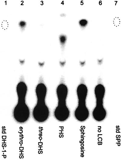
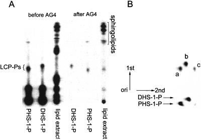
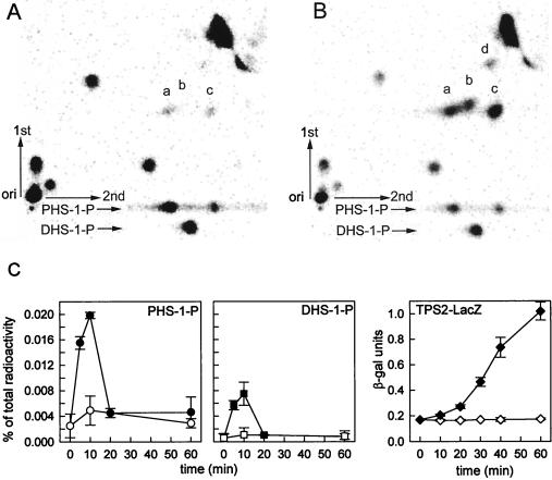
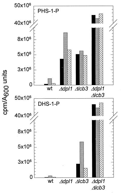
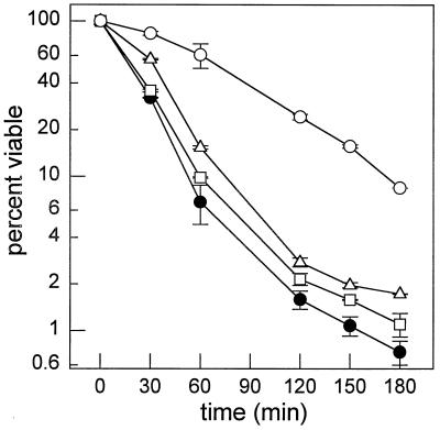
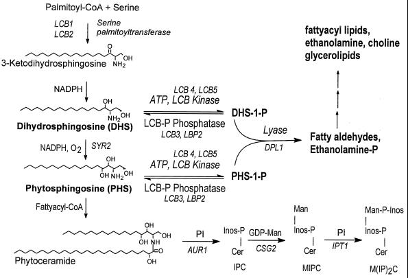
Sphingolipid long-chain bases and their phosphorylated derivatives, for example, sphingosine-1-phosphate in mammals, have been implicated as signaling molecules. The possibility that Saccharomyces cerevisiae cells also use long-chain-base phosphates to regulate cellular processes has only recently begun to be examined. Here we present a simple and sensitive procedure for analyzing and quantifying long-chain-base phosphates in S. cerevisiae cells. Our data show for the first time that phytosphingosine-1-phosphate (PHS-1-P) is present at a low but detectable level in cells grown on a fermentable carbon source at 25 degreesC, while dihydrosphingosine-1-phosphate (DHS-1-P) is only barely detectable. Shifting cells to 37 degreesC causes transient eight- and fivefold increases in levels of PHS-1-P and DHS-1-P, respectively, which peak after about 10 min. The amounts of both compounds return to the unstressed levels by 20 min after the temperature shift. These data are consistent with PHS-1-P and DHS-1-P being signaling molecules. Cells unable to break down long-chain-base phosphates, due to deletion of DPL1 and LCB3, show a 500-fold increase in PHS-1-P and DHS-1-P levels, grow slowly, and survive a 44 degreesC heat stress 10-fold better than parental cells. These and other data for dpl1 or lcb3 single-mutant strains suggest that DHS-1-P and/or PHS-1-P act as signals for resistance to heat stress. Our procedure should expedite experiments to determine how the synthesis and breakdown of these compounds is regulated and how the compounds mediate resistance to elevated temperature.


Figures

FIG. 1
FIG. 1
Synthesis of radioactive long-chain-base phosphates by soluble yeast proteins. Long-chain bases (12 μM) including dl-erythro-DHS (lane 2), l-threo-DHS (lane 3), PHS (lane 4), and d-erythro-sphingosine (lane 5) were incubated separately with [γ-32P]ATP, and soluble proteins were derived from strain MSS200 as described in Materials and Methods. The reaction mixtures were analyzed directly by TLC using solvent C. The locations of nonradioactive standards (std) for DHS-1-P (lane 1) and SPP (lane 7), identified by charring the plate, are indicated by dotted ovals.
FIG. 2
FIG. 2
Chromatographic analysis of long-chain-base phosphates (LCP-Ps). (A) DHS-1-P and PHS-1-P standards, made as described for Fig. 1, and a lipid extract from MSS200 cells were analyzed by TLC using solvent B before or after chromatography on an AG4 column. All samples were radiolabeled with 32P. (B) DHS-1-P and PHS-1-P standards, purified by chromatography on an AG4 column, were combined and separated by two-dimensional TLC using solvent B in the first dimension and solvent C in the second. Purified DHS-1-P and PHS-1-P standards were also run in the second dimension, as indicated at the bottom. Spots: a and c, PHS-1-P; b, DHS-1-P.
FIG. 3
FIG. 3
PHS-1-P and DHS-1-P transiently increase during heat stress, as determined by analysis of 32P-labeled long-chain-base phosphates present in MSS200 cells grown in PYED medium at 25°C (A) or following a shift to 37°C (B). Cells were radiolabeled, and lipids were extracted and processed as described in Materials and Methods. Lipids were separated by two-dimensional TLC with solvent B used first and solvent C used second. Purified DHS-1-P and PHS-1-P standards were also run in the second dimension as indicated at the bottom. Spots: a, c, and d, PHS-1-P; b, DHS-1-P. (C) lipid extracts were prepared at various times from cells grown at 25°C (open symbols) or following transfer to 37°C at time zero (filled symbols). The amount of radioactivity in DHS-1-P (spot a) and PHS-1-P (sum of spots b, c, and d) was quantified in each sample by using a PhosphorImager and expressed as a percentage of the counts present in the total lipid extract. The rightmost panel shows the amount of β-galactosidase activity in the cells grown at 25°C (open diamonds) or 37°C (filled diamonds). Data are the means ± standard deviations for two experiments.
FIG. 4
FIG. 4
Analysis of long-chain-base phosphates in wild-type (wt) strain MSS201 and mutant strains MSS204 (Δdpl1), MSS205 (Δlcb3), and MSS207 (Δdpl1 Δlcb3). The types and amounts of 32P-labeled long-chain-base phosphates present in log-phase cells grown at 25°C (time zero; black bars) and after 10 min (gray bars) and 40 min (cross-hatched bars) of incubation at 37°C were analyzed by two-dimensional TLC using solvent B in the first dimension and solvent C in the second dimension. Spots corresponding to PHS-1-P (top) and DHS-1-P (bottom) were quantified by PhosphorImager analysis of the TLC plate and are represented on the y axis. Purified PHS-1-P and DHS-1-P standards were run for comparison.
FIG. 5
FIG. 5
Survival during heat stress. The percentage of cells able to form colonies after incubation at 44°C for the indicated times was determined for wild-type (strain MSS201; filled circles), Δdpl1 (strain MSS204; squares), Δlcb3 (strain MSS205; triangles), and Δdpl1 Δlcb3 (strain MSS207; circles) cells. Data represent average values ± standard deviations for two separate experiments. Some error bars are covered by the symbols. A separate isolate of the Δdpl1 Δlcb3 double mutant strain gave the same result.
FIG. 6
FIG. 6
Metabolism of long-chain-base phosphates in S. cerevisiae. Known pathway intermediates, substrates, and cofactors are indicated. Genes are in italics. Abbreviations: IPC, inositol phosphorylceramide; Man, mannose; MIPC, mannose inositol-P-ceramide; M(IP)2C, mannose-(inositol-P)2-ceramide; PI, phosphatidylinositol; Cer, ceramide.

References

    1. Bornfeldt K E, Graves L M, Raines E W, Igarashi Y, Wayman G, Yamamura S, Yatomi Y, Sidhy J S, Krebs E G, Hakomori S, Ross R. Sphingosine-1-phosphate inhibits PDGF-induced chemotaxis of human arterial smooth muscle cells: spatial and temporal modulation of PDGF chemotactic signal transduction. J Cell Biol. 1995;130:193–206. - PMC - PubMed
    1. Clarke N G, Dawson R M C. Alkaline O-N transacylation. Biochem J. 1981;195:301–306. - PMC - PubMed
    1. Cross F R. ‘Marker swap’ plasmids: convenient tools for budding yeast molecular genetics. Yeast. 1997;13:647–653. - PubMed
    1. Crowther G J, Lynch D V. Characterization of sphinganine kinase activity in corn shoot microsomes. Arch Biochem Biophys. 1997;337:284–290. - PubMed
    1. De Virgilio C, Hottiger T, Dominguez J, Boller T, Wiemken A. The role of trehalose synthesis for the acquisition of thermotolerance in yeast. I. Genetic evidence that trehalose is a thermoprotectant. Eur J Biochem. 1994;219:179–186. - PubMed

Publication types

MeSH terms

LinkOut - more resources