Comparative Study
. 1999 Feb;181(4):1343-7.
doi: 10.1128/JB.181.4.1343-1347.1999.

Cloning and characterization of arylamine N-acetyltransferase genes from Mycobacterium smegmatis and Mycobacterium tuberculosis: increased expression results in isoniazid resistance

Affiliations
Comparative Study

Cloning and characterization of arylamine N-acetyltransferase genes from Mycobacterium smegmatis and Mycobacterium tuberculosis: increased expression results in isoniazid resistance

M Payton et al. J Bacteriol. 1999 Feb.

Abstract

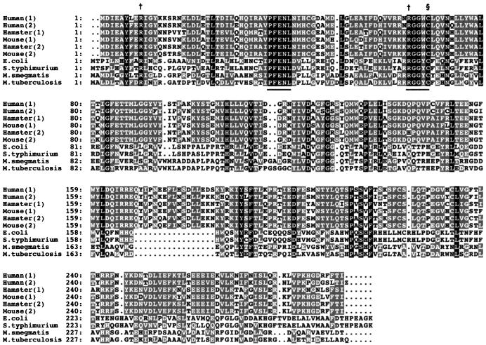
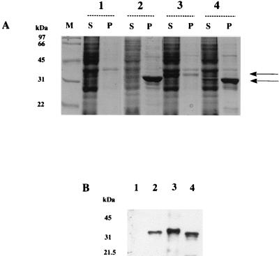
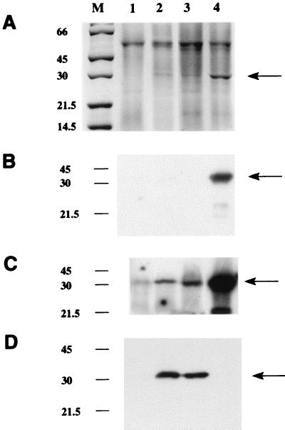
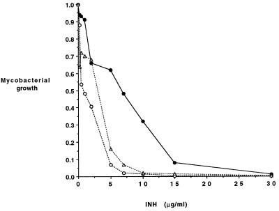
Arylamine N-acetyltransferases (NATs) are found in many eukaryotic organisms, including humans, and have previously been identified in the prokaryote Salmonella typhimurium. NATs from many sources acetylate the antitubercular drug isoniazid and so inactivate it. nat genes were cloned from Mycobacterium smegmatis and Mycobacterium tuberculosis, and expressed in Escherichia coli and M. smegmatis. The induced M. smegmatis NAT catalyzes the acetylation of isoniazid. A monospecific antiserum raised against pure NAT from S. typhimurium recognizes NAT from M. smegmatis and cross-reacts with recombinant NAT from M. tuberculosis. Overexpression of mycobacterial nat genes in E. coli results in predominantly insoluble recombinant protein; however, with M. smegmatis as the host using the vector pACE-1, NAT proteins from M. tuberculosis and M. smegmatis are soluble. M. smegmatis transformants induced to express the M. tuberculosis nat gene in culture demonstrated a threefold higher resistance to isoniazid. We propose that NAT in mycobacteria could have a role in acetylating, and hence inactivating, isoniazid.


Figures

FIG. 1
FIG. 1
Amino acid alignment of the predicted M. smegmatis and M. tuberculosis NAT against NAT homologues of eukaryotic and prokaryotic origins. Amino acids are represented by the single-letter code. Numbers refer to amino acid positions of the aligned sequences, which are ranked in order of decreasing similarity in comparison with human NAT1. Codes on the left identify the relevant NAT sequence. Human(1) is human NAT1, accession no. P18440; Human(2) is human NAT2, accession no. P11245; Hamster(1) is hamster NAT1, accession no. P50292; Mouse(1) is mouse NAT1, accession no. P50294; Hamster(2) is hamster NAT2, accession no. P50293; Mouse(2) is mouse NAT2, accession no. P50295; E. coli is E. coli NAT, accession no. P77567; S. typhimurium is S. typhimurium NAT, accession no. Q00267; M. tuberculosis is M. tuberculosis NAT, accession no. P96848, which codes for a hypothetical protein (3) and has the same sequence as the M. tuberculosis NAT described in this report; and M. smegmatis is M. smegmatis NAT, accession no. AJ006588. Amino acids showing identity in all NATs are shown in white on a black background, while residues showing conservation and similarity in more than four species of NAT are indicated in white on a grey background. A dot represents a gap introduced to maximize homology. The proposed active site cysteine is indicated (§) (32), as are the arginine residues thought to participate in the reaction (†) (6, 32). Regions of complete identity are underscored.
FIG. 2
FIG. 2
Sodium dodecyl sulfate-polyacrylamide gel electrophoresis analysis of recombinant NAT proteins in E. coli. (A) Gels were stained with Coomassie blue. E. coli cells were transformed with pET28b alone (lanes 1) or with pET28b constructs containing the coding region of nat from M. tuberculosis (lanes 2), S. typhimurium (lanes 3), or M. smegmatis (lanes 4). Lysates were used to generate supernatants (S) and pellets resuspended in the original volume of lysate (P). Individual lanes were loaded with 15 μl of each fraction. The upper and lower arrows indicate the migration of recombinant S. typhimurium and mycobacterial NATs, respectively. Lane M, molecular mass markers. (B) Western blot analysis using an antiserum (1:100,000) against recombinant S. typhimurium NAT developed by using an enhanced chemiluminescence detection system (27). Lane 1, supernatant from E. coli transformed with pET28b alone; lane 2, pellet from E. coli transformed with M. tuberculosis NAT; lane 3, supernatant from E. coli transformed with S. typhimurium NAT; lane 4, supernatant from E. coli transformed with M. smegmatis NAT.
FIG. 3
FIG. 3
Sodium dodecyl sulfate-polyacrylamide gel electrophoresis analysis of recombinant NAT proteins in M. smegmatis. Gels were loaded with 15 μl of supernatants from lysates of M. smegmatis transformed with pACE-1 alone (lane 1), with M. tuberculosis nat (lanes 2 and 3), or with M. smegmatis nat (lane 4) (M, molecular mass markers [in kilodaltons]). (A) Staining with Coomassie blue. The arrow indicates the additional band in the supernatants transformed with a nat gene. (B to D) Western blots developed by using an enhanced chemiluminescence detection system as described previously (27), where exposure time for detection of the second antibody is either 1 min (B and D) or 20 min (C). In each panel, lane 1 has been loaded with 10 μg of total protein and lanes 2 to 4 have been loaded with 600 ng of total protein. An antiserum raised against recombinant M. tuberculosis NAT protein synthesized in E. coli is shown in panel D. The antiserum used in panels B and C is the antiserum against pure S. typhimurium NAT (24). Both antisera were used at a dilution of 1:100,000.
FIG. 4
FIG. 4
Effect of expression of M. tuberculosis nat on the growth of M. smegmatis in the presence of isoniazid (INH). Results for cultures of M. smegmatis either transformed with pACE-1 alone (open circles) or with pACE-1 containing M. tuberculosis nat (solid circles) grown in minimal medium containing acetamide and cultures of pACE-1 containing M. tuberculosis nat grown in minimal medium containing glucose (triangles) are shown.

References

    1. Blattner F, Plunkett G, Bloch C, Perna N, Burland V, Riley M, Collado-Vides J, Glasner J, Rode C, Mayhew G, Gregor J, Davis N, Kirkpatrick H, Goeden M, Rose D, Mau B, Shao Y. The complete genome sequence of Escherichia coli K12. Science. 1997;277:1453–1474. - PubMed
    1. Blum M, Grant D M, McBride W, Heim M, Meyer U A. Human arylamine N-acetyltransferase genes: isolation, chromosomal localization, and functional expression. DNA Cell Biol. 1990;9:193–203. - PubMed
    1. Cole S T, Brosch R, Parkhill J, Garnier T, Churcher C, Harris D, Barrel B. Deciphering the biology of Mycobacterium tuberculosis from the complete genome sequence. Nature. 1998;393:537–544. - PubMed
    1. Cribb A E, Nakamura H, Grant D M, Miller M A, Spielberg S P. Role of polymorphic and monomorphic human arylamine N-acetyltransferases in determining sulfamethoxazole metabolism. Biochem Pharmacol. 1993;45:1277–1282. - PubMed
    1. Deguchi T. Physiology and molecular biology of arylamine N-acetyltransferases. Biomed Res. 1992;13:231–242.

Publication types

MeSH terms

Associated data

LinkOut - more resources