

The Borrelia burgdorferi 37-kilodalton immunoblot band (P37) used in serodiagnosis of early lyme disease is the flaA gene product
- PMID: 9986810
- PMCID: PMC84463
- DOI: 10.1128/JCM.37.3.548-552.1999
The Borrelia burgdorferi 37-kilodalton immunoblot band (P37) used in serodiagnosis of early lyme disease is the flaA gene product
Abstract
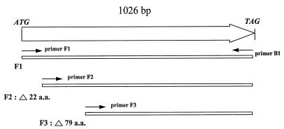
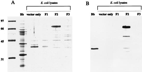
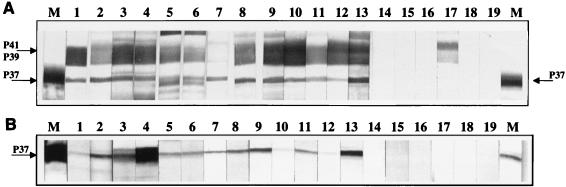
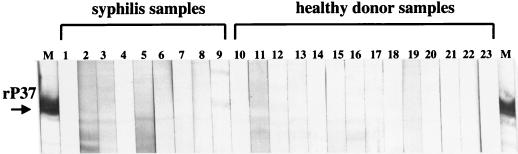
The 37-kDa protein (P37) of Borrelia burgdorferi is an antigen that elicits an early immunoglobulin M (IgM) antibody response in Lyme disease patients. The P37 gene was cloned from a B. burgdorferi genomic library by screening with antibody from a Lyme disease patient who had developed a prominent humoral response to the P37 antigen. DNA sequence analysis of this clone revealed the identity of P37 to be FlaA, an outer sheath protein of the periplasmic flagella. Recombinant P37 expression was accomplished in Escherichia coli by using a gene construct with the leader peptide deleted and fused to a 38-kDa E. coli protein. The recombinant antigen was reactive in IgM immunoblots using serum samples from patients clinically diagnosed with early Lyme disease that had been scored positive for B. burgdorferi anti-P37 reactivity. Lyme disease patient samples serologically negative for the B. burgdorferi P37 protein did not react with the recombinant. Recombinant P37 may be a useful component of a set of defined antigens for the serodiagnosis of early Lyme disease. This protein can be utilized as a marker in diagnostic immunoblots, aiding in the standardization of the present generation of IgM serologic tests.
Figures
References
-
- Association of State and Territorial Public Health Laboratory Directors and the Centers for Disease Control and Prevention. Proceedings of the Second National Conference on Serologic Diagnosis of Lyme disease, Dearborn, Mich. Washington, D.C: Association of State and Territorial Public Health Laboratory Directors; 1995. Recommendations; pp. 1–7.
-
- Bruckbauer H R, Preac-Mursic V, Fuchs R, Wilske B. Cross-reactive proteins of Borrelia burgdorferi. Eur J Clin Microbiol Infect Dis. 1992;11:224–232. - PubMed
-
- Dressler F, Whalen J A, Reinhardt B N, Steere A C. Western blotting in the serodiagnosis of Lyme disease. J Infect Dis. 1993;167:392–400. - PubMed
Publication types
MeSH terms
Substances
LinkOut - more resources
Full Text Sources
Other Literature Sources
Medical
