. 1999 Mar;37(3):615-9.
doi: 10.1128/JCM.37.3.615-619.1999.

Improved silica-guanidiniumthiocyanate DNA isolation procedure based on selective binding of bovine alpha-casein to silica particles

Affiliations

Improved silica-guanidiniumthiocyanate DNA isolation procedure based on selective binding of bovine alpha-casein to silica particles

R Boom et al. J Clin Microbiol. 1999 Mar.

Abstract

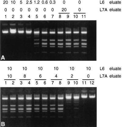
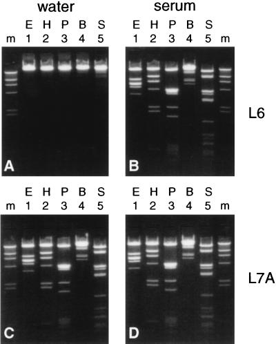
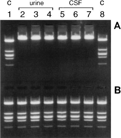
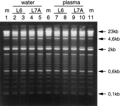
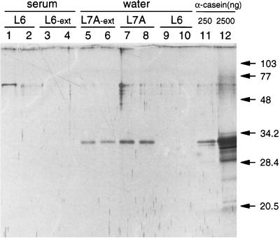
DNA purified from clinical cerebrospinal fluid and urine specimens by a silica-guanidiniumthiocyanate procedure frequently contained an inhibitor(s) of DNA-processing enzymes which may have been introduced by the purification procedure itself. Inhibition could be relieved by the use of a novel lysis buffer containing alpha-casein. When the novel lysis buffer was used, alpha-casein was bound by the silica particles in the first step of the procedure and eluted together with DNA in the last step, after which it exerted its beneficial effects for DNA-processing enzymes. In the present study we have compared the novel lysis buffer with the previously described lysis buffer with respect to double-stranded DNA yield (which was nearly 100%) and the performance of DNA-processing enzymes.


Figures

FIG. 1
FIG. 1
Phage lambda DNA was added to eluates resulting from water extractions, and DNA was digested with EcoRI in a constant 30-μl reaction volume (after adjustment with TE buffer). (A) Decreasing amounts of L6 eluate (20, 10, 5, 2.5, 1.25, 0.6, and 0.3 μl; lanes 1 to 7, respectively). Control digestions contained 20 μl of L7A eluate (lanes 8 and 9) and TE buffer only (lanes 10 and 11). (B) To a constant amount of L6 eluate (10 μl), decreasing amounts of L7A eluate were added. Lanes 1 and 2, 10 μl of L7A eluate; lanes 3 and 4, 8 μl of L7A eluate; lanes 5 and 6, 6 μl of L7A eluate; lanes 7 and 8, 4 μl of L7A eluate; lanes 9 and 10, 2 μl of L7A eluate, lanes 11 and 12, no L7A eluate.
FIG. 2
FIG. 2
Extractions were done with buffer L6 (A and B) and buffer L7A (C and D) with either water (A and C) or serum (B and D) as the input for extraction. Phage lambda DNA was added to 20 μl of the eluates, and DNA was digested with restriction enzymes. Lanes 1, EcoRI (E); lanes 2, HindIII (H); lanes 3, PvuII (P); lanes 4, BamHI (B); lanes 5, SspI (S); lanes m, length marker (HindIII digest of phage lambda DNA [500 ng]).
FIG. 3
FIG. 3
Three different urine specimens and three different CSF specimens were used as input for extraction with buffer L6 or buffer L7A. Phage lambda DNA was added to 20 μl of the eluates, and DNA was digested with EcoRI. (A) Buffer L6; (B) buffer L7A. Lanes 1 and 8, control (C) digests in TE buffer; lanes 2 to 4, urine specimens; lanes 5 to 7, CSF specimens.
FIG. 4
FIG. 4
HindIII-digested phage lambda DNA (4 μg) and a 100-bp DNA ladder (2 μg) were added to water or plasma, DNA was extracted from these mixtures with either buffer L6 or buffer L7A, and 25 μl of the eluates was electrophoresed through a 1.5% agarose gel. Lanes 1, 6, and 11, 100% recovery markers (m); lanes 2 to 5, extraction from water; lanes 2 and 3, extraction with buffer L6; lanes 4 and 5, extraction with buffer L7A; lanes 7 to 10, extraction from plasma; lanes 7 and 8, extraction with buffer L6; lanes 9 and 10, extraction with buffer L7A.
FIG. 5
FIG. 5
Extractions were done (in duplicate) with serum (lanes 1 to 4) or water (lanes 5 to 10) as input for extraction with buffer L6 (lanes 1 to 4, 9, and 10) or buffer L7A (lanes 5 to 8). In addition, in some extractions the silica particles were extensively (Ext) washed (lanes 3 to 6). Elution was with 60 μl of TE buffer; 10 μl was electrophoresed through a 15% polyacrylamide gel followed by silver staining. Marker lanes contained 250 ng (lane 11) and 2,500 ng (lane 12) of alpha-casein. Molecular masses (in kilodaltons) are indicated.

References

    1. Aaij C, Borst P. The gel electrophoresis of DNA. Biochim Biophys Acta. 1972;269:192–200. - PubMed
    1. Akrigg A, Wilkinson G W G, Oram J D. The structure of the major immediate early gene of human cytomegalovirus strain AD169. Virus Res. 1985;2:107–121. - PubMed
    1. Bernos E, Girardet J M, Humbert G, Linden G. Role of the O-phosphoserine clusters in the interaction of the bovine milk alpha s1-, beta-, kappa-caseins and the PP3 component with immobilized iron (III) ions. Biochim Biophys Acta. 1997;1337:149–159. - PubMed
    1. Blum H, Beier H, Gross H J. Improved silver staining of plant proteins, RNA and DNA in polyacrylamide gels. Electrophoresis. 1987;8:93–98.
    1. Boom, R. Unpublished data.

LinkOut - more resources