

Mycoplasma hyopneumoniae potentiation of porcine reproductive and respiratory syndrome virus-induced pneumonia
- PMID: 9986823
- PMCID: PMC84495
- DOI: 10.1128/JCM.37.3.620-627.1999
Mycoplasma hyopneumoniae potentiation of porcine reproductive and respiratory syndrome virus-induced pneumonia
Abstract
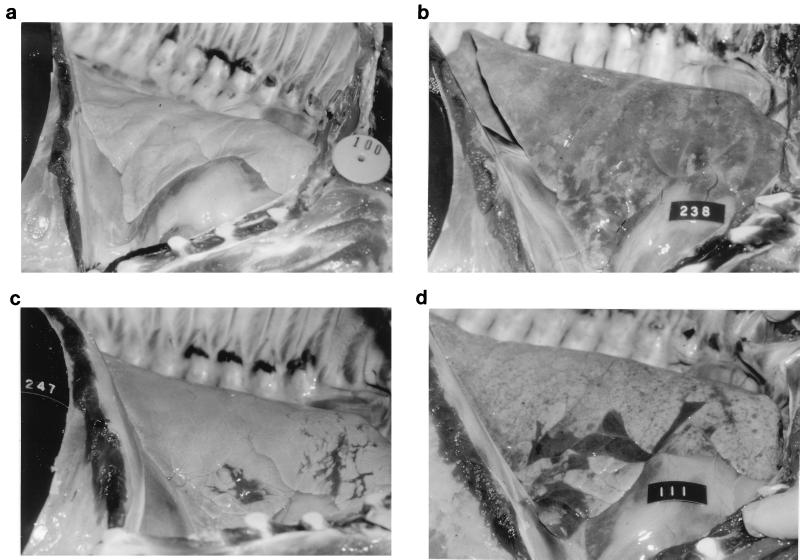
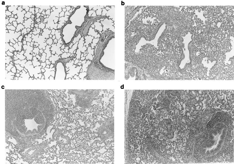
An experimental model that demonstrates a mycoplasma species acting to potentiate a viral pneumonia was developed. Mycoplasma hyopneumoniae, which produces a chronic, lymphohistiocytic bronchopneumonia in pigs, was found to potentiate the severity and the duration of a virus-induced pneumonia in pigs. Pigs were inoculated with M. hyopneumoniae 21 days prior to, simultaneously with, or 10 days after inoculation with porcine reproductive and respiratory syndrome virus (PRRSV), which induces an acute interstitial pneumonia in pigs. PRRSV-induced clinical respiratory disease and macroscopic and microscopic pneumonic lesions were more severe and persistent in M. hyopneumoniae-infected pigs. At 28 or 38 days after PRRSV inoculation, M. hyopneumoniae-infected pigs still exhibited lesions typical of PRRSV-induced pneumonia, whereas the lungs of pigs which had received only PRRSV were essentially normal. On the basis of macroscopic lung lesions, it appears that PRRSV infection did not influence the severity of M. hyopneumoniae infection, although microscopic lesions typical of M. hyopneumoniae were more severe in PRRSV-infected pigs. These results indicate that M. hyopneumoniae infection potentiates PRRSV-induced disease and lesions. Most importantly, M. hyopneumoniae-infected pigs with minimal to nondetectable mycoplasmal pneumonia lesions manifested significantly increased PRRSV-induced pneumonia lesions compared to pigs infected with PRRSV only. This discovery is important with respect to the control of respiratory disease in pigs and has implications in elucidating the potential contribution of mycoplasmas in the pathogenesis of viral infections of other species, including humans.
Figures
References
-
- Amanfu W, Weng C N, Ross R F, Barnes H J. Diagnosis of mycoplasmal pneumoniae of swine: sequential study by direct immunofluorescence. Am J Vet Res. 1984;45:1349–1352. - PubMed
-
- Bereiter M, Young T F, Joo H S, Ross R F. Evaluation of the ELISA and comparison to the complement fixation test and radial immunodiffusion enzyme assay for detection of antibodies against Mycoplasma hyopneumoniae in swine serum. Vet Microbiol. 1990;25:177–192. - PubMed
-
- Blanchard A, Montagnier L. AIDS-associated mycoplasmas. Annu Rev Microbiol. 1994;48:687–712. - PubMed
-
- Collins J E, Benfield D A, Christianson W T, Harris L, Hennings J C, Shaw D P, Goyal S M, McCullough S, Morrison R B, Joo H S, Gorcyca D, Chladek D. Isolation of swine infertility and respiratory syndrome virus (isolate ATCC VR-2332) in North America and experimental reproduction of the disease in gnotobiotic pigs. J Vet Diagn Investig. 1992;4:117–126. - PubMed
Publication types
MeSH terms
LinkOut - more resources
Full Text Sources
Other Literature Sources
