. 1999 Feb 15;189(4):673-82.
doi: 10.1084/jem.189.4.673.

Augmentation of antigen receptor-mediated responses by histamine H1 receptor signaling

Affiliations

Augmentation of antigen receptor-mediated responses by histamine H1 receptor signaling

Y Banu et al. J Exp Med. .

Abstract

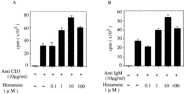
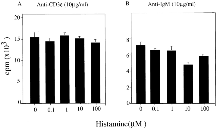
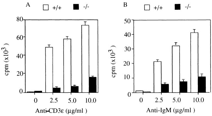
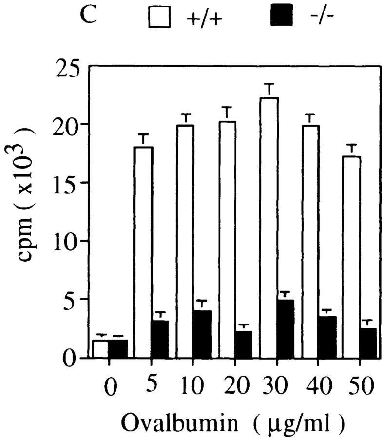
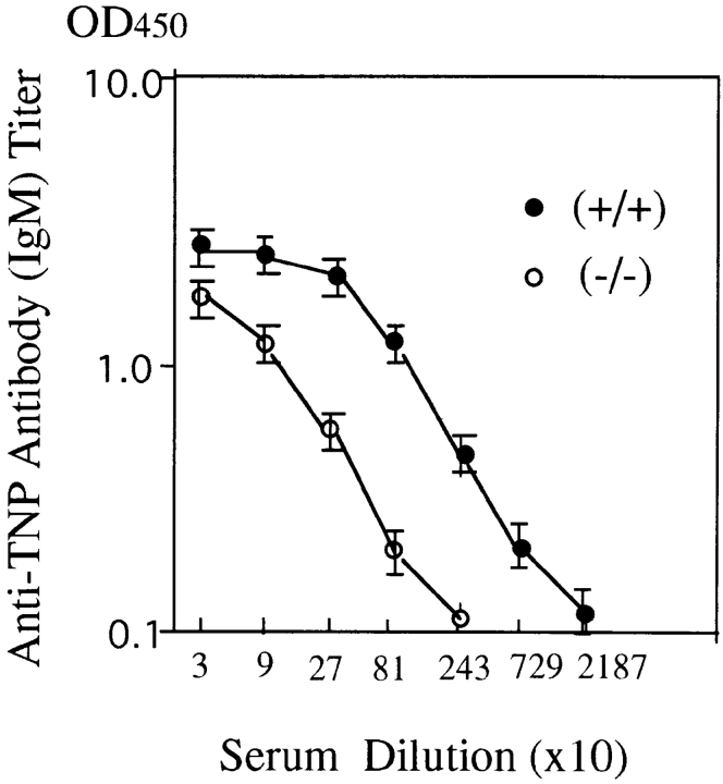
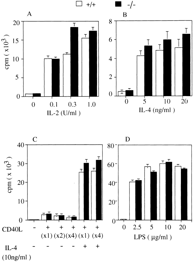
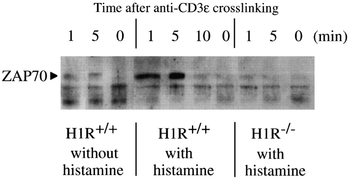
Histamine is considered one of the important mediators of immediate hypersensitivity and inflammation, and acts via G protein-coupled receptors. Here, we report that histamine may affect antigen receptor-mediated immune responses of T and B cells via a signal(s) from histamine H1 receptors (H1Rs). Histamine exhibited enhancing effects on the in vitro proliferative responses of anti-CD3epsilon- or anti-IgM-stimulated spleen T and B cells, respectively, at the culture condition that the fetal calf serum was dialyzed before culture and c-kit-positive cells were depleted from the spleen cells. In studies of histamine H1R knockout mice, H1R-deficient T cells had low proliferative responses to anti-CD3epsilon cross-linking or antigen stimulation in vitro. B cells from H1R-deficient mice were also affected, demonstrating low proliferative responses to B cell receptor cross-linking. Antibody production against trinitrophenyl-Ficoll was reduced in H1R-deficient mice. Other aspects of T and B cell function were normal in the H1R knockout mice. H1R-deficient T and B cells showed normal responses upon stimulation with interleukin (IL)-2, IL-4, CD40 ligand, CD40 ligand plus IL-4, and lipopolysaccharide. Collectively, these results imply that the signal generated by histamine through H1R augments antigen receptor-mediated immune responses, suggesting cross-talk between G protein-coupled receptors and antigen receptor-mediated signaling.


Figures

Figure 1
Figure 1
Enhancing effect of histamine on proliferative responses of c-kit–positive cell-depleted spleen T and B cells cultured in media containing dialyzed FCS. c-kit–positive cells were removed from total spleen cells and splenic B cells of wild-type C57/Bl mice were purified as described in Materials and Methods. c-kit–positive cell-depleted spleen T and B cells (5 × 10⁵ cells/ml) were cultured for 72 h in RPMI 1640 medium containing dialyzed FCS and stimulated with (A) anti-CD3ε (10 μg/ml) or (B) anti–mouse IgM (10 μg/ml) in the presence of different concentrations of histamine. The cells were pulsed for 12 h with [3H]thymidine before the end of culture. Shown is one representative experiment out of three.
Figure 2
Figure 2
Low proliferative responses of T and B cells of H1R−/− mice against anti-CD3ε and anti–mouse IgM stimulation. Total spleen cells and purified splenic B cells from H1R−/− mice were cultured in RPMI 1640 medium containing dialyzed FCS after depletion of c-kit–positive cells. Total spleen cells and purified B cells were stimulated with anti-CD3ε (A) and anti–mouse IgM (B), respectively, in the presence of different concentrations of histamine. The DNA synthesis was measured by pulsing with [3H]thymidine for 12 h before the end of culture. Shown is one representative experiment out of three.
Figure 3
Figure 3
Low proliferative responses of splenic T and B cells of histamine H1R−/− mice upon cross-linking of antigen receptors. Total spleen cells and purified B cells from unimmunized wild-type mice (+/+, white bars) and H1R−/− mice (−/−, black bars) were cultured with various concentrations of (A) anti-CD3ε antibody or (B) anti-IgM antibody for 72 h. (C) The H1R−/− and wild-type mice were immunized intraperitoneally with OVA (100 μg/ mouse) emulsified in alum as described in Materials and Methods. Mice were immunized twice in a 2-wk interval. 2 wk after the last immunization, the mice were killed and spleen cells from both wild-type and H1R−/− mice were cultured in presence of OVA (5–50 μg/ml) for 3 d. Cell proliferation was measured with [3H]thymidine incorporation by pulsing for 12 h at the end of culture. Mean CPM and SD were calculated from triplicate cultures. Shown is one representative experiment out of four.
Figure 3
Figure 3
Low proliferative responses of splenic T and B cells of histamine H1R−/− mice upon cross-linking of antigen receptors. Total spleen cells and purified B cells from unimmunized wild-type mice (+/+, white bars) and H1R−/− mice (−/−, black bars) were cultured with various concentrations of (A) anti-CD3ε antibody or (B) anti-IgM antibody for 72 h. (C) The H1R−/− and wild-type mice were immunized intraperitoneally with OVA (100 μg/ mouse) emulsified in alum as described in Materials and Methods. Mice were immunized twice in a 2-wk interval. 2 wk after the last immunization, the mice were killed and spleen cells from both wild-type and H1R−/− mice were cultured in presence of OVA (5–50 μg/ml) for 3 d. Cell proliferation was measured with [3H]thymidine incorporation by pulsing for 12 h at the end of culture. Mean CPM and SD were calculated from triplicate cultures. Shown is one representative experiment out of four.
Figure 4
Figure 4
Reduced antibody response to T cell–independent antigen, TNP-Ficoll, in H1R−/− mice. Both the H1R−/− (−/−) and wild-type mice (+/+) received one intraperitoneal immunization of TNP-Ficoll (25 μg/mouse) in 200 μl of 1× PBS. Sera were collected from three mice of each group on day 10 after immunization. Anti-TNP IgM antibody titer in serum was measured by ELISA, as described in Materials and Methods. The antibody titer was determined by measuring the optical density in 450 nm. Data are plotted as mean ± SD.
Figure 5
Figure 5
Normal proliferative responses to IL-2, IL-4, CD40L, CD40L plus IL-4, and LPS in H1R−/− mice. Total spleen cells from H1R−/− (black bars) and their wild-type littermates (white bars) were cultured with medium alone or various concentrations of IL-2 (A) or IL-4 (B). Splenic B cells were cultured with CD40L–CD8 chimeric protein (CD40L) alone or in combination with IL-4 (C) or LPS (D). Cells were cultured for 3 d and pulse-labeled with [3H]thymidine for the final 18 h. All cultures were performed in triplicate.
Figure 6
Figure 6
Level of Ig subclass in H1R−/− mice before and after immunization with the T cell–dependent antigen, OVA. (A) Levels of serum Ig subclass in unimmunized mice. Serum Ig levels in 8–10-wk-old H1R−/− mice (−/−) and their wild-type littermates were determined by isotype-specific ELISA. •, control (+/+); ○, mutant (−/−) mice. Shown are the results from five mice of each group. (B) Levels of anti-OVA antibody titers of each Ig isotype after secondary immunization. Serum samples were collected 2 wk after the second immunization. Titers of OVA-specific antibodies were determined by isotype-specific ELISA. Serum antibody levels are expressed as OD450. Shown are the results from five mice of each group. Horizontal lines indicate mean.
Figure 6
Figure 6
Level of Ig subclass in H1R−/− mice before and after immunization with the T cell–dependent antigen, OVA. (A) Levels of serum Ig subclass in unimmunized mice. Serum Ig levels in 8–10-wk-old H1R−/− mice (−/−) and their wild-type littermates were determined by isotype-specific ELISA. •, control (+/+); ○, mutant (−/−) mice. Shown are the results from five mice of each group. (B) Levels of anti-OVA antibody titers of each Ig isotype after secondary immunization. Serum samples were collected 2 wk after the second immunization. Titers of OVA-specific antibodies were determined by isotype-specific ELISA. Serum antibody levels are expressed as OD450. Shown are the results from five mice of each group. Horizontal lines indicate mean.
Figure 7
Figure 7
Tyrosine phosphorylation of ZAP-70 kinase after the cross-linking of TCR with anti-CD3ε in the absence or presence of histamine. c-kit–positive cell-depleted splenic T cells from wild-type (H1R+/+; left and center) or H1R−/− (right) mice were incubated with goat anti– mouse CD3ε antibody (10 μg/ml) with or without histamine for 0–10 min. Cell lysates were immunoprecipitated with affinity-purified goat anti–mouse ZAP-70 antibody. Immunoprecipitates were fractionated on 10% SDS-polyacrylamide gels. The proteins on the gels were transferred onto nitrocellulose membrane filter and blotted with an HRP-labeled antiphosphotyrosine mAb, PY-20. The blots were visualized with enhanced chemiluminescence reagents. The position of the band representing ZAP-70 phosphorylation is indicated by an arrow at 70 kD.

References

    1. Neer EJ. Heterotrimeric G proteins: organizers of transmembrane signaling. Cell. 1995;80:249–257. - PubMed
    1. Hamm HE, Gilchrist A. Heterotrimeric G proteins. Curr Opin Cell Biol. 1996;8:189–196. - PubMed
    1. Kehrl HJ. Heterotrimeric G protein signaling: roles in immune function and fine-tuning by RGS protein. Immunity. 1998;8:1–10. - PubMed
    1. Post GR, Brown JH. G-protein-coupled receptors and signaling pathways regulating growth responses. FASEB (Fed Am Soc Exp Biol) J. 1996;10:741–749. - PubMed
    1. Wan Y, Kurosaki T, Huang XY. Tyrosine kinase in activation of the MAP kinase cascade by G-protein coupled receptors. Nature. 1996;380:541–544. - PubMed

MeSH terms

Substances