. 1999 Feb 16;96(4):1193-200.
doi: 10.1073/pnas.96.4.1193.

Brominated 7-hydroxycoumarin-4-ylmethyls: photolabile protecting groups with biologically useful cross-sections for two photon photolysis

Affiliations

Brominated 7-hydroxycoumarin-4-ylmethyls: photolabile protecting groups with biologically useful cross-sections for two photon photolysis

T Furuta et al. Proc Natl Acad Sci U S A. .

Abstract

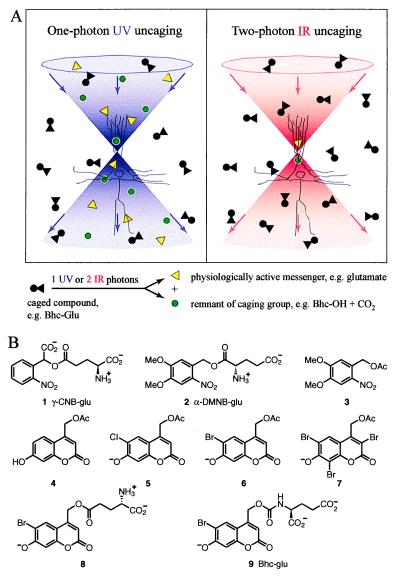
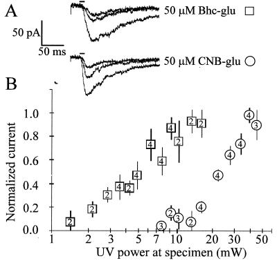
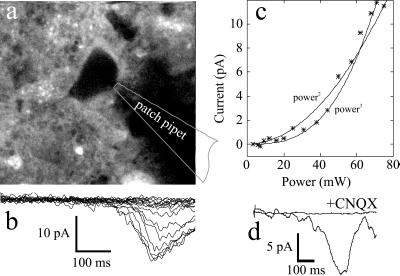
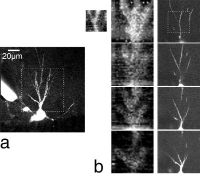
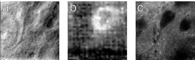
Photochemical release (uncaging) of bioactive messengers with three-dimensional spatial resolution in light-scattering media would be greatly facilitated if the photolysis could be powered by pairs of IR photons rather than the customary single UV photons. The quadratic dependence on light intensity would confine the photolysis to the focus point of the laser, and the longer wavelengths would be much less affected by scattering. However, previous caged messengers have had very small cross sections for two-photon excitation in the IR region. We now show that brominated 7-hydroxycoumarin-4-ylmethyl esters and carbamates efficiently release carboxylates and amines on photolysis, with one- and two-photon cross sections up to one or two orders of magnitude better than previously available. These advantages are demonstrated on neurons in brain slices from rat cortex and hippocampus excited by glutamate uncaged from N-(6-bromo-7-hydroxycoumarin-4-ylmethoxycarbonyl)-L-glutamate (Bhc-glu). Conventional UV photolysis of Bhc-glu requires less than one-fifth the intensities needed by one of the best previous caged glutamates, gamma-(alpha-carboxy-2-nitrobenzyl)-L-glutamate (CNB-glu). Two-photon photolysis with raster-scanned femtosecond IR pulses gives the first three-dimensionally resolved maps of the glutamate sensitivity of neurons in intact slices. Bhc-glu and analogs should allow more efficient and three-dimensionally localized uncaging and photocleavage, not only in cell biology and neurobiology but also in many technological applications.


Figures

Figure 1
Figure 1
(A) Scheme depicting the increased three-dimensional localization of uncaging with pairs of IR photons compared with single UV photons. The double cones of violet and red respectively symbolize beams of UV and IR, focused near the cell body of a schematized pyramidal neuron. Circles and triangles respectively represent the caging group and the bioactive molecule being caged. (B) Structures of caged molecules characterized in this study, showing the predominant state of ionization at pH 7.2.
Figure 2
Figure 2
Synthetic scheme for the new compounds prepared in this study. (a) Ethyl 4-chloroacetoacetate, H2SO4, rt, 6 d; (b) Br2, AcOH, rt, 1h; (c) DBU, AcOH, benzene, reflux, 1.5 h; (d) DBU, N-(tert-butoxycarbonyl)-α-tert-butyl glutamate, benzene, reflux, 1 h; (e) CF3CO2H, CH2Cl2, rt, 1 d; (f) H2O, reflux, 14 h; (g) i. DMAP, 4-nitrophenyl chloroformate, CH3CN, rt, 7 h. ii DMAP, di-tert-butyl glutamate hydrochloric acid salt, rt, 23 h; (h) CF3CO2H, CH2Cl2, rt, 2 d.
Figure 3
Figure 3
Time course of two-photon photolysis of Bhc-glu (9) at 740 nm (average power 530 mW exiting the cuvet, 172 fs pulse width), compared with breakdown of l-glutamic acid α-(3,4-dimethoxy-6-nitrobenzyl) ester (DMNB-glu, 2) with or without irradiation. ●, Percentage of Bhc-glu remaining from an initial concentration of 100 μM. Solid line, least-squares curve fit to a simple decaying exponential, which gave a time constant of 38 min. The initial rate of this photolysis run, combined with Eq. 3, gave a value of δu2 = 0.85 GM. The mean ± SD of 4 runs was 0.95 ± 0.21 GM. □, Percentage of DMNB-glu remaining under the same conditions. Dashed line, a decaying exponential of time constant 19.8 h, which had been separately measured over much longer times for the breakdown of DMNB-glu in the dark in 100 mM KMops, pH 7.2. Thus the pulsed IR laser beam does not detectably increase the rate of DMNB-glu breakdown.
Figure 4
Figure 4
Bhc-glu requires about five times less UV intensity than does CNB-glu to generate similar currents in pyramidal neurons from layers II/III of rat visual cortex. (A) Evoked current responses from uncaging 50 μM Bhc-glu at 2, 3, and 4 mW and 50 μM CNB-glu at increasing light intensities of 10, 16, and 21 mW. The horizontal bars above the traces indicate the 10 ms-long UV photolysis, 20 ms after the beginning of trace. Vertical scale, 50 pA. Horizontal scale, 50 ms. (B) Mean normalized current amplitudes (±SEM) from uncaging Bhc-glu (□, n = 4 cells) or CNB-glu (○, n = 4 cells) are plotted as a function of varying light intensities delivered to the slice, on a log scale. The numbers of cells tested at each light intensity are indicated by the numbers inside the symbols. To reduce intercell variation in response sizes, amplitudes for each cell were normalized to the average maximum response at the light intensity giving the maximal response for that cell.
Figure 5
Figure 5
Responses to two-photon uncaging of Bhc-glu. (a) Two-photon fluorescence image of a hippocampal neuron bathed in 0.5 mM Bhc-glu. (b) Whole-cell currents recorded in response to full-field raster scans like the one used for the fluorescence image (2 ms per line, 256 lines) with the beam power between 0 to 75 mW. (c) Dependence of peak current on beam power. The curves indicate fits by eye to second- and third-power functions. (d) Block of uncaging-evoked currents by a glutamate receptor antagonist (laser power 80 mW). Traces show responses at the same location in the absence and presence of 50 μM CNQX (6-cyano-7-nitroquinoxaline-2, 3-dione), an antagonist of non-N-methyl-d-aspartate receptors.
Figure 6
Figure 6
Mapping of glutamate-evoked currents. (a) Hippocampal pyramidal neuron filled with 100 μM fura-2 (image stack of 51 frames taken at 1-μm intervals, median filtered and maximum projected). (b) Maps (Left) of glutamate-evoked currents (current range is 33 pA between white and black) on dendrites taken at 4-μm depth intervals, with corresponding fluorescence images (projections of ±3 positions, right). The small image at left shows a higher-resolution scan. Laser power for uncaging images was 35 mW. Bar in a (20 μm) applies to all images. The superfusion solution contained 0.5 mM Bhc-glu and 100 μM cyclothiazide.
Figure 7
Figure 7
Glutamate-evoked current mapped at higher resolution on the soma of a cortical neuron. Transmitted-light image acquired by using the scanning laser (a), glutamate-evoked current map (b), or fluorescence image (c) in the presence of Bhc-glu. The superfusion solution contained 0.5 mM Bhc-glu. Laser power for the uncaging image was 85 mW.

References

    1. Pillai, V. N. R. (1980) Synthesis, 1–26.
    1. Adams S R, Tsien R Y. Annu Rev Physiol. 1993;55:755–784. - PubMed
    1. Marriott, G., ed. (1998) Methods Enzymol.291. - PubMed
    1. Pirrung M C. Chem Rev. 1997;97:473–488. - PubMed
    1. Denk W, Strickler J H, Webb W W. Science. 1990;248:73–76. - PubMed

Publication types

MeSH terms

LinkOut - more resources