Comparative Study
. 1999 Feb 16;96(4):1363-8.
doi: 10.1073/pnas.96.4.1363.

Structural analysis at 2.2 A of orthorhombic crystals presents the asymmetry of the allophycocyanin-linker complex, AP.LC7.8, from phycobilisomes of Mastigocladus laminosus

Affiliations
Comparative Study

Structural analysis at 2.2 A of orthorhombic crystals presents the asymmetry of the allophycocyanin-linker complex, AP.LC7.8, from phycobilisomes of Mastigocladus laminosus

W Reuter et al. Proc Natl Acad Sci U S A. .

Abstract

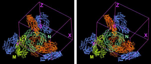
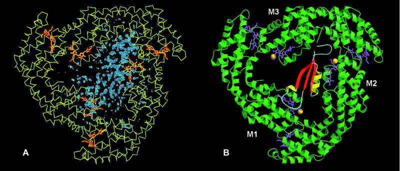
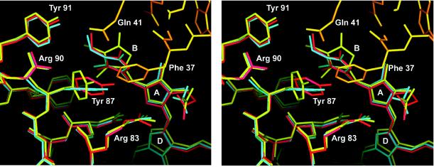
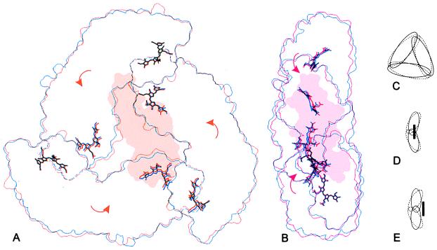
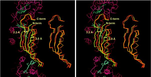
An electrophoretically purified allophycocyanin-linker complex, AP. LC7.8, from phycobilisomes of Mastigocladus laminosus has been crystallized in the orthorhombic space group P212121. Cryocrystallographic x-ray measurements enabled the structural analysis of the complex at a resolution of 2.2 A. The asymmetric unit contains two side-to-side associated "trimeric" (alphabeta)3 allophycocyanin complexes comprising the linker polypeptide in a defined orientation inside the trimer. The linker representing a protein fold related to the prosegment of procarboxypeptidase A is in contact with only two of the three beta-subunits and directly interacts with the corresponding chromophores of these proteins. In addition to a modulation of the chromophores' spectral properties, the linker polypeptide attracts the alphabeta-subcomplexes, thereby bringing the beta-chromophores closer together. These results will enable interpretations of energy-transfer mechanisms within phycobiliproteins.


Figures

Figure 1
Figure 1
Stereo view of the arrangement of AP⋅LC7.8 within the unit cell of the orthorhombic P212121 crystals. The two trimeric complexes building the asymmetric unit are shown in yellow and cyan, respectively. Their crystallographic symmetry-mates creating the entire unit cell are in orange and blue. The picture demonstrates that there are two types of contact between the two sets of molecules that can form the asymmetric unit, either side to side or back to back. The figure was prepared in main (29).
Figure 2
Figure 2
Representation of the entire AP⋅LC7.8 complex. (A) FobsFcalc Fourier map phased with the Patterson solution after exchange of those residues that differ between allophycocyanin from M. laminosus and the replacement model from S. platensis (12). Blue, FobsFcalc difference electron density; yellow, Cα plot of the α and β subunits; orange, chromophores. (B) Ribbon plot. The α/β-subunits are represented in green and the chromophores in pink. The methylated Asn-19 of the β-subunit is present in all three monomers and is shown as an orange ball. The secondary structural elements of the linker polypeptide are represented in red, yellow, and blue. The linker polypeptide interacts directly with the chromophores of only two of the three β-chromophores. Fig. 2A was prepared in main (29) and Fig. 2B was made with molscript (48) and rendered with raster3d (49).
Figure 3
Figure 3
Stereo representation of the interaction between the linker polypeptide and monomer 2. The Cα-aligned structures are shown for the symmetric, linker-free allophycocyanin (blue), monomer 3, which has no interaction with the linker polypeptide (red), and monomer 2 (yellow), which interacts with the N terminus of the long linker α-helix (orange). Water molecules are omitted for clarity. Phe-37 of the linker inserts between Tyr-87 and the pyrrole ring B of the corresponding β-subunit displacing both to opposite directions and disrupting the stacking interaction between the two aromatic ring systems, which is seen at the other two β-chromophores of the linker containing trimer. This figure was prepared in main (29).
Figure 4
Figure 4
Comparison of the molecular outlines of the symmetric, linker-free allophycocyanin (12) and the linker-containing complex (A and B) as well as an exaggerated schematic representation (C–E): The symmetric allophycocyanin (blue or dashed lines) is compared with molecule M (orange) in front view (A) and molecule N (red) in side view (B). On binding of the linker, the entire complex collapses toward the pseudo-threefold axis (C) and the α/β-ring becomes flattened (D and E) because of a rotation of the monomers as indicated by the arrows. In molecule M the linker inserts further into the trimer (D), resulting in a larger movement of the chromophores toward the pseudo-threefold axis, but with a slighter displacement of the chromophores to the right as compared with molecule N (E). These outlines were prepared by using an edge-detection filter and grasp surface representations of each molecule (50).
Figure 5
Figure 5
Comparison of the structure of the linker polypeptides of the two molecules in the asymmetric unit. On the right the linker Cα-atoms are aligned directly by an rms procedure. On the left, the linkers are shown after rms alignment of the Cα-atoms of the two trimers in the asymmetric unit. The two linker molecules are very similar. However, the linker in molecule M (yellow) is shifted by about 3 Å to the left with respect to the molecule N linker (orange). Part of the Cα-trace, as well as the chromophores of molecule N, is shown as reference in red and blue, respectively. The figure was prepared in main (29).

References

    1. Glazer A N. Annu Rev Biophys Biophys Chem. 1985;14:47–77. - PubMed
    1. Mörschel E, Riehl E. In: Membranous Structures. Harris J R, Horne R W, editors. London: Academic; 1987. pp. 210–254.
    1. Holzwarth A R. Physiol Plant. 1991;83:518–528.
    1. Sidler W. In: The Molecular Biology of Cyanobacteria. Bryant D A, editor. Dordrecht, The Netherlands: Kluwer; 1994. pp. 139–216.
    1. Bryant D A. In: Cell Culture and Somatic Cell Genetics of Plants. Bogorad L, Vasil I K, editors. San Diego: Academic; 1991. pp. 257–300.

Publication types

Associated data

LinkOut - more resources