. 1999 Feb 16;96(4):1385-90.
doi: 10.1073/pnas.96.4.1385.

A human cell-surface receptor for xenotropic and polytropic murine leukemia viruses: possible role in G protein-coupled signal transduction

Affiliations

A human cell-surface receptor for xenotropic and polytropic murine leukemia viruses: possible role in G protein-coupled signal transduction

J L Battini et al. Proc Natl Acad Sci U S A. .

Abstract

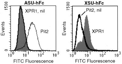
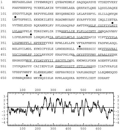
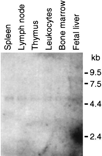
Although present in many copies in the mouse genome, xenotropic murine leukemia viruses cannot infect cells from laboratory mice because of the lack of a functional cell surface receptor required for virus entry. In contrast, cells from many nonmurine species, including human cells, are fully permissive. Using an expression library approach, we isolated a cDNA from HeLa cell RNA that conferred susceptibility to xenotropic envelope protein binding and virus infection when expressed in nonpermissive cells. The deduced product is a 696-aa multiple-membrane spanning molecule, is widely expressed in human tissues, and shares homology with nematode, fly, and plant proteins of unknown function as well as with the yeast SYG1 protein, which has been shown to interact with a G protein. This molecule also acts as a receptor for polytropic murine leukemia viruses, consistent with observed interference between xenotropic and polytropic viruses in some cell types. This xenotropic and polytropic retrovirus receptor (XPR1) is the fourth identified molecule having multiple membrane spanning domains among mammalian type C oncoretrovirus receptors and may play a role in G protein-coupled signal transduction, as do the chemokine receptors required for HIV entry.


Figures

Figure 1
Figure 1
SU-immunoadhesin binding to CHO cells expressing XPR1 or Pit2. Cells were transduced with L(XPR1)SN (XPR1, filled histograms), L(Pit2)SN (Pit2, thin lines), or the LXSN vector without an insert (nil, thick lines) and were selected in G418 to generate polyclonal populations of vector-expressing cells. The cells were detached by treatment with 1 mM EDTA, were washed with PBS, were incubated with ASU-hFc (Left) or XSU-hFc (Right) for 30 min at 37°C, were washed twice with PBS, were incubated with rabbit F(ab′)2 anti-human IgG conjugated to fluorescein isothiocyanate (Dako), and were washed before flow cytometric analysis. Dead and clumped cells were excluded by gating on forward and high-angle light scatter and exclusion of propidium iodide (1 μg/ml), and 50,000 gated events were analyzed per sample.
Figure 2
Figure 2
Amino acid sequence and hydropathy analysis of the XPR1 protein. Predicted transmembrane segments are underlined, and potential consensus N-glycosylation sites are indicated by filled circles. The glycosylation site NIT at position 248 is located in the first predicted transmembrane segment. The hydropathy plot of XPR1 was generated by the program of Kyte and Doolittle (38) by using a window of 11 residues. Positive values indicate hydrophobic region, and negative values indicate hydrophilic regions.
Figure 3
Figure 3
Analysis of XPR1 transcription in human tissues. A Northern blot of polyA+ RNA from the indicated human tissues (Multiple Tissue Northern Blot 7768-1; CLONTECH) was probed with a full length XPR1 cDNA probe at high stringency. Each lane contains ≈2 μg of polyA+ RNA adjusted to give a consistent signal when hybridized to a β-actin probe. RNA size markers are shown at right. The spot at ≈2.4 kb in the far right lane is a hybridization artifact.
Figure 4
Figure 4
Chromosomal localization of XPR1-encoding gene. Idiogram of human chromosome 1 shows linkage of XPR1 gene to D1S308E as determined by PCR mapping using the G3 Stanford Radiation Hybrid Panel. Distances in cRay between molecular markers and logarithm of odds scores for linkage between adjacent marker pairs are represented. 1 cRay ≈30 kb.

References

    1. Weiss R A. In: The Retroviridae. Levy J A, editor. Vol. 2. New York: Plenum; 1993. pp. 1–108.
    1. Hunter E. In: Retroviruses. Coffin J M, Hughes S H, Varmus H E, editors. Plainview, NY: Cold Spring Harbor Lab. Press; 1997. pp. 71–119. - PubMed
    1. Sommerfelt M A, Weiss R A. Virology. 1990;176:58–69. - PubMed
    1. Miller A D, Bonham L, Alfano J, Kiem H P, Reynolds T, Wolgamot G. J Virol. 1996;70:1804–1809. - PMC - PubMed
    1. Patience C, Takeuchi Y, Weiss R A. Nat Med. 1997;3:282–286. - PubMed

Publication types

MeSH terms

Associated data