. 1999 Feb 16;96(4):1415-20.
doi: 10.1073/pnas.96.4.1415.

Mechanism of biological synergy between cellular Src and epidermal growth factor receptor

Affiliations

Mechanism of biological synergy between cellular Src and epidermal growth factor receptor

D A Tice et al. Proc Natl Acad Sci U S A. .

Abstract

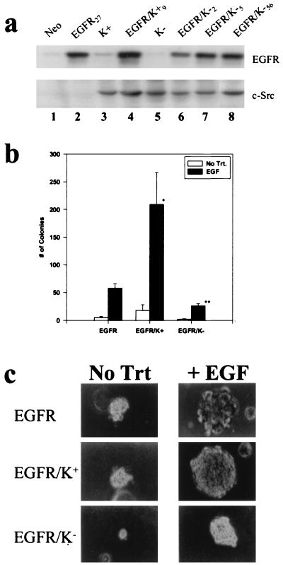
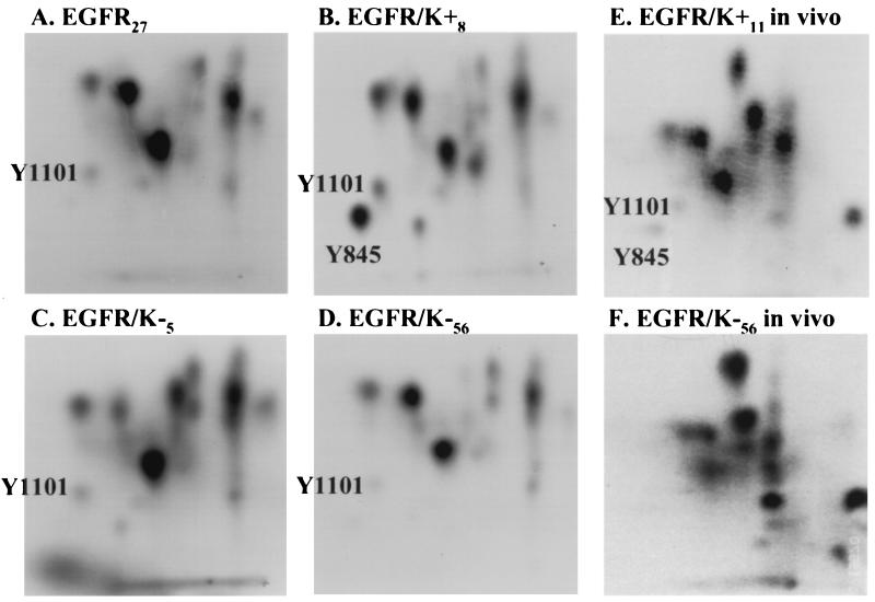
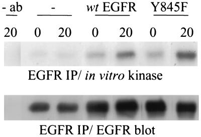
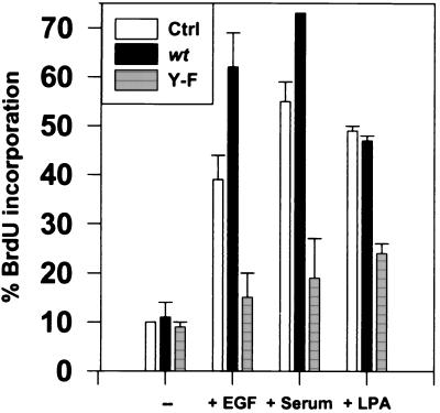
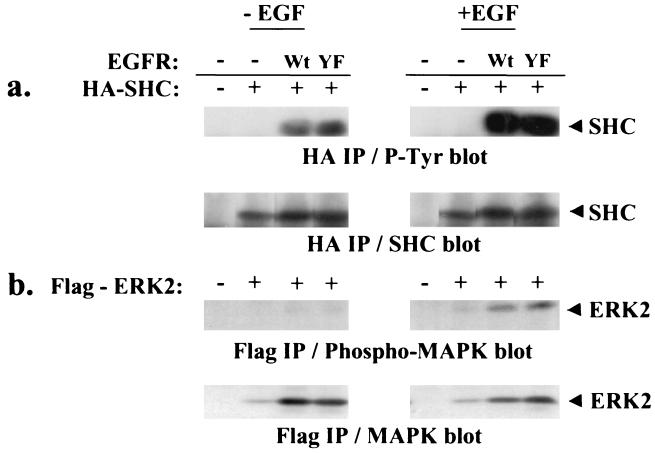
Overexpression of both cellular Src (c-Src) and the epidermal growth factor receptor (EGFR) occurs in many of the same human tumors, suggesting that they may functionally interact and contribute to the progression of cancer. Indeed, in murine fibroblasts, overexpression of c-Src has been shown to potentiate the mitogenic and tumorigenic capacity of the overexpressed EGFR. Potentiation correlated with the ability of c-Src to physically associate with the activated EGFR and the appearance of two unique in vivo phosphorylations on the receptor (Tyr-845 and Tyr-1101). Using stable cell lines of C3H10T1/2 murine fibroblasts that contain kinase-deficient (K-) c-Src and overexpressed wild-type EGFR, we show that the kinase activity of c-Src is required for both the biological synergy with the receptor and the phosphorylations on the receptor, but not for the association of c-Src with the receptor. In transient transfection assays, not only epidermal growth factor but also serum- and lysophosphatidic acid-induced DNA synthesis was ablated in a dominant-negative fashion by a Y845F mutant of the EGFR, indicating that c-Src-induced phosphorylation of Y845 is critical for the mitogenic response to both the EGFR and a G protein-coupled receptor (lysophosphatidic acid receptor). Unexpectedly, the Y845F mutant EGFR was found to retain its full kinase activity and its ability to activate the adapter protein SHC and extracellular signal-regulated kinase ERK2 in response to EGF, demonstrating that the mitogenic pathway involving phosphorylation of Y845 is independent of ERK2-activation. The application of these findings to the development of novel therapeutics for human cancers that overexpress c-Src and EGFR is discussed.


Figures

Figure 1
Figure 1
Dominant repressive effect of K− c-Src on EGF-induced soft agar colony formation. (a) Western immunoblot analysis of C3H10T½ murine fibroblast clonal cell lines stably overexpressing EGFR and either wt (K+) or kinase-deficient (K−) c-Src. (b) Values for number of colonies are the mean ± SEM of at least six experiments in which 105 cells of each clone were seeded per plate in triplicate. Three clones of each cell type were averaged. ∗, P < 0.04 and ∗∗, P < 0.002 compared with EGFR. (c) Photomicrographs of representative fields of soft agar colonies formed from the indicated cell lines were taken after 2 weeks of growth. Trt, treatment. (×200.)
Figure 2
Figure 2
K− c-Src associates with the EGFR. Confluent, serum-starved cells were untreated or treated with EGF (100 ng/ml) for 15 min, and extracts were immunoprecipitated with either chicken c-Src-specific mAb EC10 (designated +) or a negative control mouse IgG (−). (a) Immunocomplexes were subjected to an in vitro kinase assay using [γ-32P]ATP, and phosphorylated products were analyzed by SDS/PAGE and autoradiography. The region of the autoradiograph around the 170-kDa products is shown. (b) The amount of c-Src in each precipitate was visualized by Western immunoblot analysis with EC10 mAb. K− c-Src in EGFR/K− clones was verified to be catalytically inactive in the immune complex kinase assay (data not shown).
Figure 3
Figure 3
Y845 is not phosphorylated in EGFR complexed with K− c-Src. (AD) The 170-kDa bands that were phosphorylated in vitro (as in Fig. 2) in c-Src (B–D) or receptor (A) immunocomplexes prepared from the indicated cell lines were excised, digested with trypsin, resolved by two-dimensional electrophoresis/chromatography, and subjected to autoradiography. The positions of peptides containing Y845 and Y1101, which were identified previously (15), are indicated. (E and F) Receptor immunoprecipitates from the indicated cell lines that had been metabolically labeled with 32Pi were analyzed as in A–D. Equal cpm were loaded in A–D and in E vs. F. The apparent increase in tyrosine phosphorylation in F is due to a slightly darker exposure compared with E to emphasize the complete ablation of Y845 phosphorylation. The appearance of darker or novel spots in F was not reproduced in repeated experiments.
Figure 4
Figure 4
Phosphorylation of Y845 is not essential for EGFR autokinase activity. COS-7 cells were transiently transfected with either vector alone (−), or plasmids encoding Y845F or wt EGFR and stimulated with EGF for 5 min. The EGFR was immunoprecipitated from extracts and subjected to an in vitro kinase assay for 0 or 20 min. The reaction was stopped with the addition of sample buffer, and the products were resolved by SDS/PAGE and transferred to a membrane. After autoradiography, EGFR was detected by Western immunoblotting and visualized by using enhanced chemiluminescence (ECL; Amersham).
Figure 5
Figure 5
Phosphorylation of Y845 is essential for EGFR function. K+ cells were transfected with plasmid DNA encoding Y845F (Y-F) or wt EGFR, cultured for 2 days, serum starved for 30 hr, and left untreated or treated with 40 ng/ml EGF, 10% fetal bovine serum, or 10 μM LPA for 18 hr. Results are expressed as the mean percent ± SEM of cells expressing EGFR that were positive for BrdUrd incorporation. Thirty-five to 100 cells were analyzed for each variable in three independent experiments.
Figure 6
Figure 6
Y845F mutant receptor retains its ability to phosphorylate SHC and activate MAPK. COS-7 cells were transfected with plasmid DNA encoding HA-SHC or Flag-ERK2 and either Y845F or wt EGFR, cultured for 2 days, serum starved overnight, and left untreated or treated with 100 ng/ml EGF for 10 min. Extracts were immunoprecipitated with 12CA5 anti-HA antibody or anti-Flag M2 affinity gel and resolved by SDS/PAGE. The amount of tyrosine phosphorylated HA-SHC (a) and Flag-ERK2 (b) was observed by Western immunoblot analysis. The amount of wt EGFR or Y845F EGFR expressed in the populations of cells was the same as that shown for Fig. 4.

References

    1. Ottenhoff-Kalff A E, Rijksen G, van Beurden E A, Hennipman A, Michels A A, Staal G E. Cancer Res. 1992;52:4773–4778. - PubMed
    1. Khazaie K, Schirrmacher V, Lichtner R B. Cancer Metastasis Rev. 1993;12:255–274. - PubMed
    1. Banker N, Evers B M, Hellmich M R, Townsend C J. Surg Oncol. 1996;5:201–210. - PubMed
    1. Biscardi, J. S., Tice, D. A. & Parsons, S. J. (1999) Adv. Cancer Res. 76, in press. - PubMed
    1. Harris J R, Lippmann M E, Veronesi U, Willett W. N Engl J Med. 1992;327:473–480. - PubMed

Publication types

MeSH terms