

doi: 10.1073/pnas.96.4.1469.
Organization of an echinoderm Hox gene cluster
Affiliations
- PMID: 9990047
- PMCID: PMC15486
- DOI: 10.1073/pnas.96.4.1469
Item in Clipboard
Organization of an echinoderm Hox gene cluster
Proc Natl Acad Sci U S A.
.
Abstract
The Strongylocentrotus purpuratus genome contains a single ten-gene Hox complex >0.5 megabase in length. This complex was isolated on overlapping bacterial artificial chromosome and P1 artificial chromosome genomic recombinants by using probes for individual genes and by genomic walking. Echinoderm Hox genes of Paralog Groups (PG) 1 and 2 are reported. The cluster includes genes representing all paralog groups of vertebrate Hox clusters, except that there is a single gene of the PG4-5 types and only three genes of the PG9-12 types. The echinoderm Hox gene cluster is essentially similar to those of the bilaterally organized chordates, despite the radically altered pentameral body plans of these animals.
Figures
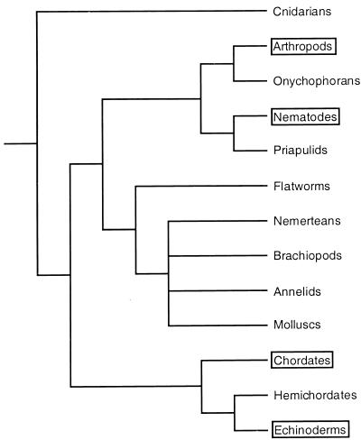
Phylogenetic tree for Metazoa, including
representative protostome phyla and the three phyla that constitute the
deuterostomes. Molecular phylogenies divide the protostomes into two
great clades, namely the ecdysozoans (here arthropods to priapulids)
and the lophotrochozoans (here flatworms to molluscs); deuterostomes
consist of hemichordates and echinoderms, which are sister groups, plus
chordates (vertebrate and invertebrate) (–44). The only phyla in
which Hox gene clusters have been structurally
characterized at the genome level are boxed (see text for references).
The echinoderm box refers to the present work.
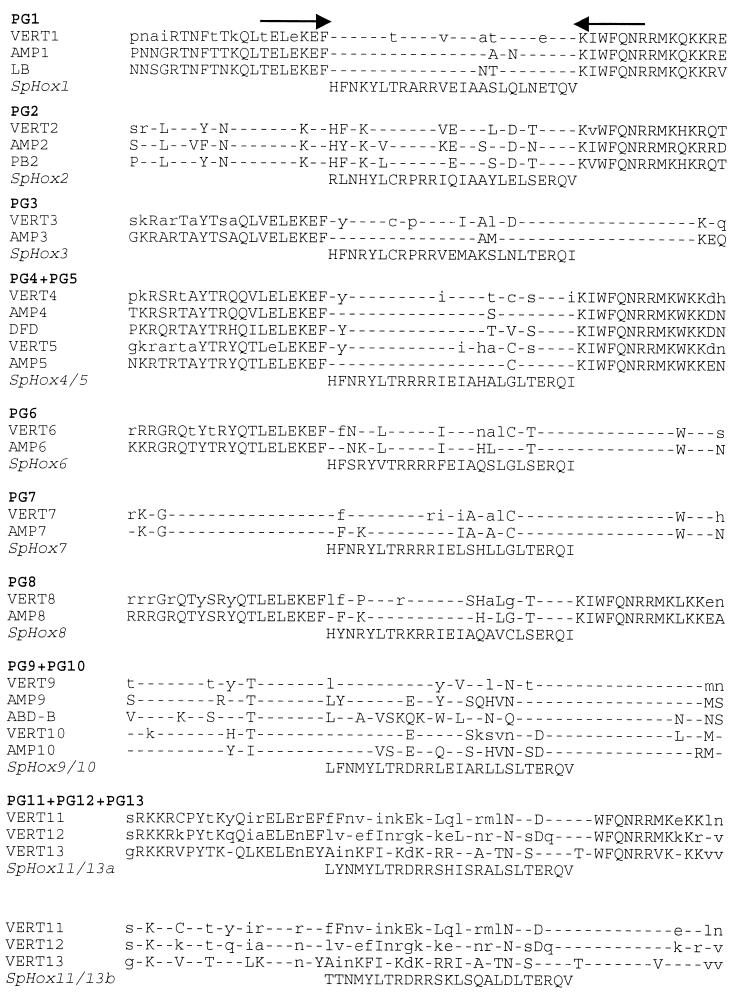
Alignment of vertebrate, amphioxus,
Drosophila, and S. purpuratus homeodomain
sequences. The S. purpuratus homeodomain sequences are
shown flanked by arrows representing the positions of the PCR fragments
used in our screens (18). Homeodomain sequences for some genes were
obtained by other methods or were from a combination of sources, e.g.,
clones isolated by genomic walking or cDNA clones, as described in the
text. In vertebrate homeodomain consensus sequences (VERT), uppercase
letters indicate a residue conserved in all known vertebrate sequences
of that paralog group, e.g., all mouse and human PG1 genes (24).
Lowercase letters indicate a residue found in the majority but not all
vertebrate sequences of each paralog group, i.e., comparing the
multiple vertebrate sequences available for each Paralog Group (there
is only a single amphioxus gene from each Paralog Group). Dashes
indicate amino acid identity at that position between the S.
purpuratus genes and all vertebrate genes as well as
Drosophila and amphioxus genes of that paralog group.
Amphioxus sequences [AMP, from Branchiostoma (3)] are
shown below the vertebrate consensus sequences.
Drosophila sequences included in the comparison are
Labial (LB), Proboscipedia (PB),
Deformed (DFD), and Abdominal B (ABD-B).
Sequences are compiled from ref. .
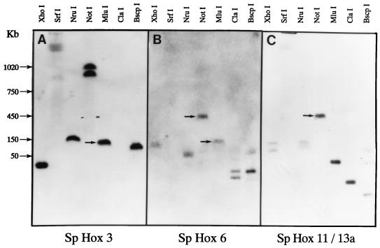
Genome blot hybridizations carried out on
pulsed-field electrophoretic displays of S. purpuratus
genomic DNA. The DNA was obtained from sperm of a single individual.
Seven different restriction enzymes were used for the blots in each
panel, as indicated. Arrows indicate common bands revealed by probes
for more than one gene.
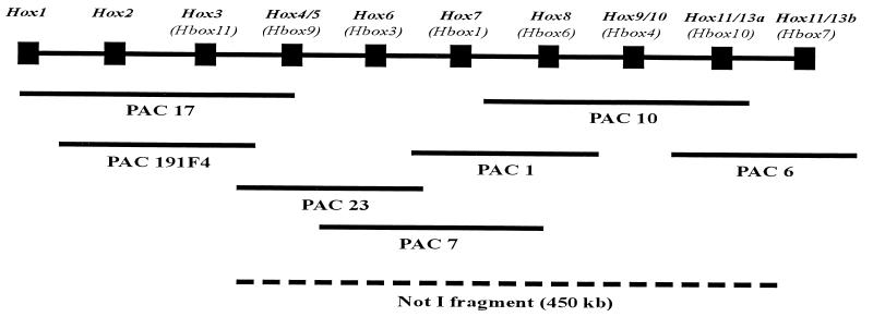
Organization of the S. purpuratus
Hox gene cluster. The diagram is not to scale, as the
intergenic distances within the cluster have not been determined. The
sequence of Hox genes within the cluster was inferred
from their locations within PAC and BAC genomic inserts (see text) and
the overlaps amongst clones containing each gene. For brevity, only one
set of PAC genomic clones is shown, though each genomic region was
analyzed on the basis of overlaps of multiple independent PAC and BAC
clones. The correct names of the Hox genes with respect
to their paralogous affinities with vertebrate Hox genes
appear at the top of the diagram, and beneath in parentheses are
designations found in earlier literature describing isolations of
Hox homeodomains or cDNAs in various laboratories (see
text for references). The dashed line indicates the span of the 450-kb
fragment indicated in Fig. 3, which includes all the genes from
SpHox4/5 to SpHox11/13a.
References
-
- Lewis E B. Nature (London) 1978;276:565–570. - PubMed
-
- McGinnis W, Krumlauf R. Cell. 1992;68:283–302. - PubMed
-
- Garcia-Fernandez J, Holland P W H. Nature (London) 1996;370:563–566. - PubMed
-
- Balavoine G. C R Acad Sci. 1997;329:83–94. - PubMed
-
- Adoutte, A., Balavoine, G., Lartillot, N. & de Rosa, R. Trends Genet., in press. - PubMed
Publication types
MeSH terms
Grants and funding
LinkOut - more resources
Full Text Sources
Miscellaneous
