. 1999 Feb 16;96(4):1541-6.
doi: 10.1073/pnas.96.4.1541.

GA-binding protein factors, in concert with the coactivator CREB binding protein/p300, control the induction of the interleukin 16 promoter in T lymphocytes

Affiliations

GA-binding protein factors, in concert with the coactivator CREB binding protein/p300, control the induction of the interleukin 16 promoter in T lymphocytes

N Bannert et al. Proc Natl Acad Sci U S A. .

Abstract

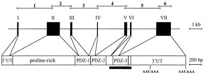
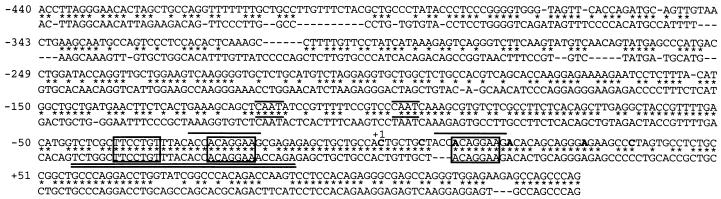
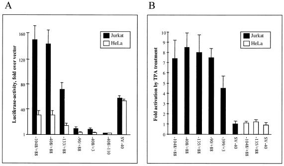
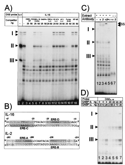
Interleukin 16 (IL-16) is a chemotactic cytokine that binds to the CD4 receptor and affects the activation of T cells and replication of HIV. It is expressed as a large 67-kDa precursor protein (pro-IL-16) in lymphocytes, macrophages, and mast cells, as well as in airway epithelial cells from asthmatics after challenge with allergen. This pro-IL-16 is subsequently processed to the mature cytokine of 13 kDa. To study the expression of IL-16 at the transcriptional level, we cloned the human chromosomal IL-16 gene and analyzed its promoter. The human IL-16 gene consists of seven exons and six introns. The 5' sequences up to nucleotide -120 of the human and murine IL-16 genes share >84% sequence homology and harbor promoter elements for constitutive and inducible transcription in T cells. Although both promoters lack any TATA box, they contain two CAAT box-like motifs and three binding sites of GA-binding protein (GABP) transcription factors. Two of these motifs are part of a highly conserved and inducible dyad symmetry element shown previously to control a remote IL-2 enhancer and the CD18 promoter. In concert with the coactivator CREB binding protein/p300, which interacts with GABPalpha, the binding of GABPalpha and -beta to the dyad symmetry element controls the induction of IL-16 promoter in T cells. Supplementing the data on the processing of pro-IL-16, our results indicate the complexity of IL-16 expression, which is tightly controlled at the transcriptional and posttranslational levels in T lymphocytes.


Figures

Figure 1
Figure 1
Genomic organization of the human IL-16 gene. The seven exons are shown as black boxes and projected on the IL-16 mRNA. UT, untranslated 5′ and 3′ sequences; AAUAAA, poly(A) addition motifs. The fragments encoding the proline-rich region and the three PDZ domains of pro-IL-16 protein are indicated. The black bar below denotes the sequence coding for the mature IL-16 protein. The complete gene of approximately 12.8 kb was cloned and sequenced as six overlapping fragments depicted above the gene structure. The use of an alternative splice acceptor in intron 5, three nucleotides downstream of the regular site, generates a transcript with a missing alanine codon (not shown). This splice acceptor is predominantly used in mice because we were unable to find cDNAs encoding this alanine in murine PBMCs (19).
Figure 2
Figure 2
Comparison of 5′ sequences of human and murine IL-16 genes. The published start points of transcription in human cells are shown in bold (4). Nucleotide +1 corresponds to the first transcription start site of the human gene. Identical nucleotides in both sequences are indicated by ∗. Deletions indicated by dashes were introduced to obtain the highest degree of homology. The CAAT box-like sequences around positions −98 and −118 and direct repeats around +12 and −25 are overlined, the dyad symmetry element, DSE, is double-underlined, and the GABP-binding sites are boxed.
Figure 3
Figure 3
Promoter activity of 5′ sequences from the human IL-16 gene in Jurkat T cells and HeLa cells. The numbers below indicate the sequences of DNA fragments cloned in front of a luciferase reporter gene. The activity of a pGL3 vector (Promega) containing the SV40 early promoter is also shown. (A) Deletion analysis of the 5′ IL-16 DNA. Relative luciferase activities are given as fold activity above the activity of the promoter-less pGL3-Basic vector. The standard deviations represent the mean values from four independent experiments. (B) TPA-mediated induction of 5′ segments of IL-16 gene. Differences of luciferase activities are shown between uninduced cells and cells treated with 10 nM TPA and indicated as “Fold activation.” The standard deviations represent the mean values from three independent experiments.
Figure 4
Figure 4
GABP factors bind to the Ets-like sequence motifs of IL-16 promoter. (A) EMSAs using nuclear proteins from Jurkat cells and the DSE from the IL-16 promoter as probe. Two micrograms of nuclear protein from noninduced Jurkat cells was incubated either with the DSEIL-2 (lane 1) or DSEIL-16 (lanes 2–18) as probes (see B) followed by electrophoresis on a native 4% polyacrylamide gel. For competition, 25 and 50 ng of the following oligonucleotides were added: lanes 3 and 4, DSEIL-16; lanes 5 and 6, DSEIL-16 mutated in both Ets motifs; lanes 7 and 8, IL-16 Ets motif at position +12; lanes 9 and 10, IL-2 ERE-A motif (see B); lanes 11 and 12, distal NF-AT site from the murine IL-2 promoter (see ref. 22); lanes 13 and 14, AP-1 binding site from the IL-2 promoter (22); lanes 15 and 16, Ets site from the SV40 enhancer (20); lanes 17 and 18, consensus NF-κB site. (B) Sequences of DSEs from the IL-16 promoter and distal IL-2 enhancer (20). The palindromic organization of Ets-related elements, EREs, is indicated in gray. (C) Supershift EMSAs with Abs raised against GABPα and -β. In lanes 1–5, 1 μg of nuclear protein from Jurkat cells was incubated with the DSEIL-16 probe, alone or with 1 μg of Ab raised against GABPα (lane 2), -β (lane 3), or with both Abs (lane 4). As a control, a Pu.1-specific Ab was added in lane 5. In lanes 6 and 7, the DSEIL-16 probe was incubated with Abs alone. SS, complexes supershifted by GABP Abs. (D) Binding of recombinant GABPα and -β to the DSEIL-16. Bacterially expressed GABPα or -β (2.5–20 ng) was incubated with a DSEIL-16 probe as indicated. I–III indicate DNA–protein complexes identical in mobility to those generated with nuclear proteins from Jurkat cells (A).
Figure 5
Figure 5
GABPα and -β stimulate, in concert with p300, the activity of IL-16 promoter. (A) GABPα and -β stimulate the IL-16 promoter. Jurkat cells were cotransfected with a luciferase reporter gene controlled by the IL-16 promoter (from −408 to +88), SRSPA-based vectors expressing GABPα or -β, or both, as indicated. The cells were left untreated or stimulated with TPA (10 ng/ml) for 24 h. (B) The GABP-binding sites within the IL-16 promoter are of crucial importance for its activity. Human 293 cells were transfected with a wild-type IL-16 promoter/luciferase gene construct or mutated IL-16 promoter/luciferase constructs alone or together with vectors expressing GABPα and -β. CmDm, mutations in the ERE-C and ERE-D motifs (see Fig. 4B); INIm, mutations within the +12 GABP site which overlaps the transcriptional initiation site. (C) CBP/p300 enhance the activity of the IL-16 promoter in a dose-dependent manner. 293 cells were cotransfected with a luciferase reporter gene controlled by the IL-16 promoter (from −408 to +88) and 0–50 ng of a p300 expression vector as indicated. (D) Interaction between GABPα and p300. Equal amounts of bacterially expressed glutathione S-transferase/p300 fusion proteins spanning N-terminal, middle, or C-terminal portions of p300 immobilized on glutathione agrose beads were incubated with 500 ng of bacterially expressed GABPα, -β or -α+β. The bound proteins were eluted and immunodetected by using a mix of GABPα+β-specific Abs. As a control for p300/GABP interaction, a glutathione S-transferase fusion protein containing the N-terminal transacting domain of NF-ATc (TADNF-ATc, 113–205) was used. In lane 13, 50 μg of nuclear proteins from 293 cells were fractionated. The appearance of a weak GABPα band in lane 11 is the result of an accidental contamination of the GABPβ protein preparation with GABPα. The appearance of a second GABPα band in many lanes is probably a GABPα cleavage product. The lowest strong band is most likely the result of the crossreactivity of GABP Abs with a bacterial protein binding unspecifically to p300.

References

    1. Center D M, Cruikshank W. J Immunol. 1982;128:2563–2568. - PubMed
    1. Bazan J F, Schall T J. Nature (London) 1996;381:29–30. - PubMed
    1. Bannert N, Baier M, Werner A, Kurth R. Nature (London) 1996;381:30. - PubMed
    1. Baier M, Bannert N, Werner A, Lang K, Kurth R. Proc Natl Acad Sci USA. 1997;94:5273–5277. - PMC - PubMed
    1. Doyle D A, Lee A, Lewis J, Kim E, Sheng M, MacKinnon R. Cell. 1996;85:1067–1076. - PubMed

Publication types

MeSH terms

Associated data