

Association of HFE protein with transferrin receptor in crypt enterocytes of human duodenum
- PMID: 9990067
- PMCID: PMC15523
- DOI: 10.1073/pnas.96.4.1579
Association of HFE protein with transferrin receptor in crypt enterocytes of human duodenum
Abstract
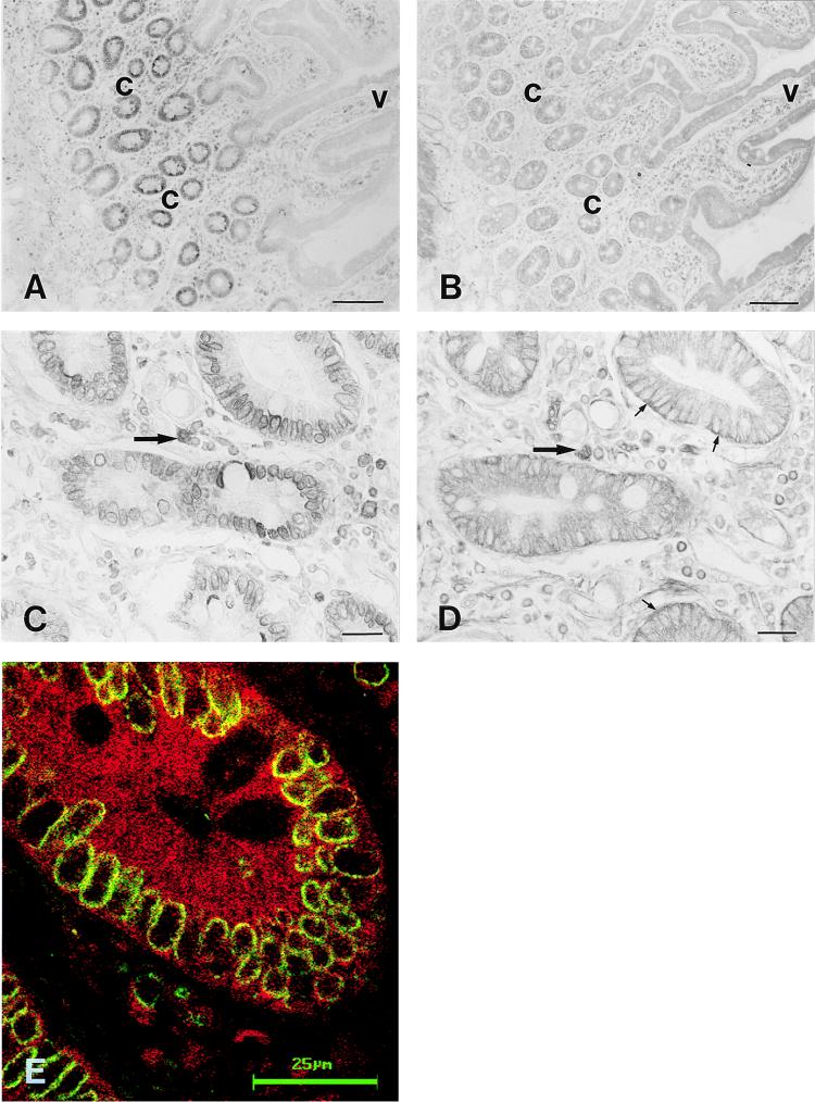
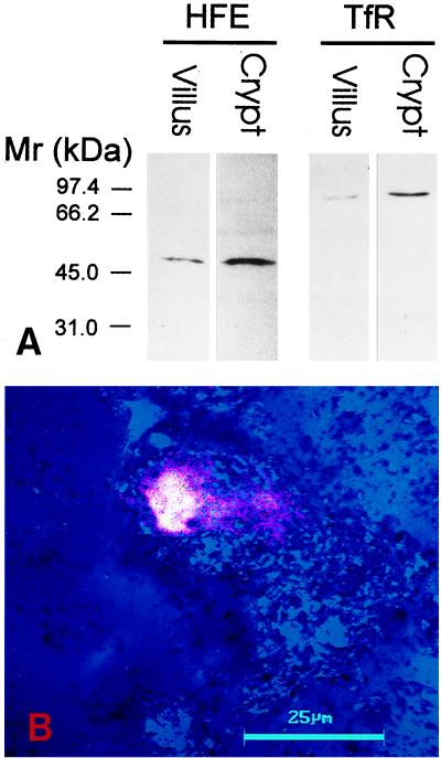
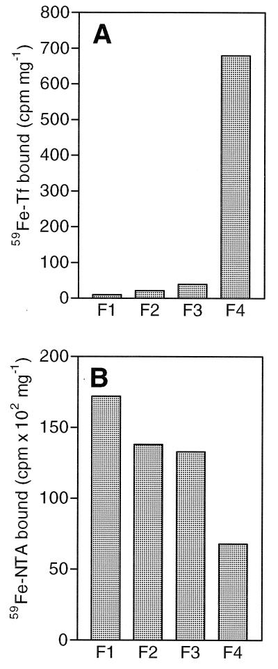
In hereditary hemochromatosis (HH), intestinal absorption of dietary iron is increased, leading to excessive iron accumulation in tissues and resultant organ damage. The HFE protein, which is defective in HH, normally is expressed in crypt enterocytes of the duodenum where it has a unique, predominantly intracellular localization. In placenta, the HFE protein colocalizes with and forms a stable association with the transferrin receptor (TfR), providing a link between the HFE protein and iron transport. In the present study, we examined the relationship of the HFE protein to the TfR in enterocytes of the human duodenum and measured the uptake of transferrin-bound iron and ionic iron by isolated crypt and villus enterocytes. Immunocytochemistry showed that the HFE protein and TfR both are expressed in the crypt enterocytes. Western blots showed that, as was the case in human placenta, the HFE protein in crypt enterocytes is physically associated with the TfR and with beta2-microglobulin. The crypt cell fraction exhibited dramatically higher transferrin-bound iron uptake than villus cells. On the other hand, the villus cells showed 2-3 times higher uptake of ionic iron than crypt cells. We propose that the HFE protein modulates the uptake of transferrin-bound iron from plasma by crypt enterocytes and participates in the mechanism by which the crypt enterocytes sense the level of body iron stores. Impairment of this function caused by HFE gene mutations in HH could provide a paradoxical signal in crypt enterocytes that programs the differentiating enterocytes to absorb more dietary iron when they mature into villus enterocytes.
Figures

References
-
- Cartwright G E, Edwards C Q, Kravitz K, Skolnick M, Amos D B, Johnson A, Buskjaer L. N Engl J Med. 1979;301:175–179. - PubMed
-
- Cox T M, Lord D K. Eur J Haematol. 1989;42:113–125. - PubMed
-
- Edwards C Q, Griffen L M, Goldgar D, Drummond C, Skolnick M H, Kushner J P. N Engl J Med. 1988;318:1355–1362. - PubMed
-
- Bacon B R, Tavill A S. In: Hepatology: A Textbook of Liver Disease. 3rd Ed. Zakim D, Boyer T D, editors. Philadelphia: Saunders; 1996. pp. 1439–1472.
-
- Feder J N, Gnirke A, Thomas W, Tsuchihashi Z, Ruddy D A, Basava A, Dormishian F, Domingo R, Jr, Ellis M C, Fullan A, et al. Nat Genet. 1996;13:399–408. - PubMed
Publication types
MeSH terms
Substances
Grants and funding
LinkOut - more resources
Full Text Sources
Other Literature Sources
Molecular Biology Databases
