. 1999 Feb 1;13(3):270-83.
doi: 10.1101/gad.13.3.270.

The SCFbeta-TRCP-ubiquitin ligase complex associates specifically with phosphorylated destruction motifs in IkappaBalpha and beta-catenin and stimulates IkappaBalpha ubiquitination in vitro

Affiliations

The SCFbeta-TRCP-ubiquitin ligase complex associates specifically with phosphorylated destruction motifs in IkappaBalpha and beta-catenin and stimulates IkappaBalpha ubiquitination in vitro

J T Winston et al. Genes Dev. .

Erratum in

  • Genes Dev 1999 Apr 15;13(8):1050

Abstract

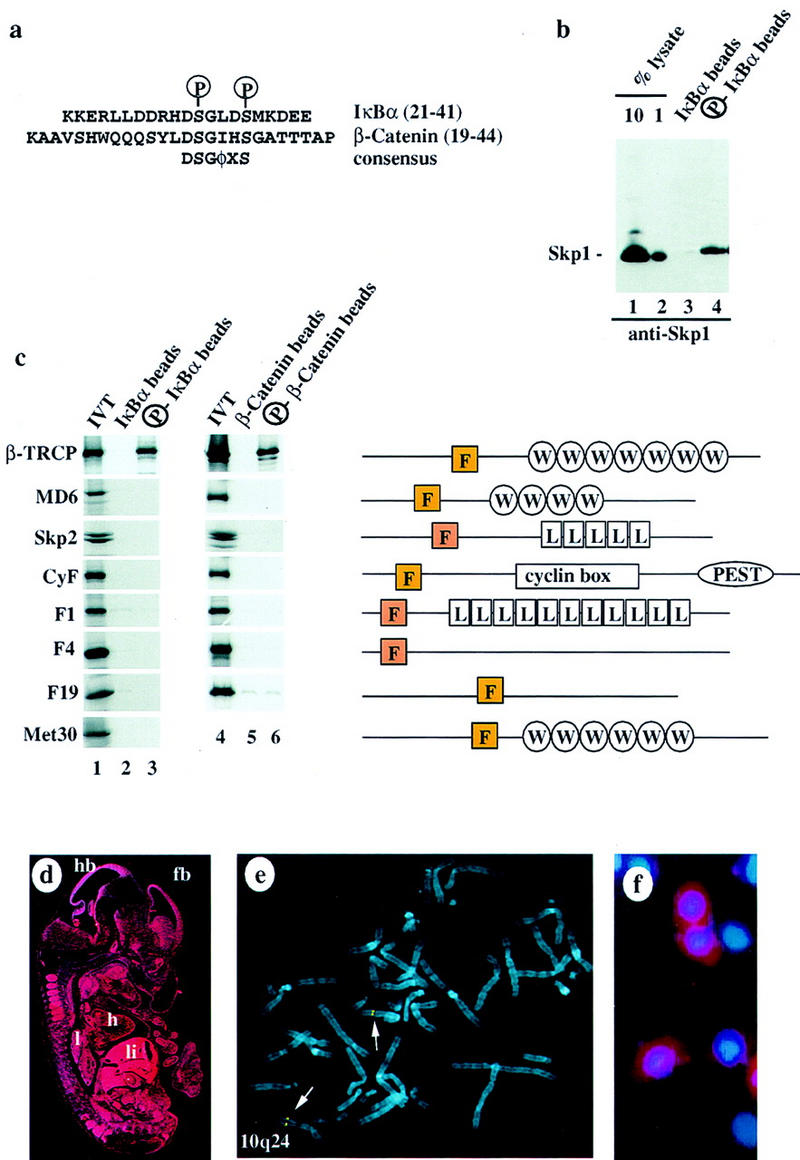
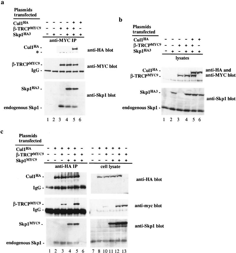
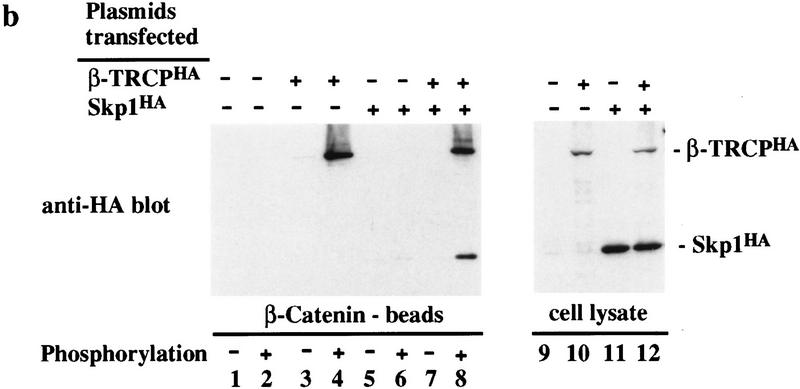
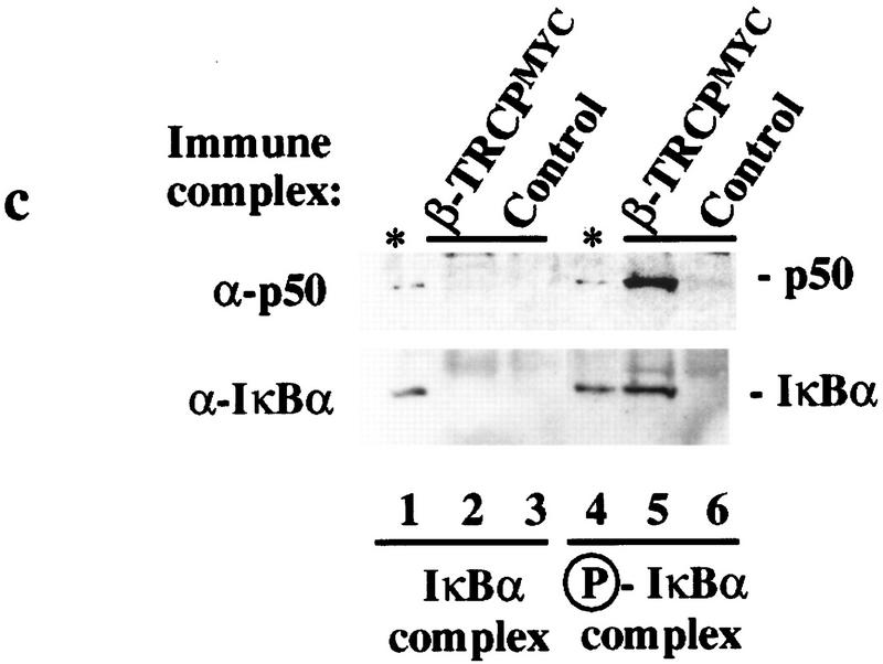
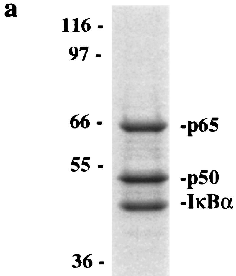
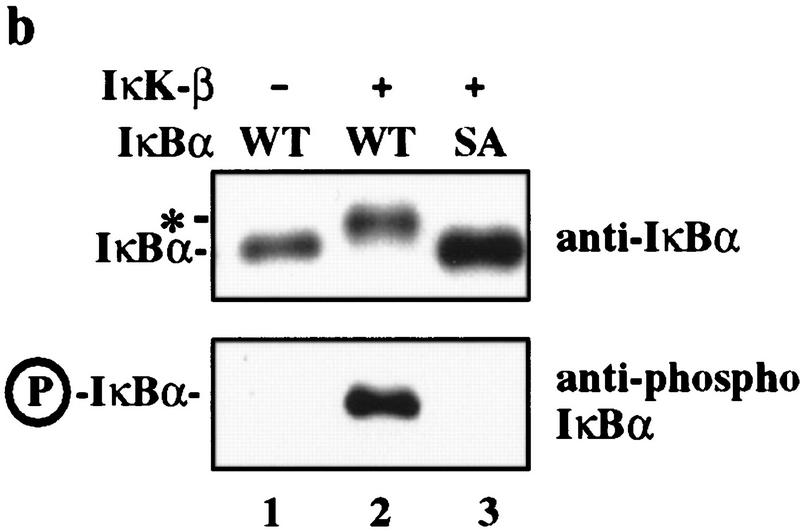
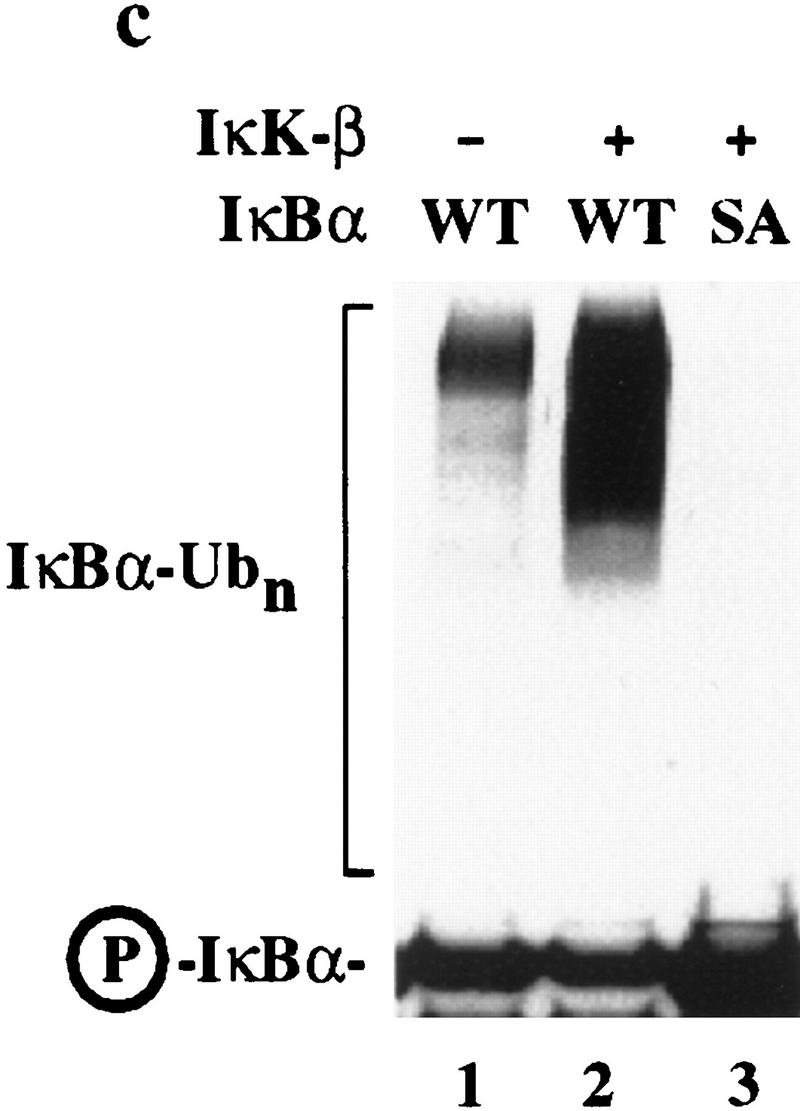
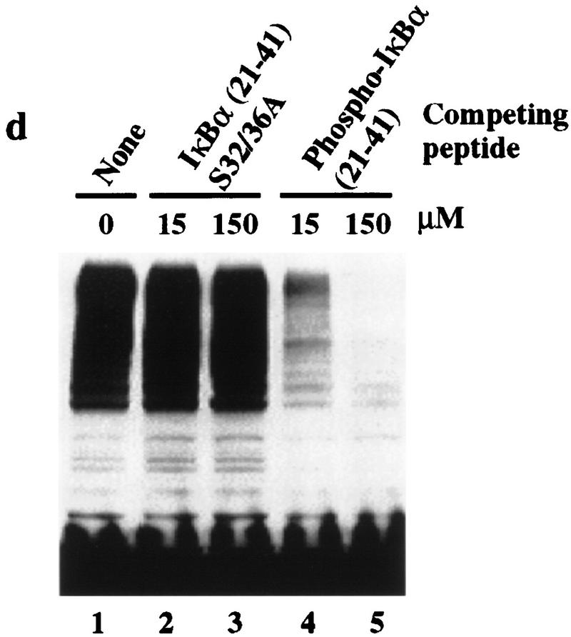
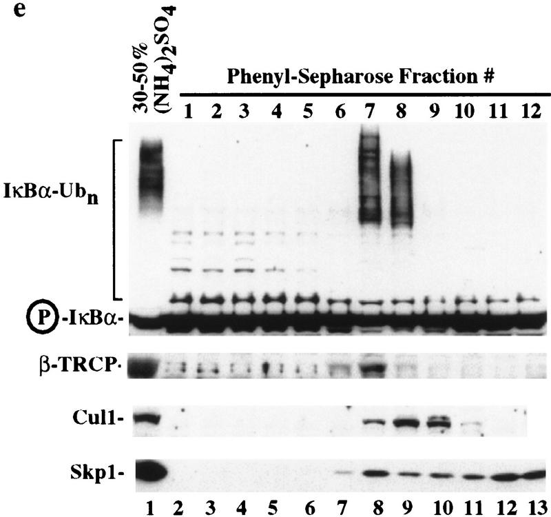
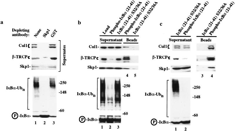
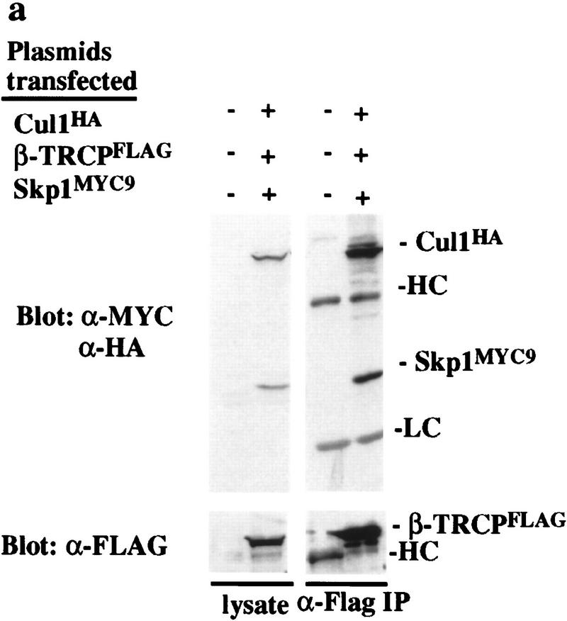
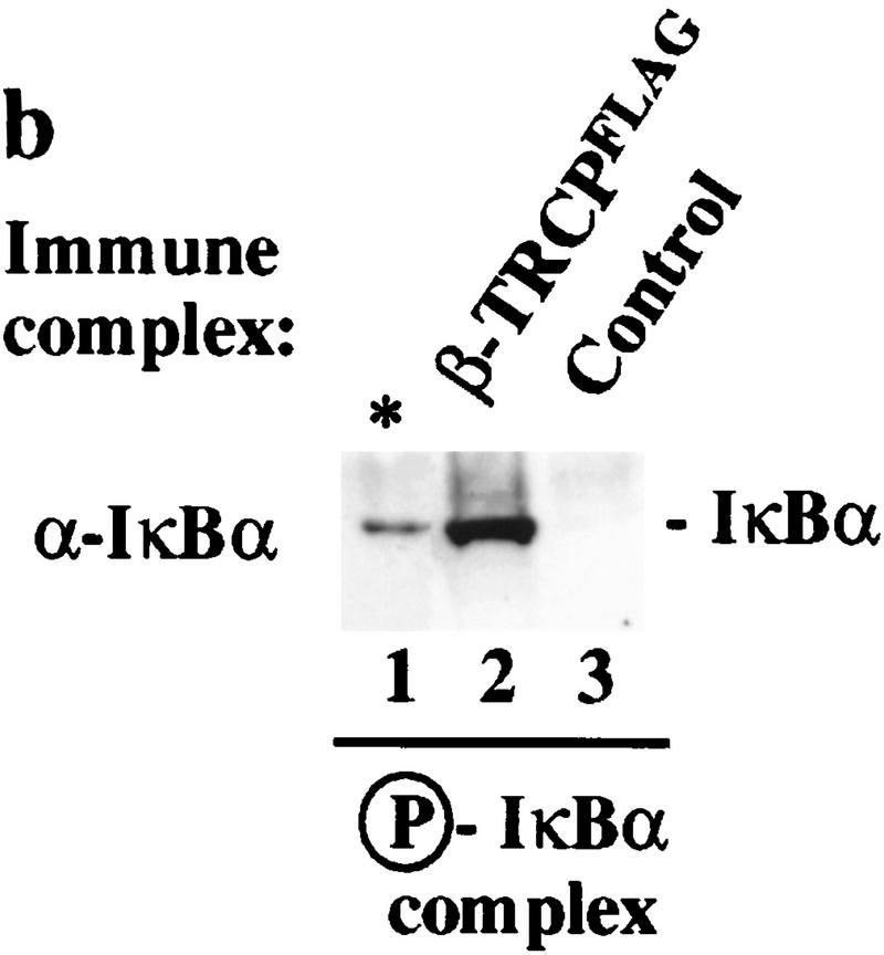
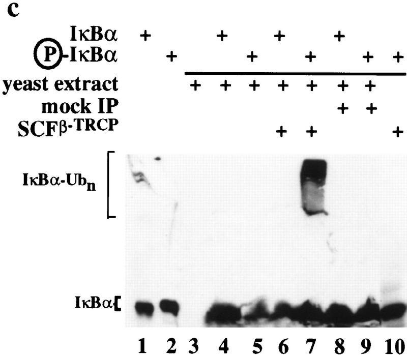
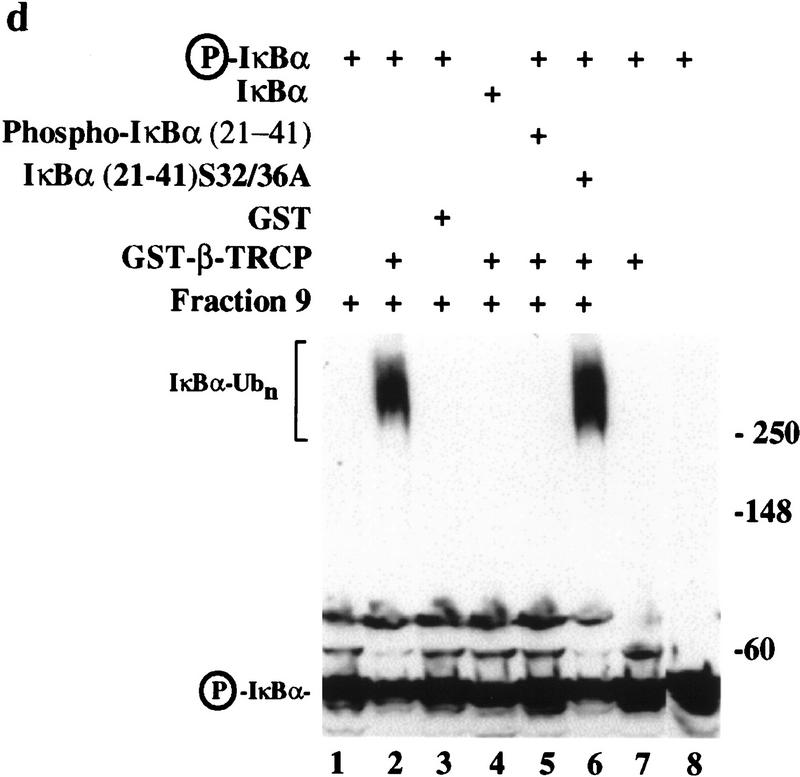
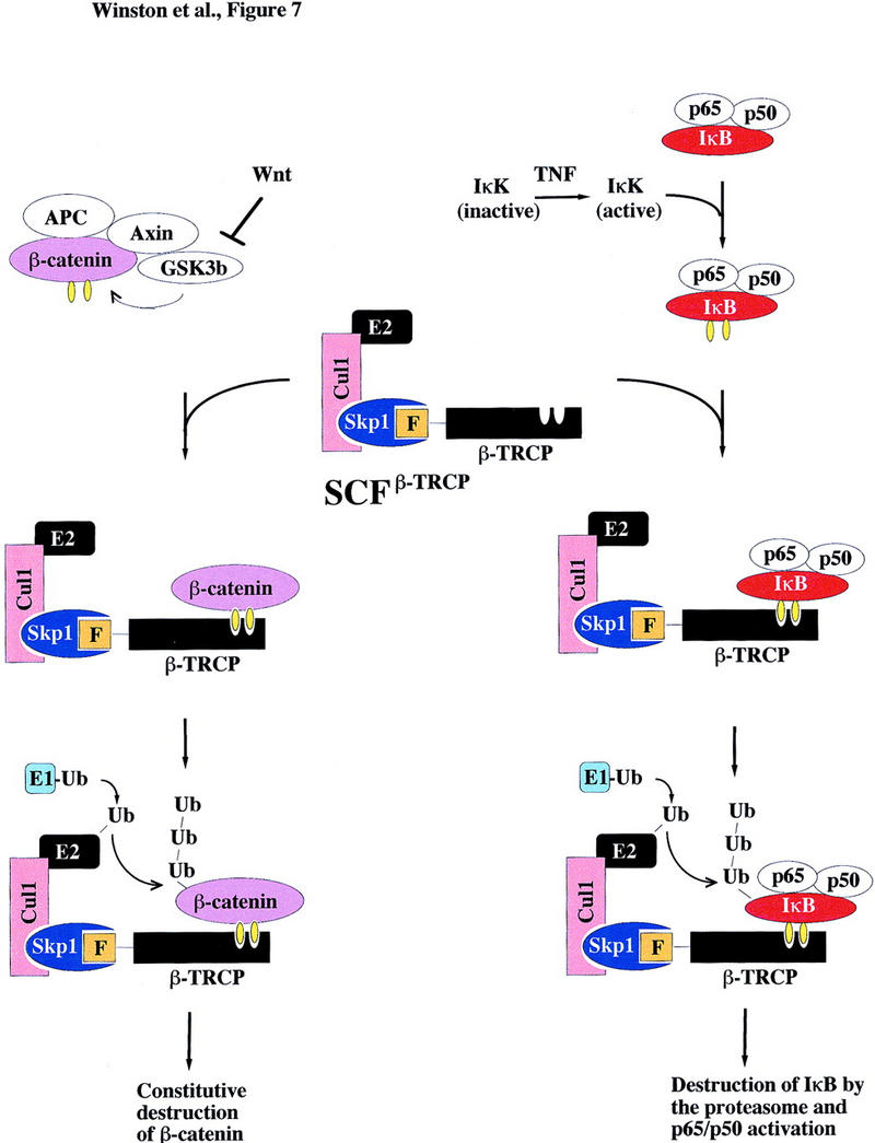
Ubiquitin-mediated proteolysis has a central role in controlling the intracellular levels of several important regulatory molecules such as cyclins, CKIs, p53, and IkappaBalpha. Many diverse proinflammatory signals lead to the specific phosphorylation and subsequent ubiquitin-mediated destruction of the NF-kappaB inhibitor protein IkappaBalpha. Substrate specificity in ubiquitination reactions is, in large part, mediated by the specific association of the E3-ubiquitin ligases with their substrates. One class of E3 ligases is defined by the recently described SCF complexes, the archetype of which was first described in budding yeast and contains Skp1, Cdc53, and the F-box protein Cdc4. These complexes recognize their substrates through modular F-box proteins in a phosphorylation-dependent manner. Here we describe a biochemical dissection of a novel mammalian SCF complex, SCFbeta-TRCP, that specifically recognizes a 19-amino-acid destruction motif in IkappaBalpha (residues 21-41) in a phosphorylation-dependent manner. This SCF complex also recognizes a conserved destruction motif in beta-catenin, a protein with levels also regulated by phosphorylation-dependent ubiquitination. Endogenous IkappaBalpha-ubiquitin ligase activity cofractionates with SCFbeta-TRCP. Furthermore, recombinant SCFbeta-TRCP assembled in mammalian cells contains phospho-IkappaBalpha-specific ubiquitin ligase activity. Our results suggest that an SCFbeta-TRCP complex functions in multiple transcriptional programs by activating the NF-kappaB pathway and inhibiting the beta-catenin pathway.


Figures

Figure 1
Figure 1
The F-box protein β-TRCP associates with phosphorylated destruction motifs in IκBα and β-catenin. (a) Sequences of the IκBα (p21) and β-catenin peptides used in this study. The positions of phosphorylation in the peptides are shown as is the consensus sequence for the destruction motif. (φ) Hydrophobic amino acid. (b) Phosphorylation-specific association of the IκBα destruction motif with Skp1 in vitro. HeLa cell proteins (600 μg) were incubated with phosphorylated or unphosphorylated IκBα peptides (p21) immobilized on beads. Bound proteins were immunoblotted with anti-Skp1 antibodies. Approximately 1% of the Skp1 contained in these lysates remained bound to the phosphorylated IκBα beads. (c) β-TRCP specifically associated with phosphorylated IκBα and β-catenin destruction motifs. A panel of [35S]methionine-labeled in vitro-translated F-box proteins was used in binding reactions with IκBα (lanes 13) or β-catenin (lanes 46) peptide beads. Bound proteins were analyzed by SDS-PAGE and autoradiography. (Right) The domain structures of each F-box protein. (d) The pattern of expression of β-TRCP at day 11.5 during mouse development was determined by in situ hybridization. The dark-field signal from the β-TRCP riboprobe is shown in red. (hb) Hindbrain; (fb) forebrain; (h) heart; (l) lung; (li) liver. (e) Chromosomal localization of β-TRCP. A bacmid containing human β-TRCP DNA was hybridized to metaphase chromosomes (blue) and detected using fluorescein. The position of hybridization (yellow) is 10q24 (indicated by arrows). (f) β-TRCP is localized in the cytoplasm. HeLa cells were transiently transfected with pCMV–HA–β-TRCP and subcellular localization determined after 48 hr by indirect immunofluorescence. Anti-HA localization, red; nuclei stained with DAPI, blue.
Figure 2
Figure 2
β-TRCP associates with Skp1 and Cul1 in tissue culture cells. 293T cells were transfected with the indicated plasmids and lysates (0.5 μg of protein/250 μl) used for immunoprecipitation as described in Materials and Methods. Immune complexes or crude lysates from each transfection were analyzed for the presence of Skp1, Cul1, and β-TRCP by immunoblotting. (a) Anti-β-TRCPMyc immune complexes. Blots were probed first for β-TRCP, stripped, and probed for Skp1 and Cul1. The bands indicated by the asterisk indicate the position of β-TRCP whose antibody was not efficiently stripped from the blot. (b) Crude cell lysates (50 μg) corresponding to extracts used in a. (c) Anti-Cul1HA immune complexes (lanes 16) and corresponding cell lysates (50 μg) (lanes 713). The positions of both epitope-tagged and endogenous Skp1 are shown.
Figure 3
Figure 3
Association of SCFβ-TRCP with IκBα and β-catenin destruction motifs and with the IκBα/NF-κB complex. (a,b) Cell lysates (0.3 μg of protein/150 μl) from Fig. 2 were used in IκBα (a) and β-catenin (b) peptide bead binding reactions as described in Materials and Methods. Bound proteins were analyzed by immunoblotting with the indicated antibodies. (c) Phosphorylation-dependent association of β-TRCPMyc with the IκBα/p50/p65 complex in vitro. β-TRCPMyc immune complexes (lanes 2,5) corresponding to those in Fig. 2a (lane 3) or control complexes (lanes 3,6) corresponding to those in Fig. 2a (lane 1) were used in binding reactions with either IκBα/p50/p65 or IκK-β phosphorylated IκBα/p50/p65 complexes (see Materials and Methods). Bound proteins were separated by SDS-PAGE and immunoblotted using anti-p50 or anti-IκBα antibodies. The asterisk (lanes 1,4) indicates the positions of 15% of the input IκBα complexes used in the binding reaction.
Figure 3
Figure 3
Association of SCFβ-TRCP with IκBα and β-catenin destruction motifs and with the IκBα/NF-κB complex. (a,b) Cell lysates (0.3 μg of protein/150 μl) from Fig. 2 were used in IκBα (a) and β-catenin (b) peptide bead binding reactions as described in Materials and Methods. Bound proteins were analyzed by immunoblotting with the indicated antibodies. (c) Phosphorylation-dependent association of β-TRCPMyc with the IκBα/p50/p65 complex in vitro. β-TRCPMyc immune complexes (lanes 2,5) corresponding to those in Fig. 2a (lane 3) or control complexes (lanes 3,6) corresponding to those in Fig. 2a (lane 1) were used in binding reactions with either IκBα/p50/p65 or IκK-β phosphorylated IκBα/p50/p65 complexes (see Materials and Methods). Bound proteins were separated by SDS-PAGE and immunoblotted using anti-p50 or anti-IκBα antibodies. The asterisk (lanes 1,4) indicates the positions of 15% of the input IκBα complexes used in the binding reaction.
Figure 4
Figure 4
IκBα–ubiquitin ligase activity from human cells cofractionates with β-TRCP. (a) IκBα/p50/p65 complexes were purified to near homogeneity from insect cells as described in Materials and Methods. Proteins were separated by SDS-PAGE and stained with Coomassie blue. (b) Phosphorylation of the IκBα/p50/p65 complex by IκK-β. The IκBα complex (lanes 1,2) or a nonphosphorylatable IκBα mutant (S32/36A) complex (lane 3) was incubated in the presence of ATP and IκK-β as indicated in Materials and Methods. Products were analyzed by immunoblotting with anti-IκBα antibodies to detect a mobility shift accompanying phosphorylation that is absent in the nonphosphorylatable mutant (top) or with antibodies that specifically detect the Ser-32-phosphorylated form of IκBα (bottom). (c) Ubiquitination of IκBα complexes by crude cell lysates was performed as described in Materials and Methods. Phosphorylation leads to a ∼10- to 20-fold increase in ubiquitin conjugates relative to the unphosphorylated complex, whereas no activity is observed with the nonphosphorylatable IκBα complexes. (d) Inhibition of IκBα ubiquitination by phosphorylated IκBα destruction motif peptides but not by nonphosphorylatable destruction motif peptides (p19). Ubiquitination reactions were performed with crude cell extracts and IκK-β phosphorylated IκBα complexes in the presence or absence of phosphorylated or nonphosphorylatable destruction motif peptides. Specific inhibition of ubiquitination was observed with the phosphorylated peptide. (e) Cofractionation of β-TRCP with endogenous IκBα–ubiquitin ligase activity. Crude extracts from THP.1 cells were precipitated with ammonium sulfate. Solubilized proteins containing ubiquitin ligase activity were fractionated using a phenyl–Sepharose column and activity in each fraction determined as described in Materials and Methods. Aliquots of column fractions were assayed for β-TRCP, Cul1, and Skp1 by immunoblotting. Fractions containing β-TRCP, Cul1, and Skp1 (fractions 7,8) contain IκBα–ubiquitin ligase activity.
Figure 4
Figure 4
IκBα–ubiquitin ligase activity from human cells cofractionates with β-TRCP. (a) IκBα/p50/p65 complexes were purified to near homogeneity from insect cells as described in Materials and Methods. Proteins were separated by SDS-PAGE and stained with Coomassie blue. (b) Phosphorylation of the IκBα/p50/p65 complex by IκK-β. The IκBα complex (lanes 1,2) or a nonphosphorylatable IκBα mutant (S32/36A) complex (lane 3) was incubated in the presence of ATP and IκK-β as indicated in Materials and Methods. Products were analyzed by immunoblotting with anti-IκBα antibodies to detect a mobility shift accompanying phosphorylation that is absent in the nonphosphorylatable mutant (top) or with antibodies that specifically detect the Ser-32-phosphorylated form of IκBα (bottom). (c) Ubiquitination of IκBα complexes by crude cell lysates was performed as described in Materials and Methods. Phosphorylation leads to a ∼10- to 20-fold increase in ubiquitin conjugates relative to the unphosphorylated complex, whereas no activity is observed with the nonphosphorylatable IκBα complexes. (d) Inhibition of IκBα ubiquitination by phosphorylated IκBα destruction motif peptides but not by nonphosphorylatable destruction motif peptides (p19). Ubiquitination reactions were performed with crude cell extracts and IκK-β phosphorylated IκBα complexes in the presence or absence of phosphorylated or nonphosphorylatable destruction motif peptides. Specific inhibition of ubiquitination was observed with the phosphorylated peptide. (e) Cofractionation of β-TRCP with endogenous IκBα–ubiquitin ligase activity. Crude extracts from THP.1 cells were precipitated with ammonium sulfate. Solubilized proteins containing ubiquitin ligase activity were fractionated using a phenyl–Sepharose column and activity in each fraction determined as described in Materials and Methods. Aliquots of column fractions were assayed for β-TRCP, Cul1, and Skp1 by immunoblotting. Fractions containing β-TRCP, Cul1, and Skp1 (fractions 7,8) contain IκBα–ubiquitin ligase activity.
Figure 5
Figure 5
Depletion of IκBα–ubiquitin ligase activity by anti-Skp1 antibodies and destruction motif peptides correlates with removal of β-TRCP. (a) Ubiquitin ligase activity from phenyl–Sepharose fractions 7 and 8 was depleted with antibodies against Skp1 or GST and the supernatant assayed for ubiquitination activity toward phosphorylated IκBα (bottom). The levels of Skp1, Cul1, and β-TRCP in the supernatants from the depleted fractions were determined by immunoblotting (top). (b,c) Endogenous β-TRCP associates with phosphorylated IκBα destruction motif peptides during depletion of IκBα–ubiquitin ligase activity. Crude cell extracts (b) or active fractions from a phenyl–Sepharose column (c) were incubated with beads containing either phosphorylated or nonphosphorylatable IκBα peptides and the supernatants assayed for ubiquitin ligase activity (bottom panels). The levels of Skp1, Cul1, and β-TRCP in the supernatant and associated with destruction motif peptides were determined by immunoblotting (top). β-TRCP is associated with the phosphorylated destruction motif peptides and is substantially depleted from active ubiquitin ligase fractions.
Figure 6
Figure 6
Stimulation of IκBα–ubiquitin ligase activity by SCFβ-TRCP in vitro. (a) Flag-tagged SCFβ-TRCP was prepared after transient transfection in 293T cells by immunoprecipitation along with a Flag immune complex from mock-transfected cells. Immune complexes were analyzed for the presence of Cul1HA, Skp1Myc, and β-TRCPFlag by immunoblotting (lanes 3,4). Crude lysates used for immunoprecipitation are shown as controls (lanes 1,2). (b) β-TRCPFlag immune complexes associate with phosphorylated IκBα in vitro. Immune complexes (10 μl beads) from (a) were incubated with 15 nm phosphorylated IκBα/NF-κB complexes in a total volume of 100 μl. Washed beads were subjected to SDS-PAGE and IκBα determined by immunobloting. The asterisk indicates a sample containing 15% of the input IκBα complex. (c) Stimulation of IκBα–ubiquitin ligase activity by SCFβ-TRCP in vitro. Yeast extracts (supplemented with E1, ubiquitin, and an ATP-regenerating system) were incubated with unphosphorylated or phosphorylated IκBα/NF-κB complexes (25 nm) in the presence of 10 μl of control immune complexes (lanes 8,9) or β-TRCPFlag immune complexes (lanes 6,7). After 90 min, reaction mixtures were separated by SDS-PAGE and IκBα detected by immunoblotting with anti-IκBα antibodies. As controls, untreated IκBα complexes (lanes 1,2), supplemented yeast lysates (lane 3), and an SCFβ-TRCP immune complex reaction mixture containing all components except the yeast extract (lane 10) were also included. (d) Reconstitution of IκBα ubiquitination activity in mammalian extracts by addition of purified GST–β-TRCP. Reaction mixtures, prepared as described in Materials and Methods, contained E1, ubiquitin, ATP, HQ unbound as a source of E2 activity, and other components as indicated (lanes 16). Control reactions (lanes 7,8) lacked phenyl–Sepharose fraction 9. After 90 min, products were analyzed by SDS-PAGE and immunoblotting with anti-IκBα antibodies.
Figure 6
Figure 6
Stimulation of IκBα–ubiquitin ligase activity by SCFβ-TRCP in vitro. (a) Flag-tagged SCFβ-TRCP was prepared after transient transfection in 293T cells by immunoprecipitation along with a Flag immune complex from mock-transfected cells. Immune complexes were analyzed for the presence of Cul1HA, Skp1Myc, and β-TRCPFlag by immunoblotting (lanes 3,4). Crude lysates used for immunoprecipitation are shown as controls (lanes 1,2). (b) β-TRCPFlag immune complexes associate with phosphorylated IκBα in vitro. Immune complexes (10 μl beads) from (a) were incubated with 15 nm phosphorylated IκBα/NF-κB complexes in a total volume of 100 μl. Washed beads were subjected to SDS-PAGE and IκBα determined by immunobloting. The asterisk indicates a sample containing 15% of the input IκBα complex. (c) Stimulation of IκBα–ubiquitin ligase activity by SCFβ-TRCP in vitro. Yeast extracts (supplemented with E1, ubiquitin, and an ATP-regenerating system) were incubated with unphosphorylated or phosphorylated IκBα/NF-κB complexes (25 nm) in the presence of 10 μl of control immune complexes (lanes 8,9) or β-TRCPFlag immune complexes (lanes 6,7). After 90 min, reaction mixtures were separated by SDS-PAGE and IκBα detected by immunoblotting with anti-IκBα antibodies. As controls, untreated IκBα complexes (lanes 1,2), supplemented yeast lysates (lane 3), and an SCFβ-TRCP immune complex reaction mixture containing all components except the yeast extract (lane 10) were also included. (d) Reconstitution of IκBα ubiquitination activity in mammalian extracts by addition of purified GST–β-TRCP. Reaction mixtures, prepared as described in Materials and Methods, contained E1, ubiquitin, ATP, HQ unbound as a source of E2 activity, and other components as indicated (lanes 16). Control reactions (lanes 7,8) lacked phenyl–Sepharose fraction 9. After 90 min, products were analyzed by SDS-PAGE and immunoblotting with anti-IκBα antibodies.
Figure 7
Figure 7
Schematic representation of the proposed pathways controlling ubiquitin-mediated proteolysis of IκBα and βcatenin. β-TRCP, an F-box protein, is a component of an SCF–ubiquitin ligase. In response to appropriate signals (i.e., TNFα), the IκK complex is activated and phosphorylates IκBα in complexes with NF-κB on Ser-32 and Ser-36. This complex is then recognized by β-TRCP in an SCF complex, facilitating ubiquitination by an E1- and E2-dependent mechanism. βCatenin, in complexes with APC, axin, and GSK3β, is phosphorylated on Ser-33 and Ser-37. This phosphorylated β-catenin can then associate with SCFβ-TRCP, resulting in ubiquitination. It is not clear at present whether β-catenin alone or the APC/β-catenin complex is the relevant target. Yellow ovals indicate phosphorylation.

References

    1. Aberle H, Bauer A, Stappert J, Kispert A, Kemler R. β-Catenin is a target for the ubiquitin-proteasome pathway. EMBO J. 1997;16:3797–3804. - PMC - PubMed
    1. Alkalay I, Yaron A, Hatzubai A, Orian A, Ciechanover A, Ben-Neriah Y. Stimulation-dependent I kappa B alpha phosphorylation marks the NF-kappa B inhibitor for degradation via the ubiquitin-proteasome pathway. Proc Natl Acad Sci. 1995;92:10599–10603. - PMC - PubMed
    1. Bai C, Sen P, Mathias N, Hofmann K, Goebl M, Harper JW, Elledge SJ. SKP1 connects cell cycle regulation to the ubiquitin proteolysis machinery through a novel motif, the F-box. Cell. 1996;86:263–274. - PubMed
    1. Baeuerle PA, Baltimore D. NF-κB: Ten years after. Cell. 1996;87:13–20. - PubMed
    1. Beg AA, Baltimore D. An essential role for NF-κB in preventing TNFα-induced cell death. Science. 1996;274:782–784. - PubMed

Publication types

MeSH terms

Substances

Associated data