. 1999 Feb 1;13(3):334-44.
doi: 10.1101/gad.13.3.334.

The PERIANTHIA gene encodes a bZIP protein involved in the determination of floral organ number in Arabidopsis thaliana

Affiliations

The PERIANTHIA gene encodes a bZIP protein involved in the determination of floral organ number in Arabidopsis thaliana

C F Chuang et al. Genes Dev. .

Abstract

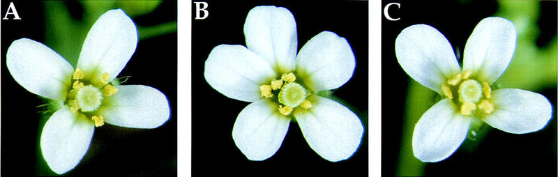
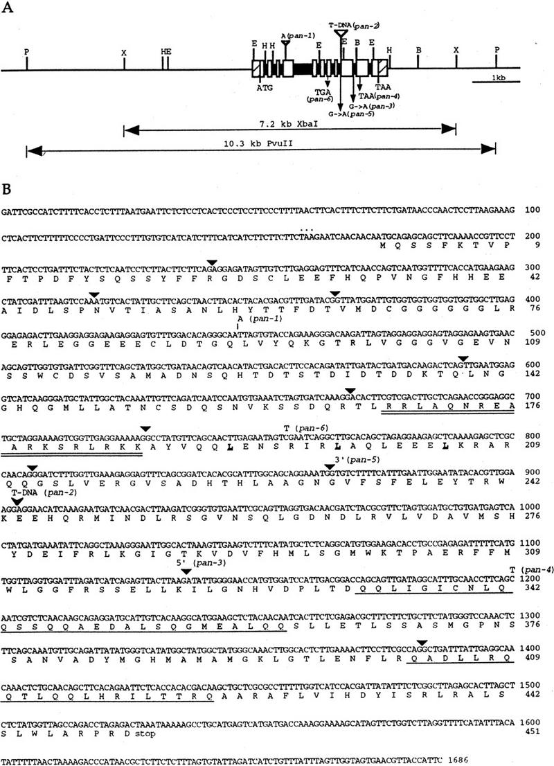
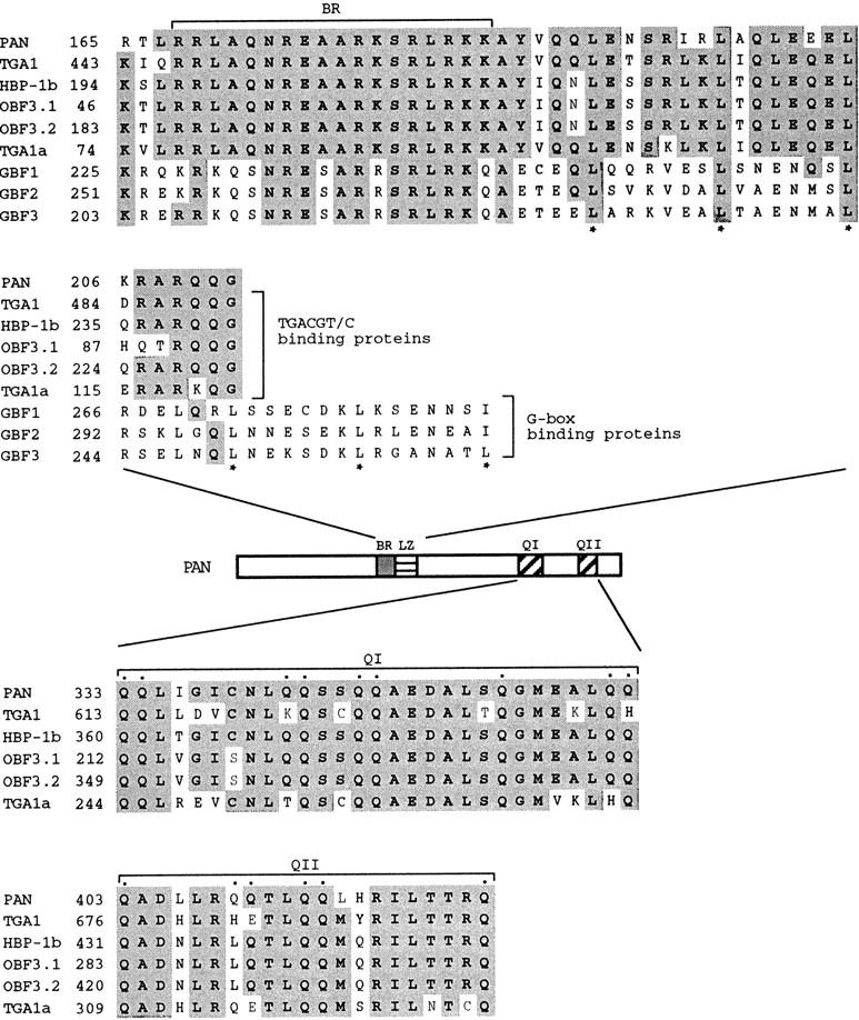
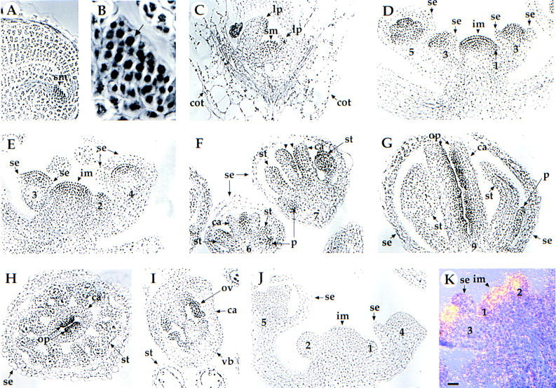
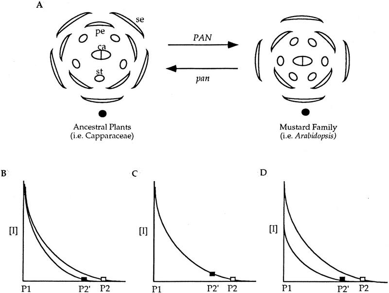
Mutations in the PERIANTHIA (PAN) gene of Arabidopsis thaliana specifically transform flowers from tetramerous to largely pentamerous, which is a characteristic of flowers of ancestral plants. We have cloned the PAN gene and here we show that it encodes a member of the basic region/leucine zipper class of transcription factors. Immunohistochemical analysis shows that the encoded protein is present in the apical meristem, the floral meristem, each whorl of organ primordia, and in ovule primordia during wild-type flower development. PAN expression occurs independently of genes affecting floral meristem identity, floral meristem size, or floral organ number. The near absence of a phenotype in transgenic plants overexpressing PAN and the contrast between the broad expression of PAN and the specificity of its mutant phenotype suggest that its activity may be regulated post-translationally or by the presence of partner proteins. Based on these results and on data reported previously, we propose models for the role of PAN in the evolution of flower pattern in the mustard family.


Figures

Figure 1
Figure 1
(A) Wild-type flower. (B) pan-1 flower. (C) pan-1 flower with a 7.2-kb XbaI genomic fragment containing the PAN gene. (A) Wild-type flowers have four sepals, four petals, six stamens, and two carpels. (B) Most pan mutant flowers have five sepals, five petals, five stamens, and two carpels. (C) pan-1 plants carrying the transgenic wild-type PAN gene produce flowers indistinguishable from wild type.
Figure 2
Figure 2
Genomic structure of the PAN gene, and sequence of the PAN cDNA. (A) Structure and restriction map of the PAN genomic region. The open boxes and solid boxes represent exons and introns, respectively. The hatched boxes represent the 5′ and 3′ untranslated regions. The genomic regions used for complementation of pan mutant phenotype are indicated below. ATG and TAA indicate the positions of the putative translational start and stop codons, respectively. Mutations in the six pan alleles are also shown. Restriction sites are indicated as follows: (B) BamHI; (E) EcoRI; (H) HindIII; (P) PvuII; (X) XbaI. (B) The cDNA and deduced amino acid sequence of the PAN gene. The predicted amino acid sequence for the longest open reading frame is shown directly below the nucleotide sequence. Numbers to the right of the sequence refer to the positions of nucleotide and amino acid residues. Triangles indicate the positions of introns. The basic region is doubly underlined and the leucines in the leucine zipper are in bold and underlined. The glutamine-rich regions are singly underlined. The inframe stop codon preceding the first methionine is indicated by dots above the nucleotide sequence. One nucleotide insertion in pan-1 is shown above the nucleotide sequence. The position of the T-DNA insertion in pan-2 allele is indicated by an arrowhead. The pan-3 mutation occurs at the splice donor site of the ninth intron. The pan-5 allele is mutated in the splice acceptor site of the eighth intron. Point mutations resulting in premature translational stop sites in pan-4 and pan-6 are shown above the nucleotide sequence.
Figure 3
Figure 3
Similarity between PAN and other plant bZIP proteins. (Top) Comparison of the PAN bZIP regions to other plant bZIP proteins: the Arabidopsis protein TGA1 (Schindler et al. 1992a), the wheat protein HBP-1b (Tabata et al. 1991), the maize protein OBF 3.1 and OBF 3.2 (Foley et al. 1993), the tobacco protein TGA1a (Katagiri et al. 1989), and the Arabidopsis proteins GBF1, GBF2, and GBF3 (Schindler et al. 1992b). Asterisks (*) indicate leucine residues within the leucine zipper domain. (Middle) Schematic presentation of PAN, indicating the positions of the basic region (BR), the leucine zipper domain (LZ), and the two glutamine-rich regions (QI and QII). (Bottom) Sequence alignments within the carboxy-terminal glutamine-rich domains of PAN and the TGACGT/C-binding proteins shown in the top. Glutamine residues within the glutamine-rich regions of PAN are indicated by dots. Identical amino acids are shown as shaded boxes. Numbers to the left refer to the amino acid positions within individual proteins.
Figure 4
Figure 4
Localization of PAN protein and distribution of PAN RNA in wild-type (A–I, K) and pan mutant (J) plants. (A) Section through the shoot apical meristem (sm) in the mature embryo. Seeds were imbibed at 4°C for 2 days and transferred to constant fluorescent light at 20°C for 1 day. PAN is highly expressed in the shoot apical meristem (arrowhead). (B) High magnification view of the cells in the shoot apical meristem shown in A. The arrow indicates that the PAN protein is localized primarily to the nucleus (arrow). (C) Longitudinal section through a shoot apical meristem of 6-day-old seedling. PAN protein is detected in the shoot apical meristem (sm) and leaf primordia (lp), but not in the cotyledons (cot). (D,E) Longitudinal section of an inflorescence meristem (im) with stage 2–5 flowers. PAN protein is detected at high levels throughout the apical region of the inflorescence meristem (arrowhead), throughout the floral meristem of the stage 2 flower, the central dome of the floral meristem of stage 3–4 flowers, and the inner three whorls of organ primordia in the stage 5 flower. (se) Sepal. (F) Longitudinal section of stage 6–7 flowers. PAN protein becomes restricted in the base of petal (p) primordia, the adaxial side of stamen (st) primordia, and the adaxial side of carpel (c) primordia (arrowhead). Longitudinal section (G) and cross section (H) of a stage 9 flower. PAN protein remains at high levels in the ovule primordia (op). (I) Cross section of a stage 12 flower. Expression level of PAN protein becomes weaker in the ovule (ov). The PAN protein is not expressed at levels above background in sections of inflorescences from pan-1 (J) and pan-2 (data not shown). (K) In situ hybridization of a PAN antisense probe with a longitudinal section of an inflorescence meristem (im) with stage 1–3 flowers. Expression of PAN RNA is detected throughout the apical region of the inflorescence meristem and throughout the floral meristem of stage 1–2 flowers. Expression in the stage 3 flower is limited to regions interior to the sepals (se). In the bright field/dark field double exposures, the silver grains representing PAN expression were made to appear yellow. The number indicated corresponds to the stages of floral development as described by Smyth et al. (1990). All panels shown at the same magnification. Bar, 20 μm.
Figure 5
Figure 5
Expression of the PAN protein in lfy, ap1, and ap2 mutants. (A–B) lfy-6. (A) PAN protein is expressed in the inflorescence meristem (im) and the center of abnormal young flowers. The stages of flowers (3 and 4) and the floral bud (f) in the axil of the bract (b) are indicated. (B) PAN protein is detected in the secondary floral buds (sf) in the axils of the first-whorl organs (1w) of an old lfy-6 flower. (g) Gynoecium. (C–E) ap1-1. (C) Early expression in the inflorescence meristem (im) and in the stage 2, 3, and 6 floral buds is normal (compared with Figs. 4D–F). (st) Stamen. (D) PAN protein is expressed normally in the ovule primordia (op) at stage 9 flowers (cf. with Fig. 4G). (c) Carpel. (E) Secondary floral buds (sf) in the axil of the first-whorl organs (1w) show PAN protein expression at levels comparable to the early flower primordia arising on the inflorescence apex (Figs. 4D,E). (F–G) ap2-2. PAN protein expression is largely normal in the inflorescence meristem (im) and young floral buds (stage 2–4). At later stages, PAN protein is detected in the ovule primordia (arrowhead) in both of the first-whorl carpels (1c) and the central gyneocium (g). The number indicated corresponds to the developmental stage of each flower (Smyth et al. 1990). Bar, 20 μm.
Figure 6
Figure 6
Expression of PAN protein in clv1-4, clv3-2, ett-3, wig-1, and tsl-1 mutants. (A–B) clv1-4. PAN is expressed in the inflorescence meristem (im), the floral meristem (fm, A), and the fifth-whorl (5w) of clv1-4 flowers (B). PAN protein expression is normal in the inflorescence meristem and the floral meristem in clv3-2 (C), ett-3 (D), wig-1 (E), and tsl-1 (F) flowers. (se) Sepal; (p) petal; (st) stamen; (c) carpel. The number indicated corresponds to the developmental stage of each flower (Smyth et al. 1990). Bar, 20 μm.
Figure 7
Figure 7
Possible models for PAN in regulating floral organ number. We postulate that there is an inhibitory mechanism controlling the relative positions of first and second whorl floral organ primordia in ancestral plants (i.e., Capparaceae) of the mustard family (i.e., Arabidopsis) and PAN participates in the pre-existing inhibitory pathway to establish tetramerous flowers in the mustard family. (A) Introduction of PAN into the morphogen pathway leads to more distant spacing of organ primordia and thus fewer organs. When PAN activity is reduced or eliminated (pan), plants produce flowers with a pattern resembling the ancestral flower form. (B–D) The inhibitor concentration [I] is plotted as function of distance. The diffusing inhibitor decays with time and thus distance. P1, the first primordium. (█) pan; (□) PAN. (B) PAN might facilitate inhibitor diffusion or persistence to allow the inhibitor to spread more widely, resulting in the formation of the second primordium (P2) at a more distant position than in pan (P2′). (C) PAN might enhance inhibitor reception, leading to formation of the next primordium at a lower inhibitor concentration than in pan. (D) PAN might increase inhibitor production, indirectly leading to its wider diffusion. (se) Sepal; (pe) petal; (st) stamen; (ca) carpel; (solid ovals) inflorescence meristem; (□) and (█) the lowest concentration in the inhibitory field allowing the new primordium to arise in PAN and pan floral meristems, respectively.

References

    1. Armstrong GA, Weisshaar B, Hahlbrock K. Homodimeric and heterodimeric leucine zipper proteins and nuclear factors from parsley recognize diverse promoter elements with ACGT cores. Plant Cell. 1992;4:525–537. - PMC - PubMed
    1. Bechtold N, Ellis J, Pelletier G. In planta Agrobacterium mediated gene transfer by infiltration of adult Arabidopsis thaliana plants. CR Acad Sci. 1993;316:1194–1199.
    1. Bouchez D, Tokuhisa JG, Llewellyn DJ, Dennis ES, Ellis JG. The ocs-element is a component of the promoters of several T-DNA and plant viral genes. EMBO J. 1989;8:4197–4204. - PMC - PubMed
    1. Bowman JL, editor. Arabidopsis: An atlas of morphology and development. New York, NY: Springer-Verlag; 1994. Flowers: Introduction; pp. 135–145.
    1. Bowman JL, Smyth DR, Meyerowitz EM. Genes directing flower development in Arabidopsis. Plant Cell. 1989;1:37–52. - PMC - PubMed

Publication types

MeSH terms

Associated data