. 1999 Feb 1;13(3):357-70.
doi: 10.1101/gad.13.3.357.

Amino-terminal sequences of sigmaN (sigma54) inhibit RNA polymerase isomerization

Affiliations

Amino-terminal sequences of sigmaN (sigma54) inhibit RNA polymerase isomerization

W Cannon et al. Genes Dev. .

Abstract

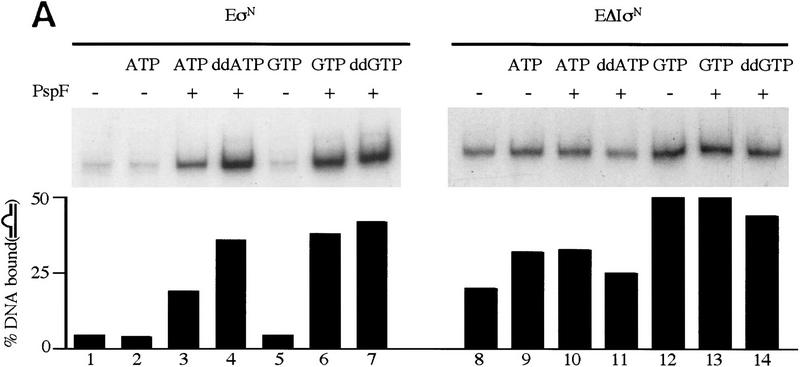
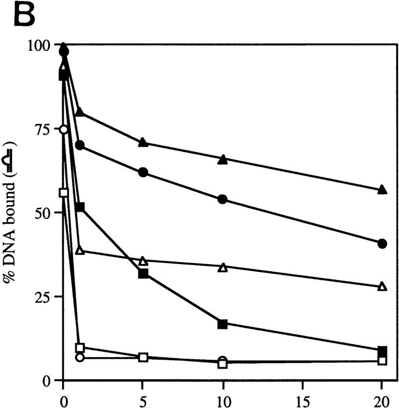
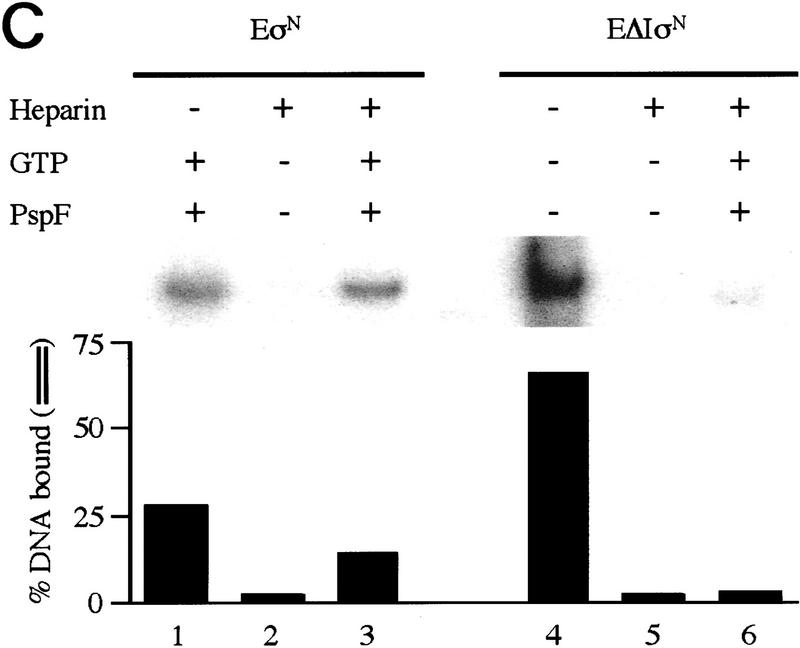
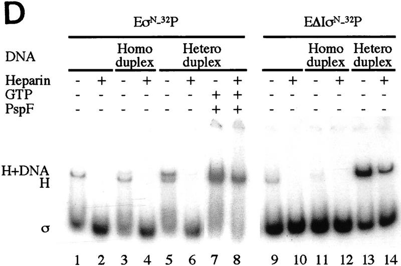
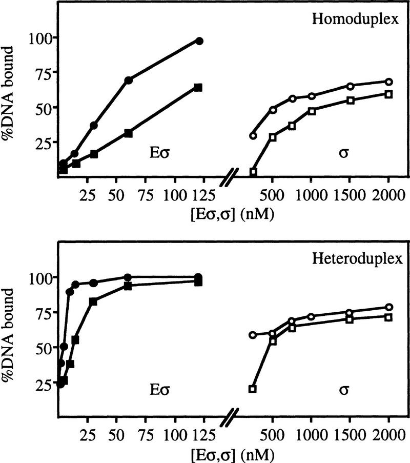
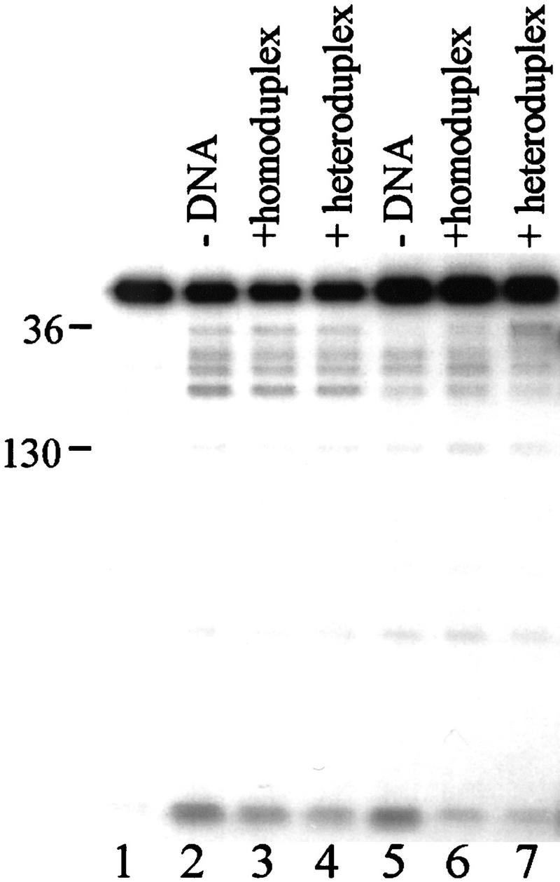
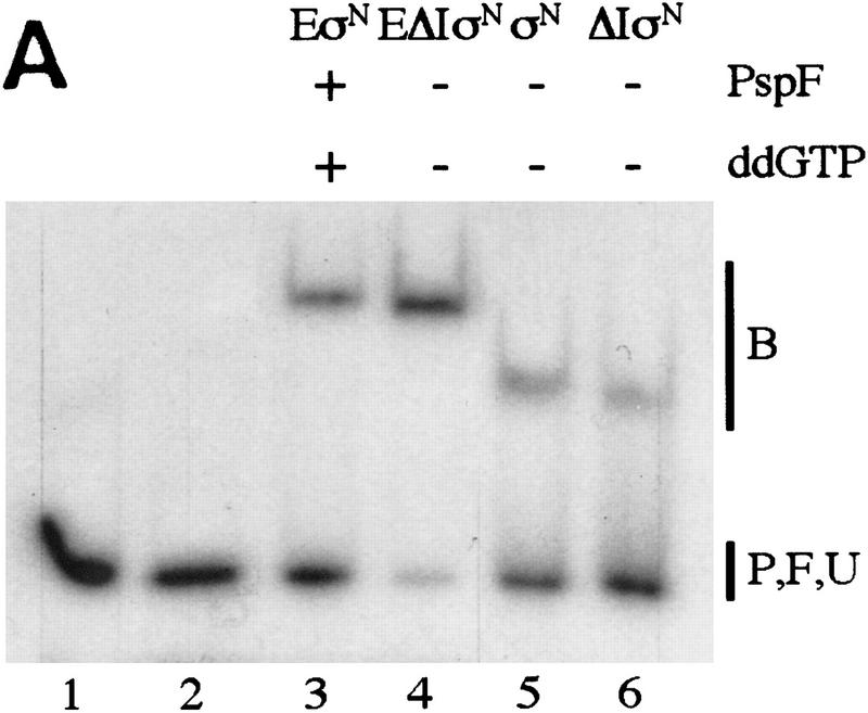
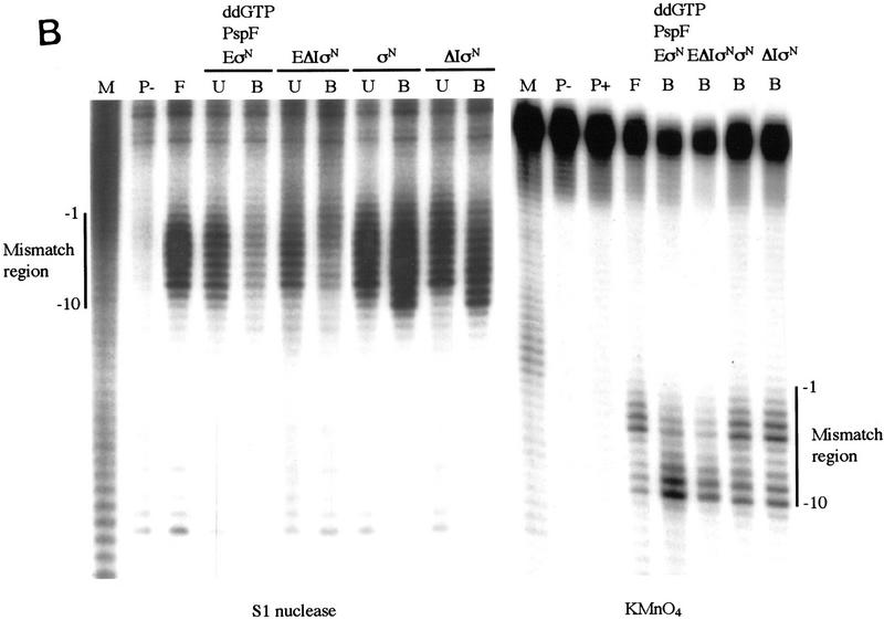
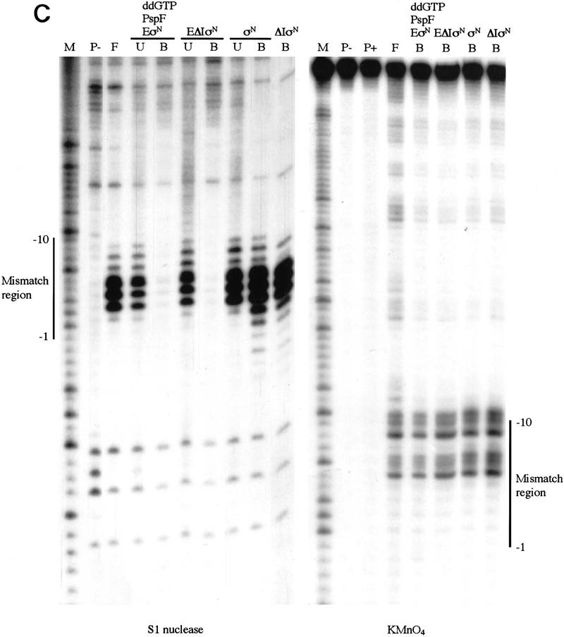
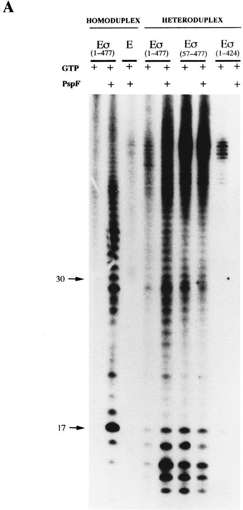
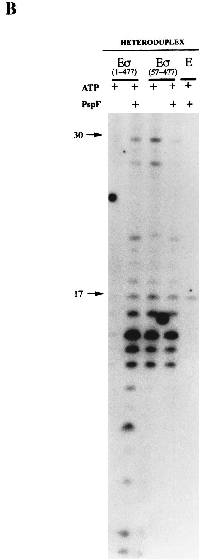
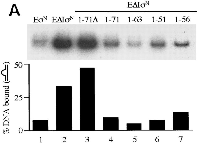
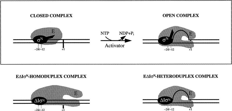
In bacteria, association of the specialized sigmaN protein with the core RNA polymerase subunits forms a holoenzyme able to bind promoter DNA, but unable to melt DNA and initiate transcription unless acted on by an activator protein. The conserved amino-terminal 50 amino acids of sigmaN (Region I) are required for the response to activators. We have used pre-melted DNA templates, in which the template strand is unpaired and accessible for transcription initiation, to mimic a naturally melted promoter and explore the function of Region I. Our results indicate that one activity of Region I sequences is to inhibit productive interaction of holoenzyme with pre-melted DNA. On pre-melted DNA targets, either activation of sigmaN-holoenzyme or removal of Region I allowed efficient formation of complexes in which melted DNA was sequestered by RNA polymerase. Like natural pre-initiation complexes formed on conventional DNA templates through the action of activator, such complexes were heparin-resistant and transcriptionally active. The inhibitory sigmaN Region I domain functioned in trans to confer heparin sensitivity to complexes between Region I-deleted holoenzyme and pre-melted promoter DNA. Evidence that Region I senses the conformation of the promoter was obtained from protein footprint experiments. We suggest that one function for Region I is to mask a single-strand DNA-binding activity of the holoenzyme. On the basis of extended DNA footprints of Region I-deleted holoenzyme, we also propose that Region I prevents RNA polymerase isomerization, a conformational change necessary for access to and the subsequent stable association of holoenzyme with melted DNA.


Figures

Figure 1
Figure 1
Gel mobility-shift assays on R. meliloti nifH promoter DNA. (A) Effect of activator and/or nucleotides on heparin-resistant complex formation between σN- and ΔIσN-holoenzymes (EσN and EΔIσN) and heteroduplex 1 DNA (see Table 1). Holoenzymes were at 100 nm, activator PspFΔHTH (lanes 3,4,6,7,10,13,14) at 4 μm, nucleotides at 1 mm and end-labeled DNA at 16 nm. Prior to gel loading, heparin (100 μg/ml) was added for 5 min. Reactions included salmon sperm DNA (455 ng/μl), which helped to reduce background smears. The percentage of DNA shifted is indicated in the histogram. (B) Stability of holoenzyme complexes on heteroduplex DNA. Following holoenzyme binding to DNA and incubation with PspFΔHTH and ddGTP where indicated, samples were taken prior to (time 0) and after addition of heparin for 1, 5, 10, and 20 min, and immediately loaded onto running native gels. Holoenzymes (EσN, EΔIσN) alone (□,█), plus ddGTP (○,●), and plus ddGTP and PspFΔHTH (▵,▴). (C) Formation of heparin-resistant complexes with homoduplex DNA. Holoenzymes were at 250 nm, end-labeled 88-mer homoduplex DNA at 16 nm, PspFΔHTH at 4 μm and GTP at 4 mm (lanes 1,3,6). Prior to gel loading, heparin (lanes 2,3,5,6) was added for 5 min. The level of initial complexes (lanes 1,4) was unaffected by GTP plus PspFΔHTH (data not shown). (D) Region I sequences confer heparin sensitivity to holoenzyme. Holoenzymes (50 nm) containing end-labeled σ proteins (EσN-32P, EΔIσN-32P) were incubated with or without 50 nm 88-mer homoduplex (lanes 3,4,11,12) and heteroduplex 1 (lanes 58,13,14), challenged with heparin for 5 min where indicated and loaded onto native gels. PspFΔHTH (4 μm) and GTP (4 mm) were added for activation (lanes 7,8). (H) Holoenzyme; (H+DNA) holoenzyme–DNA complex; (σ) free sigma.
Figure 1
Figure 1
Gel mobility-shift assays on R. meliloti nifH promoter DNA. (A) Effect of activator and/or nucleotides on heparin-resistant complex formation between σN- and ΔIσN-holoenzymes (EσN and EΔIσN) and heteroduplex 1 DNA (see Table 1). Holoenzymes were at 100 nm, activator PspFΔHTH (lanes 3,4,6,7,10,13,14) at 4 μm, nucleotides at 1 mm and end-labeled DNA at 16 nm. Prior to gel loading, heparin (100 μg/ml) was added for 5 min. Reactions included salmon sperm DNA (455 ng/μl), which helped to reduce background smears. The percentage of DNA shifted is indicated in the histogram. (B) Stability of holoenzyme complexes on heteroduplex DNA. Following holoenzyme binding to DNA and incubation with PspFΔHTH and ddGTP where indicated, samples were taken prior to (time 0) and after addition of heparin for 1, 5, 10, and 20 min, and immediately loaded onto running native gels. Holoenzymes (EσN, EΔIσN) alone (□,█), plus ddGTP (○,●), and plus ddGTP and PspFΔHTH (▵,▴). (C) Formation of heparin-resistant complexes with homoduplex DNA. Holoenzymes were at 250 nm, end-labeled 88-mer homoduplex DNA at 16 nm, PspFΔHTH at 4 μm and GTP at 4 mm (lanes 1,3,6). Prior to gel loading, heparin (lanes 2,3,5,6) was added for 5 min. The level of initial complexes (lanes 1,4) was unaffected by GTP plus PspFΔHTH (data not shown). (D) Region I sequences confer heparin sensitivity to holoenzyme. Holoenzymes (50 nm) containing end-labeled σ proteins (EσN-32P, EΔIσN-32P) were incubated with or without 50 nm 88-mer homoduplex (lanes 3,4,11,12) and heteroduplex 1 (lanes 58,13,14), challenged with heparin for 5 min where indicated and loaded onto native gels. PspFΔHTH (4 μm) and GTP (4 mm) were added for activation (lanes 7,8). (H) Holoenzyme; (H+DNA) holoenzyme–DNA complex; (σ) free sigma.
Figure 2
Figure 2
Binding of σ proteins (σ) and their holoenzymes (Eσ) to 88-mer homoduplex and heteroduplex 1 DNA. Binding reactions were essentially as in Fig. 1A, except that 1.6 nm DNA was used and heparin and nonspecific DNA were omitted. (□) σN, (○) ΔIσN, (█) σN-holoenzyme, (●) ΔIσN-holoenzyme.
Figure 3
Figure 3
Region I sequences are sensitive to promoter DNA conformation. End-labeled σN (lanes 14) or holoenzyme prepared with end-labeled σN (lanes 57) were incubated for 10 min without DNA (lanes 2,5), with homoduplex DNA (lanes 3,6) or with heteroduplex DNA (lanes 4,7) and then cleaved with endoproteinase Glu-C (Casaz and Buck 1997). Numbers on the left indicate the amino acid at which cleavage has occurred.
Figure 4
Figure 4
Premelted sequences are sequestered in stable complexes but accessible to KMnO4. (A) Gel mobility-shift assays of heteroduplex 1 DNA complexes. Binding reactions were conducted as in Fig. 1A, treated with S1 nuclease or KMnO4 (see Material and Methods) and loaded onto running native gels. (B) Bound DNA (lanes 36); (P) undigested probe (lane 1); (F) digested probe (lane 2); (U) unbound DNA (lanes 36). (B,C) The DNA bands were isolated from native gels described in A, and both strands analyzed on sequencing gels [(B) top strand; (C) bottom strand]. The mismatch region between −10 and −1 is indicated. Markers (M) were generated by chemical cleavage of the DNA with piperidine following partial methylation with dimethylsulfate. Probe alone or cleaved with piperidine (without treatment with KMnO4) are lanes marked P− and P+, respectively. Footprints of ΔIσN-holoenzyme plus PspFΔHTH and ddGTP were the same as ΔIσN-holoenzyme alone (not shown).
Figure 4
Figure 4
Premelted sequences are sequestered in stable complexes but accessible to KMnO4. (A) Gel mobility-shift assays of heteroduplex 1 DNA complexes. Binding reactions were conducted as in Fig. 1A, treated with S1 nuclease or KMnO4 (see Material and Methods) and loaded onto running native gels. (B) Bound DNA (lanes 36); (P) undigested probe (lane 1); (F) digested probe (lane 2); (U) unbound DNA (lanes 36). (B,C) The DNA bands were isolated from native gels described in A, and both strands analyzed on sequencing gels [(B) top strand; (C) bottom strand]. The mismatch region between −10 and −1 is indicated. Markers (M) were generated by chemical cleavage of the DNA with piperidine following partial methylation with dimethylsulfate. Probe alone or cleaved with piperidine (without treatment with KMnO4) are lanes marked P− and P+, respectively. Footprints of ΔIσN-holoenzyme plus PspFΔHTH and ddGTP were the same as ΔIσN-holoenzyme alone (not shown).
Figure 5
Figure 5
Holoenzyme complexes lacking Region I are transcriptionally active on heteroduplex DNA without activator. Holoenzymes (100 nm) with GTP or ATP (1 mm) in the presence or absence of PspFΔHTH (4 μm) were used to transcribe the 88-mer homoduplex and heteroduplex 1 (16 nm) templates. Core RNAP and holoenzyme containing the DNA nonbinding 1–424 σ derivative (Cannon et al. 1995b) were used as controls. The location of the DNA size markers are shown (arrows). (A) [α-32P]UTP-labeled transcripts with GTP as the hydrolyzable nucleotide used by activator. (B) [γ-32P]GTP-labeled transcripts with ATP as the hydrolyzable nucleotide used by activator.
Figure 5
Figure 5
Holoenzyme complexes lacking Region I are transcriptionally active on heteroduplex DNA without activator. Holoenzymes (100 nm) with GTP or ATP (1 mm) in the presence or absence of PspFΔHTH (4 μm) were used to transcribe the 88-mer homoduplex and heteroduplex 1 (16 nm) templates. Core RNAP and holoenzyme containing the DNA nonbinding 1–424 σ derivative (Cannon et al. 1995b) were used as controls. The location of the DNA size markers are shown (arrows). (A) [α-32P]UTP-labeled transcripts with GTP as the hydrolyzable nucleotide used by activator. (B) [γ-32P]GTP-labeled transcripts with ATP as the hydrolyzable nucleotide used by activator.
Figure 6
Figure 6
Region I-containing fragments confer heparin sensitivity in trans. (A) Different amino-terminal peptides of σN: 1–51, 1–63, 1–71, 1–71Δ (15 μm) and 1–56 (2.5 μm) were added to ΔIσN (200 nm) prior to core RNAP addition (100 nm) and heteroduplex 1 DNA (16 nm) was added last in a binding reaction. After incubation, heparin was added for 5 min and samples loaded onto native gels. The percentage of heparin-resistant DNA complexes is indicated. (B) Effect of Region I on heparin stability. Gel mobility shifts were conducted with ΔIσN-holoenzyme (100 nm), GTP (1 mm) and heteroduplex 1 DNA (16 nm; █) and Region I peptide (1–56, 2 μm) was added before ΔIσN-holoenyme assembly (▴) or after holoenzyme–heteroduplex complex formation (●). σN-Holoenzyme plus heteroduplex DNA, in which most complexes are heparin sensitive (see Fig. 1B), was used as a control (□). Samples were taken prior to (time 0) and after the addition of heparin for 1, 5, 10, 20 min.
Figure 6
Figure 6
Region I-containing fragments confer heparin sensitivity in trans. (A) Different amino-terminal peptides of σN: 1–51, 1–63, 1–71, 1–71Δ (15 μm) and 1–56 (2.5 μm) were added to ΔIσN (200 nm) prior to core RNAP addition (100 nm) and heteroduplex 1 DNA (16 nm) was added last in a binding reaction. After incubation, heparin was added for 5 min and samples loaded onto native gels. The percentage of heparin-resistant DNA complexes is indicated. (B) Effect of Region I on heparin stability. Gel mobility shifts were conducted with ΔIσN-holoenzyme (100 nm), GTP (1 mm) and heteroduplex 1 DNA (16 nm; █) and Region I peptide (1–56, 2 μm) was added before ΔIσN-holoenyme assembly (▴) or after holoenzyme–heteroduplex complex formation (●). σN-Holoenzyme plus heteroduplex DNA, in which most complexes are heparin sensitive (see Fig. 1B), was used as a control (□). Samples were taken prior to (time 0) and after the addition of heparin for 1, 5, 10, 20 min.
Figure 7
Figure 7
S1 nuclease footprints on linear DNA. Linear end-labeled templates prepared by primer extension were used. (A) σN and holoenzyme (amino acids 1–477); (B) ΔIσN and holoenzyme (amino acids 57–477). (Solid line) The extent of protection seen with holoenzyme (H) or σ (S); (broken line) weaker protection; (star) −10 cutting lost on holoenzyme binding. Markers were chemical cleavage G reactions. σ Proteins were at 1 μm, holoenzymes at 100 nm, activator PspFΔHTH at 4 μm, and GTP at 1 mm. Compared to σN-holoenzyme, the ΔIσN-holoenzyme displays an extended footprint in the direction of transcription (A vs. B). Activation of the σN-holoenzyme by PspFΔHTH (A) results in an extended footprint with increased protection from −5 to +7. The extended footprint to +20 observed with ΔIσN-holoenzyme is independent of activator and nucleotide. Reproducible footprints and cutting of promoter template DNA was observed with several different batches of S1 nuclease. A control with core RNAP did not produce a S1 footprint.
Figure 7
Figure 7
S1 nuclease footprints on linear DNA. Linear end-labeled templates prepared by primer extension were used. (A) σN and holoenzyme (amino acids 1–477); (B) ΔIσN and holoenzyme (amino acids 57–477). (Solid line) The extent of protection seen with holoenzyme (H) or σ (S); (broken line) weaker protection; (star) −10 cutting lost on holoenzyme binding. Markers were chemical cleavage G reactions. σ Proteins were at 1 μm, holoenzymes at 100 nm, activator PspFΔHTH at 4 μm, and GTP at 1 mm. Compared to σN-holoenzyme, the ΔIσN-holoenzyme displays an extended footprint in the direction of transcription (A vs. B). Activation of the σN-holoenzyme by PspFΔHTH (A) results in an extended footprint with increased protection from −5 to +7. The extended footprint to +20 observed with ΔIσN-holoenzyme is independent of activator and nucleotide. Reproducible footprints and cutting of promoter template DNA was observed with several different batches of S1 nuclease. A control with core RNAP did not produce a S1 footprint.
Figure 8
Figure 8
Gel mobility-shift, footprinting, and transcription assays (see Figs. 1, 4, and 7) show activator and nucleotide hydrolysis are required to efficiently form stable heparin-resistant σN-holoenzyme complexes on homoduplex (open complex, in which DNA is melted) and heteroduplex DNA. In contrast, ΔIσN-holoenzyme bypasses the activator requirement for stable complex formation on heteroduplex DNA but fails on homoduplex, regardless of the presence of activator. We propose that the inhibition of DNA melting associated with Region I (Wang et al. 1995, 1997a) is due to prevention of polymerase isomerization, necessary to reveal a single-strand DNA-binding activity in the holoenzyme required for its stable association with melted DNA (Fig. 4). The thermodynamic barrier to open complex formation (Wedel and Kustu 1995) at σN-dependent promoters is suggested to be associated with energetically unfavorable DNA strand separation.

References

    1. Austin S, Dixon RA. The prokaryotic enhancer binding protein NtrC has ATPase activity which is phosphorylation and DNA dependent. EMBO J. 1992;11:2219–2228. - PMC - PubMed
    1. Buck M, Cannon W. Specific binding of the transcription factor σ-54 to promoter DNA. Nature. 1992;358:422–424. - PubMed
    1. Burgess R, Travers A, Dunn J, Bautz E. Factor stimulating transcription by RNA polymerase. Nature. 1969;221:43–46. - PubMed
    1. Cannon W, Claverie-Martin F, Austin S, Buck M. Identification of a DNA-contacting surface in the transcription factor σ54 (σN) Mol Microbiol. 1994;11:227–236. - PubMed
    1. Cannon W, Austin S, Moore M, Buck M. Identification of close contacts between σ54 (σN) protein and promoter DNA in closed promoter complexes. Nucleic Acids Res. 1995a;23:351–356. - PMC - PubMed

Publication types

LinkOut - more resources