. 2020 Apr;40(3):421-435.
doi: 10.1007/s10571-019-00745-w. Epub 2019 Oct 28.

Inhibition of PDE4 Attenuates TNF-α-Triggered Cell Death Through Suppressing NF-κB and JNK Activation in HT-22 Neuronal Cells

Affiliations

Inhibition of PDE4 Attenuates TNF-α-Triggered Cell Death Through Suppressing NF-κB and JNK Activation in HT-22 Neuronal Cells

Jiao Xiao et al. Cell Mol Neurobiol. 2020 Apr.

Abstract

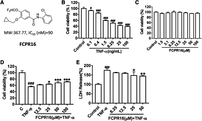
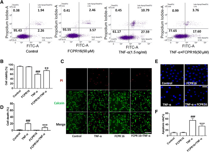
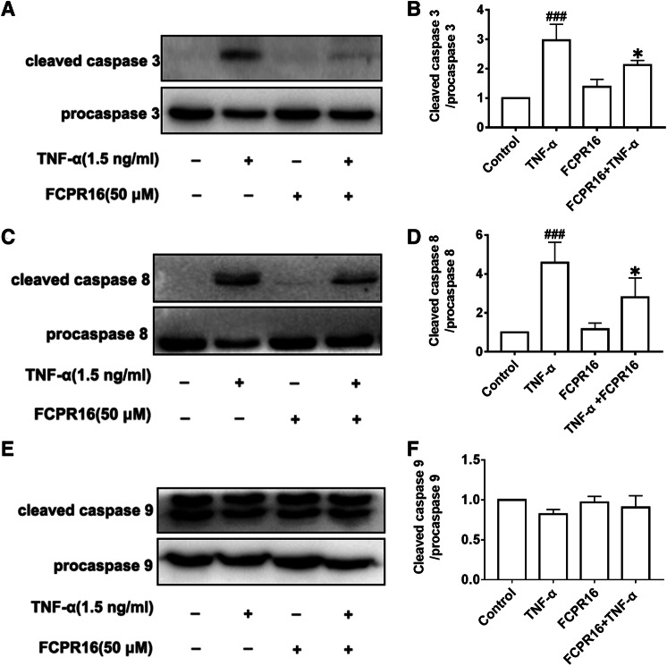
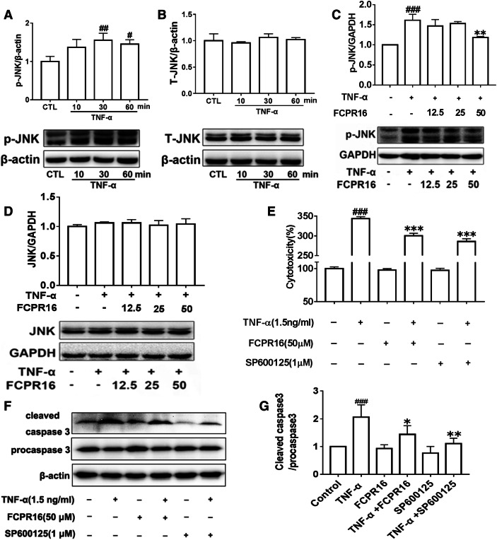
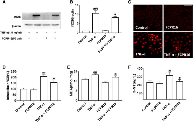
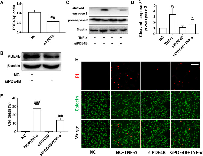
Tumor necrosis factor-α (TNF-α) is a critical pro-inflammatory cytokine regulating neuroinflammation. At high concentrations, it is toxic to neurons, and such damage is positively correlated with acute and chronic neurological diseases. Our previous studies showed that inhibition of phosphodiesterase 4 (PDE4) attenuated the production of TNF-α induced by lipopolysaccharides in microglial cells. However, whether PDE4 inhibition can block the neurotoxic effects of TNF-α in neuronal cells is unknown. In this study, we investigated the protective effects of FCPR16, a novel PDE4 inhibitor, against TNF-α-induced cellular apoptosis in HT-22 hippocampal neuronal cells. We demonstrated that FCPR16 dose-dependently increased the viability of HT-22 cells exposed to TNF-α insult. Propidium iodide/calcein staining and flow cytometry analysis showed that FCPR16 decreased cell apoptosis triggered by TNF-α. Western blot analysis showed that FCPR16 decreased the level of cleaved caspase 3 and caspase 8, but had no effect on caspase 9. Mechanistically, FCPR16 blocked the TNF-α-induced phosphorylation of c-Jun N-terminal kinase (JNK) in HT-22 cells, and inhibition of JNK showed a similar protective effect as FCPR16. Furthermore, FCPR16 decreased the translocation of nuclear factor-κB (NF-κB) p65 from the cytosol into the nucleus. In addition, FCPR16 decreased the expression of inducible nitric oxide synthase and the production of reactive oxygen species in HT-22 cells exposed to TNF-α. Moreover, knockdown of PDE4B by specific small interfering RNA reduced the apoptosis of HT-22 cells treated with TNF-α. Taken together, our findings suggest that FCPR16 promotes the survival of neuronal cells exposed to TNF-α by suppressing the activation of JNK and NF-κB.

Keywords: FCPR16; JNK; NF-κB; Neuroinflammation; Phosphodiesterase 4.


Conflict of interest statement

The authors declare that they have no conflict of interest.

Figures

Fig. 1
Fig. 1
FCPR16 attenuates cell injury triggered by TNF-α in HT-22 neuronal cells. a Chemical structure of FCPR16. b HT-22 cells were incubated with TNF-α for 24 h at different concentrations (0.1–100 ng/mL). Cell viability was detected using the CCK-8 assay. c HT-22 cells were incubated with FCPR16 (1.5–100 μM) for 24 h. Cell viability was detected by the CCK-8 assay. d HT-22 cells were pre-treated with FCPR16 (12.5–100 μM) for 1 h and then exposed to TNF-α (1.5 ng/mL) for 24 h, followed by the CCK-8 assay. e HT-22 cells were pre-treated with FCPR16 (6.25–50 μM) for 1 h and then exposed to TNF-α (1.5 ng/mL) for 24 h, followed by the LDH release assay. Results are presented as the mean ± SD and represent three independent experiments (n = 3). #p < 0.05, ###p < 0.001 compared with the control group; *p < 0.05, **p < 0.01, ***p < 0.001 compared with the TNF-α-treated group
Fig. 2
Fig. 2
FCPR16 rescues neuronal cells from TNF-α-induced apoptosis. a HT-22 cells pre-treated with 50 μM FCPR16 for 1 h were exposed to TNF-α (1.5 ng/mL) for 12 h, and the apoptosis of HT-22 cells was determined by flow cytometry. b Quantitative analysis of the ratio of cells. The cell apoptotic ratio was expressed as the proportion of early apoptosis and late apoptosis to total cells. c HT-22 cells were pre-incubated with FCPR16 for 1 h, followed by treatment with TNF-α (1.5 ng/mL) for 12 h. Then HT-22 cells were incubated with PI and calcein-AM. The live cells are presented in green, while dead cells are presented in red. (scale bar = 200 μm). d The cell death rate was calculated by counting PI positive HT-22 cells. e HT-22 cells were exposed to TNF-α (1.5 ng/mL) in the absence and presence of FCPR16 for 12 h, and apoptotic cells were determined by the Hoechst assay (Bar = 100 μm). f Apoptotic cells were quantified by counting from the photomicrographs with densely condensed chromatin. The apoptotic rate was presented as a percentage of the total number. Results were obtained from three independent cultures and shown as the mean ± SD (n = 3). ###p < 0.001 versus the control group. **p < 0.01, ***p < 0.001 versus the TNF-α-treated group
Fig. 3
Fig. 3
Effects of FCPR16 on apoptosis-related protein expression in HT-22 cells. a, c, e HT-22 cells pre-treated with 50 μM FCPR16 for 1 h were exposed to TNF-α (1.5 ng/mL) for 12 h. The levels of caspases 3, 8, and 9 were detected by Western blotting. b, d, f Quantification of cleaved caspase 3/pro-caspase 3, cleaved caspase 8/pro-caspase 8, and cleaved caspase 9/pro-caspase 9. Results are presented as the mean ± SD from three independent experiments (n = 3). ###p < 0.01 versus the control group; *p < 0.05 versus the TNF-α group
Fig. 4
Fig. 4
FCPR16 suppresses the activation JNK induced by TNF-α. a, b HT-22 cells were treated with TNF-α (1.5 ng/mL) for various times (0–60 min). The levels of phosphorylated JNK (p-JNK) and total JNK (T-JNK) were detected by Western blotting. c, d HT-22 cells were pre-treated with various concentrations of FCPR16 (12.5–50 μM) for 1 h, and then cells were exposed to TNF-α (1.5 ng/mL) for 30 min. The levels of the p-JNK and T-JNK were detected by Western blotting. e After treatment with FCPR16 or JNK inhibitor SP600125 (1 μM) for 1 h, HT-22 cells were exposed to TNF-α (1.5 ng/mL) for 24 h. Cytotoxicity was detected by the LDH assay. f Cells were treated as described above, the level of cleaved caspase 3 was detected by Western blotting. g Quantification of cleaved caspase 3/pro-caspase 3. The results are presented as the mean ± SD from three independent experiments (n = 3). #p < 0.05, ##p < 0.01, ###p < 0.001 versus the control group; **p < 0.05, ***p < 0.001 versus the TNF-α group
Fig. 5
Fig. 5
FCPR16 markedly blocks the nuclear translocation of NF-κB p65 in HT-22 neuronal cells stimulated by TNF-α. a HT-22 cells pre-treated with 50 μM FCPR16 were incubated with TNF-α for 1 h. The translocation of NF-κB p65 was detected by immunofluorescence straining (bar = 50 μm). b, c NF-κB p65 protein level in cell nucleus was analyzed by Western blotting, and the corresponding quantification data were accounted from three independent experiments by densitometry of the blots. d, e NF-κB p65 protein level in whole cells was detected by Western blotting and the corresponding quantification data were accounted the same as described above. Data are represented as the mean ± SD from three independent experiments (n = 3). ###p < 0.001 versus the control group. *p < 0.05 versus the TNF-α group
Fig. 6
Fig. 6
Effects of FCPR16 on levels of iNOS, and oxidative stress in HT-22 cells. a, b After pre-treatment with FCPR16 (50 μM) for 1 h, HT-22 cells were incubated with TNF-α (1.5 ng/mL) for 12 h. The expression level of iNOS was detected by Western blotting. c, d After treatment with 50 μM FCPR16 for 1 h, HT-22 cells were incubated with or without 1.5 ng/mL TNF-α for 2 h. The level of ROS in HT-22 cells was determined by CellROX Deep Red Reagent (bar = 100 μm). e After treatment with 50 μM FCPR16 for 1 h, HT-22 cells were incubated with or without 1.5 ng/mL TNF-α for 12 h. The level of MDA was detected by a lipid peroxidation MDA assay Kit. f The level of 3-NT was detected by a 3-Nitro-Tyrosine (3-NT) Elisa Kit. Data are represented as the mean ± SD from three independent experiments (n = 3). ##p < 0.01, ###p < 0.001 versus the control group. *p < 0.05 versus the TNF-α group
Fig. 7
Fig. 7
Knockdown of PDE4B expression attenuates cell death caused by TNF-α in HT-22 cells. a, b HT-22 cells were transfected with specific PDE4B siRNA for 24 h. Silence efficiency was detected by Western blotting. c HT-22 cells were transfected with PDE4B siRNA, and then cells were stimulated with or without TNF-α (1.5 ng/mL) for 12 h. The level of cleaved caspase 3 was detected by Western blotting. d Quantification of cleaved caspase 3/pro-caspase 3. e, f HT-22 cells were exposed to TNF-α (1.5 ng/mL) for 12 h after transfection with PDE4B siRNA. After incubation with TNF-α for 12 h, cells were incubated with PI and calcein-AM, and fluorescence was observed by a microscope. Green staining (calcein-AM) indicates live cells, and red staining (PI) indicates dead cells (bar = 200 μm). The cell death rate was evaluated by counting dead (red) HT-22 cells. Results are presented as the mean ± SD from three independent experiments (n = 3). ###p < 0.001, ##p < 0.01 versus the control group; *p < 0.05, **p < 0.01 versus the TNF-α group

References

    1. Aggarwal BB (2003) Signalling pathways of the TNF superfamily: a double-edged sword. Nat Rev Immunol 3(9):745–756 - PubMed
    1. Annibaldi A, Meier P (2018) Checkpoints in TNF-induced cell death: implications in inflammation and cancer. Trends Mol Med 24(1):49–65 - PubMed
    1. Belarbi K, Jopson T, Tweedie D, Arellano C, Luo W, Greig NH, Rosi S (2012) TNF-alpha protein synthesis inhibitor restores neuronal function and reverses cognitive deficits induced by chronic neuroinflammation. J Neuroinflamm 9:23 - PMC - PubMed
    1. Bolger GB (2017) The PDE4 cAMP-specific phosphodiesterases: targets for drugs with antidepressant and memory-enhancing action. Adv Neurobiol 17:63–102 - PubMed
    1. Borghi A, Verstrepen L, Beyaert R (2016) TRAF2 multitasking in TNF receptor-induced signaling to NF-kappaB, MAP kinases and cell death. Biochem Pharmacol 116:1–10 - PubMed

MeSH terms

Substances

LinkOut - more resources