. 2024 Feb 21;12(1):35.
doi: 10.1186/s40168-024-01753-9.

Virome diversity shaped by genetic evolution and ecological landscape of Haemaphysalis longicornis

Affiliations

Virome diversity shaped by genetic evolution and ecological landscape of Haemaphysalis longicornis

Run-Ze Ye et al. Microbiome. .

Abstract

Background: Haemaphysalis longicornis is drawing attentions for its geographic invasion, extending population, and emerging disease threat. However, there are still substantial gaps in our knowledge of viral composition in relation to genetic diversity of H. longicornis and ecological factors, which are important for us to understand interactions between virus and vector, as well as between vector and ecological elements.

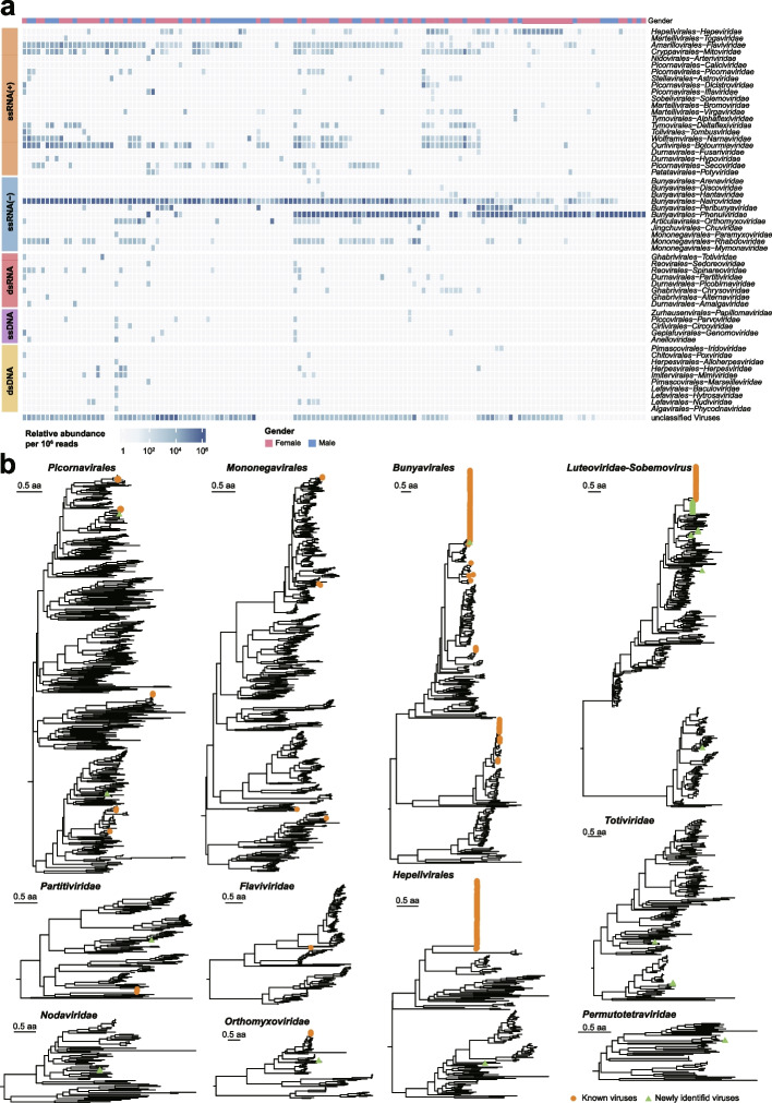
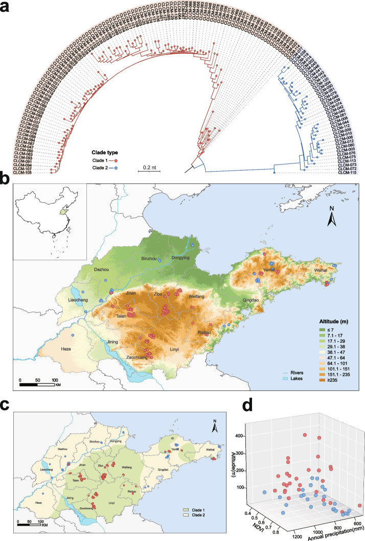
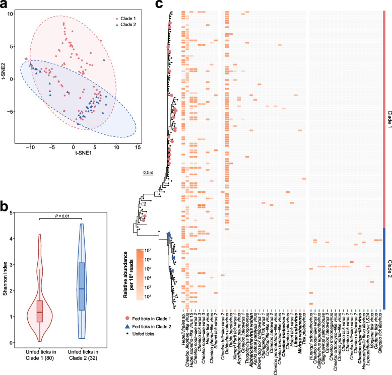
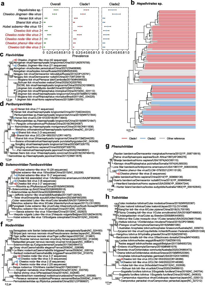
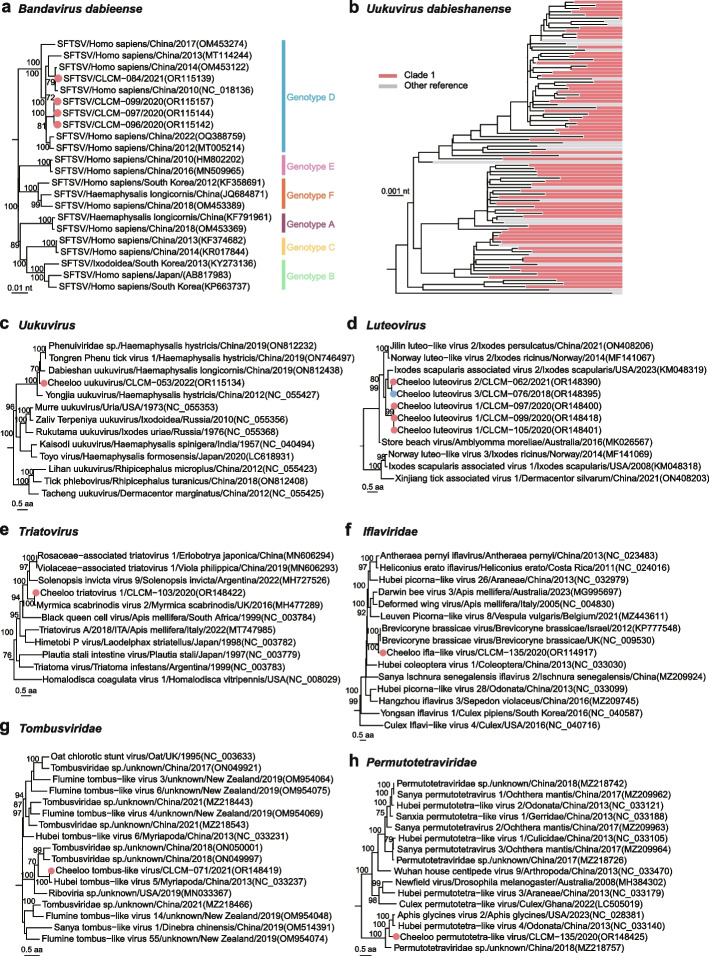
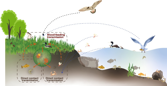
Results: We conducted the meta-transcriptomic sequencing of 136 pools of H. longicornis and identified 508 RNA viruses of 48 viral species, 22 of which have never been reported. Phylogenetic analysis of mitochondrion sequences divided the ticks into two genetic clades, each of which was geographically clustered and significantly associated with ecological factors, including altitude, precipitation, and normalized difference vegetation index. The two clades showed significant difference in virome diversity and shared about one fifth number of viral species that might have evolved to "generalists." Notably, Bandavirus dabieense, the pathogen of severe fever with thrombocytopenia syndrome was only detected in ticks of clade 1, and half number of clade 2-specific viruses were aquatic-animal-associated.

Conclusions: These findings highlight that the virome diversity is shaped by internal genetic evolution and external ecological landscape of H. longicornis and provide the new foundation for promoting the studies on virus-vector-ecology interaction and eventually for evaluating the risk of H. longicornis for transmitting the viruses to humans and animals. Video Abstract.


Conflict of interest statement

The authors declare no competing interests.

Figures

Fig. 1
Fig. 1
Virome diversity of H. longicornis. a Virus relative abundance profile across H. longicornis libraries. Each cell in the heat map represents the normalized number of reads belonging to the given virus order or family according to Kraken2 annotation. All families were grouped according to Baltimore classification, including ssRNA( +), ssRNA( −), dsRNA, ssDNA, dsDNA, and unclassified viruses. The gender of each sample was indicated in the corresponding colors on the top. b Phylogenetic trees were constructed on the basis of RdRp protein for RNA viruses. Newly identified viruses in this study are labeled with green solid triangle; known viruses are labeled with orange solid circles. Families without assembled contigs containing the RdRp domain were not displayed in b, although the viral reads were detected in a
Fig. 2
Fig. 2
Genetic clade and distribution of H. longicornis in relation to ecological factors. a Phylogenetic tree of H. longicornis based on mitochondrial variants. The red and blue branches represent genetic clade 1 and clade 2, respectively. b Spatial distribution of tick collection. The background colors range from green to brown, indicating gradual elevation. The red and blue points represent collection sites of clade 1 and clade 2, respectively. c Spatial clustering of each H. longicornis clade based on the inverse distance weighting (IDW) analysis. The green-shaded areas are the clusters of ticks in clade 1, and the yellow-shaded areas are the clusters of ticks in clade 2. d Three-dimensional scatter plot of tick samples in relation to the three ecological variables. The red and blue points represent clade 1 and clade 2, respectively
Fig. 3
Fig. 3
Virus diversity in genetic clades of H. longicornis. a Between-group clustering of viromes between genetic clades of H. longicornis by t-SNE analysis. b Shannon indexes of tick viromes between genetic clades of unfed H. longicornis (nclade1 = 80, nclade2 = 32). Boxplot elements: center line, median; box limits, upper and lower quartiles; whiskers (error bars), the highest and lowest points within 1.5 interquartile range of the upper and lower quartiles. The P-value was calculated using a two-sided Wilcoxon rank-sum test. c Abundance of each virus identified in this study. The fed tick samples in clade 1 are indicted by red solid circles, and those in clade 2 are indicated by blue solid triangles. The viruses shown in bold is only present in fed tick samples. Each cell in the heat map represents the normalized abundance of viral reads
Fig. 4
Fig. 4
Phylogenetic analysis of viruses in both clades of H. longicornis. a The prevalence with 95% confidence interval (CI) of each virus. The virus species name shown in red are newly discovered in this study. b Phylogeny of viruses in the family Hepelivirales sp. based on amino acid sequence of RNA-dependent RNA polymerase (RdRp) gene. Tree s are colored according to the genetic clade of libraries which viral sequences collected from: The red strips indicate tick samples from clade 1, the blue strips represent ticks from clade 2, and the gray strips are reference sequences. c Phylogeny of viruses in the group of Jingmenvirus based on amino acid sequence of VP1b protein. d Phylogeny of viruses in the family Peribunyaviridae based on amino acid sequence of RdRp protein. e Phylogeny of viruses between the families Solemoviridae and Tombusviridae based on amino acid sequence of RdRp protein. f Phylogeny of viruses in the family Nodaviridae based on amino acid sequence of capsid protein. g Phylogeny of viruses in the family Phenuiviridae based on amino acid sequence of nucleoprotein. h Phylogeny of viruses in the family Totiviridae based on amino acid sequence of RdRp protein. Viruses detected in this study are marked by red solid circles for ticks in clade 1 and blue solid circles for ticks in clade 2
Fig. 5
Fig. 5
Phylogenetic analysis of clade 1-specific viruses. a Phylogenetic tree of Bandavirus dabieense based on nucleotide sequences of RdRp gene. b Phylogeny of Uukuvirus dabieshanense based on nucleotide sequence of RNA-dependent RNA polymerase (RdRp) gene. The red strips indicate tick samples from clade 1 and the gray strips are reference sequences. c Phylogeny of viruses in the genus of Uukuvirus based on amino acid sequence of RdRp protein. d Phylogeny of viruses in the genus Luteovirus based on amino acid sequence of RdRp protein. e Phylogeny of viruses in the genus Triatovirus based on amino acid sequence of RdRp protein. f Phylogeny of viruses in the genus Iflaviridae based on amino acid sequence of RdRp protein. g Phylogeny of viruses in the family Tombusviridae based on amino acid sequence of RdRp protein. h Phylogeny of viruses in the family Permutotetraviridae based on amino acid sequence of RdRp protein. Viruses in this study are marked by solid circles
Fig. 6
Fig. 6
Phylogenetic analysis of aquatic-animal-associated viruses in clade 2. a Phylogeny of viruses in the subfamily of Alpharhabdovirinae based on amino acid sequence of RNA-dependent RNA polymerase (RdRp) protein. b Phylogeny of viruses in the order of Mononegavirales based on amino acid sequence of RdRp protein. c Phylogeny of viruses in the order of Mononegavirales based on amino acid sequence of glycoprotein. d Phylogeny of viruses in the family of Orthomyxoviridae based on amino acid sequence of PB1 protein. e Phylogeny of viruses in the family of Partitiviridae based on amino acid sequence of RdRp protein. f Phylogeny of viruses between the family Solemoviridae and Tombusviridae based on amino acid sequence of RdRp protein. g Phylogeny of viruses between the family Nodaviridae and Solemoviridae based on amino acid sequence of coat protein. h Phylogeny of viruses in the family of Virgaviridae based on amino acid sequence of RdRp protein. i Phylogeny of viruses in the family of Nodaviridae based on amino acid sequence of RdRp protein. Viruses in this study are marked in bold. The animal hosts of viruses are labeled with cartoon images
Fig. 7
Fig. 7
Ecological schematic diagram of H. longicornis in clade 2. The red arrows indicate the path of the virus spreading

Similar articles

Cited by

References

    1. Tanne JH. New tick seen in nine US states is an emerging disease threat, warns CDC. BMJ. 2018;363:k5191. doi: 10.1136/bmj.k5191. - DOI - PubMed
    1. Nwanade CF, Wang M, Li S, Yu Z, Liu J. The current strategies and underlying mechanisms in the control of the vector tick, Haemaphysalis longicornis: implications for future integrated management. Ticks Tick Borne Dis. 2022;13(2):101905. doi: 10.1016/j.ttbdis.2022.101905. - DOI - PubMed
    1. Rainey T, Occi JL, Robbins RG, Egizi A. Discovery of Haemaphysalis longicornis (Ixodida: Ixodidae) parasitizing a sheep in New Jersey, United States. J Med Entomol. 2018;55(3):757–759. doi: 10.1093/jme/tjy006. - DOI - PubMed
    1. Jia N, Wang J, Shi W, Du L, Sun Y, Zhan W, et al. Large-scale comparative analyses of tick genomes elucidate their genetic diversity and vector capacities. Cell. 2020;182(5):1328–40.e13. doi: 10.1016/j.cell.2020.07.023. - DOI - PubMed
    1. Zhao L, Li J, Cui X, Jia N, Wei J, Xia L, et al. Distribution of Haemaphysalis longicornis and associated pathogens: analysis of pooled data from a China field survey and global published data. Lancet Planet Health. 2020;4(8):e320–e329. doi: 10.1016/S2542-5196(20)30145-5. - DOI - PubMed

Publication types

LinkOut - more resources