. 2023 Oct;10(29):e2302298.
doi: 10.1002/advs.202302298. Epub 2023 Aug 7.

Loss of ZBED6 Protects Against Sepsis-Induced Muscle Atrophy by Upregulating DOCK3-Mediated RAC1/PI3K/AKT Signaling Pathway in Pigs

Affiliations

Loss of ZBED6 Protects Against Sepsis-Induced Muscle Atrophy by Upregulating DOCK3-Mediated RAC1/PI3K/AKT Signaling Pathway in Pigs

Huan Liu et al. Adv Sci (Weinh). 2023 Oct.

Abstract

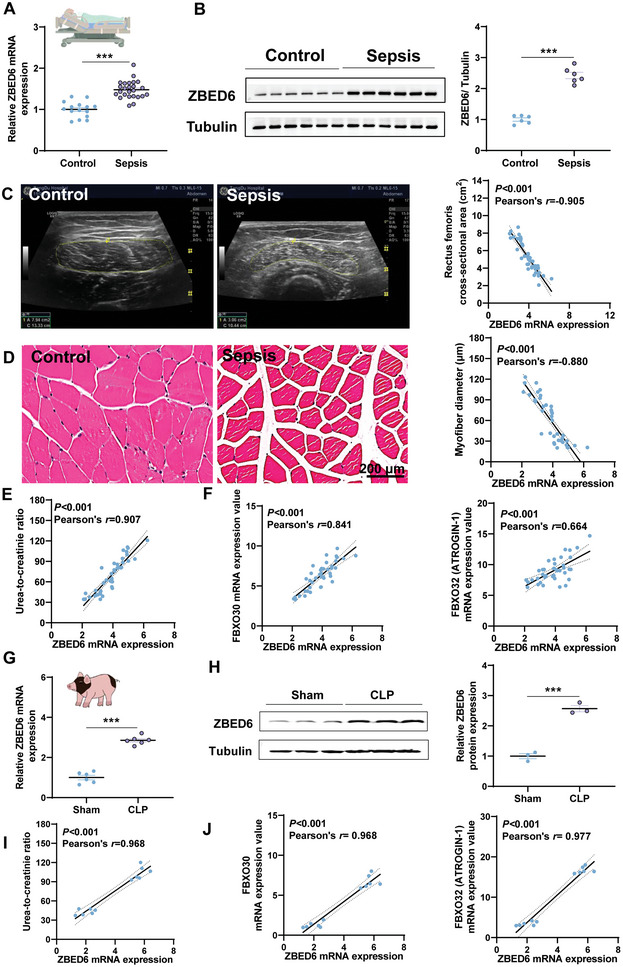
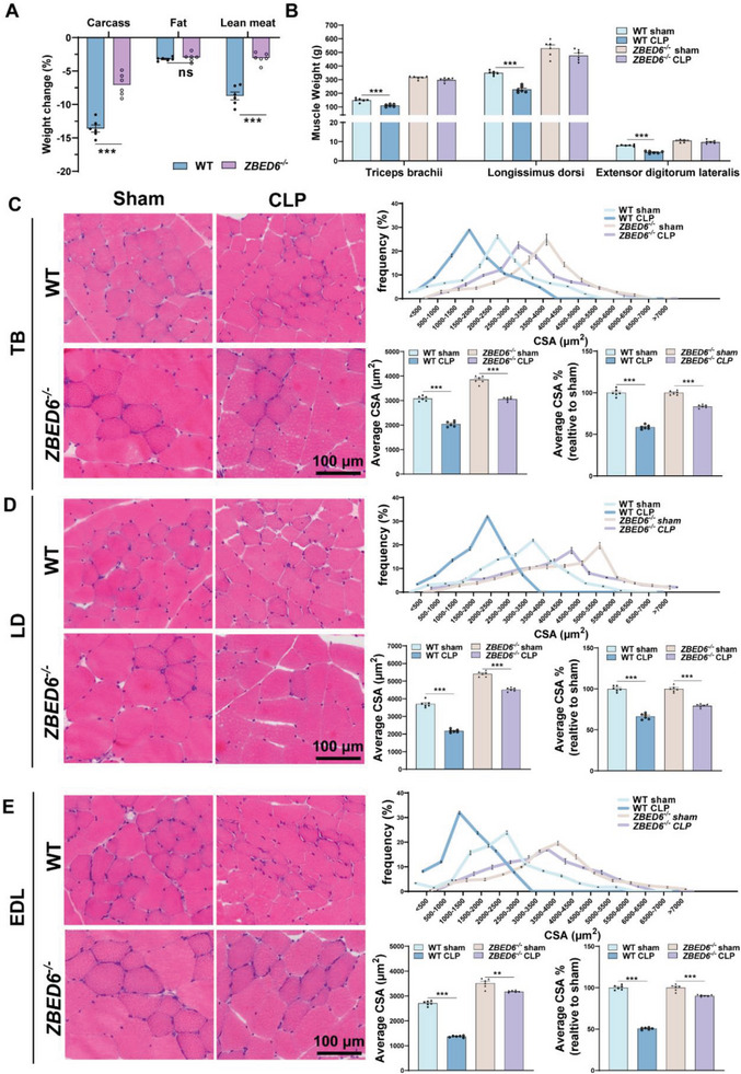
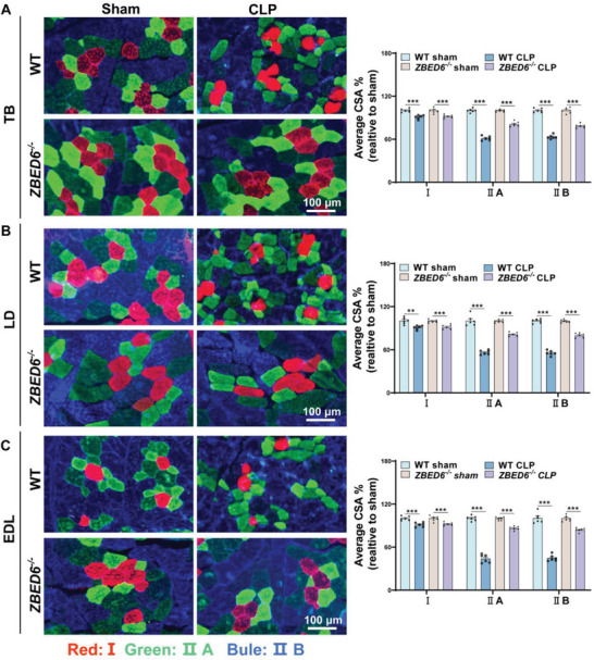
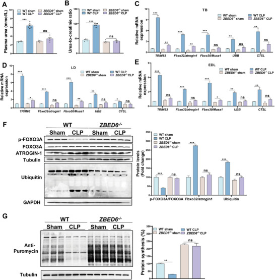
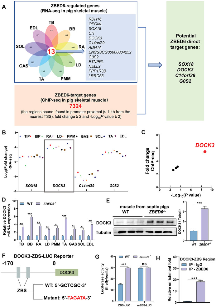
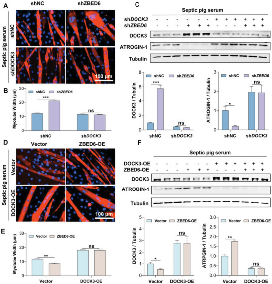
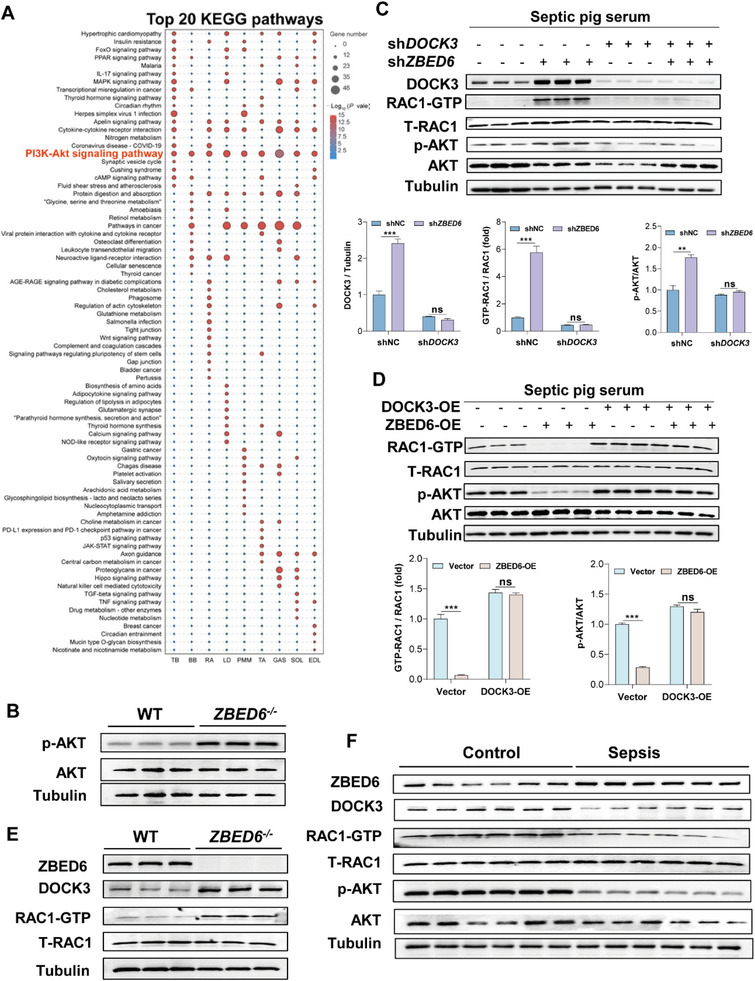
Sepsis-induced muscle atrophy often increases morbidity and mortality in intensive care unit (ICU) patients, yet neither therapeutic target nor optimal animal model is available for this disease. Here, by modifying the surgical strategy of cecal ligation and puncture (CLP), a novel sepsis pig model is created that for the first time recapitulates the whole course of sepsis in humans. With this model and sepsis patients, increased levels of the transcription factor zinc finger BED-type containing 6 (ZBED6) in skeletal muscle are shown. Protection against sepsis-induced muscle wasting in ZBED6-deficient pigs is further demonstrated. Mechanistically, integrated analysis of RNA-seq and ChIP-seq reveals dedicator of cytokinesis 3 (DOCK3) as the direct target of ZBED6. In septic ZBED6-deficient pigs, DOCK3 expression is increased in skeletal muscle and myocytes, activating the RAC1/PI3K/AKT pathway and protecting against sepsis-induced muscle wasting. Conversely, opposite gene expression patterns and exacerbated muscle wasting are observed in septic ZBED6-overexpressing myotubes. Notably, sepsis patients show increased ZBED6 expression along with reduced DOCK3 and downregulated RAC1/PI3K/AKT pathway. These findings suggest that ZBED6 is a potential therapeutic target for sepsis-induced muscle atrophy, and the established sepsis pig model is a valuable tool for understanding sepsis pathogenesis and developing its therapeutics.

Keywords: DOCK3; ZBED6; muscle atrophy; sepsis.


Conflict of interest statement

The authors declare no conflict of interest.

Figures

Figure 1
Figure 1
Upregulation of ZBED6 in sepsis‐induced muscle loss for humans and pigs. A) mRNA expression of ZBED6 in muscle biopsies of 25 sepsis patients with muscle loss and 15 orthopedic controls. B) Immunoblot and densitometric analysis of ZBED6 in rectus femoris muscle tissues of sepsis patients and controls (n = 6). Representative images are shown. C) The rectus femoris cross‐sectional area (RF‐CSA) was measured using ultrasound imaging in sepsis patients and orthopedic controls. The area was outlined by a dotted line. Scatterplots showing correlations of ZBED6 transcript with RF‐CSA in sepsis patients and orthopedic controls. D) H&E staining of histological cross sections in rectus femoris muscle tissues of sepsis patients and controls. Representative images are shown. Scale bar = 200 µm. The mean minimum Feret diameter of muscle fibers from sepsis patients and controls were correlated with ZBED6 transcript expression. E) Scatterplots showing correlations of ZBED6 transcript with urea‐to‐creatinine ratio in sepsis patients and controls. F) Scatterplots showing correlations of ZBED6 transcript with the transcripts of FBXO30 and FBXO32 in the rectus femoris muscle tissues of sepsis patients and those of controls. G) Bama pigs were subjected to sham or modified CLP surgery (n = 6). mRNA expression of ZBED6 in skeletal muscles of sham and modified CLP pigs 14 days post‐surgery. (H) Immunoblot and densitometric analysis of ZBED6 in skeletal muscle from sham and modified CLP pigs (n = 3). I) Scatterplots showing correlations of ZBED6 transcript with urea‐to‐creatinine ratio in the sham and modified CLP pigs. J) Scatterplots showing correlations of ZBED6 transcript with FBXO32 and FBXO30 in skeletal muscle of sham and modified CLP pigs. Data are expressed as mean ± SEM; *p < 0.05, **p < 0.01, ***p < 0.001.
Figure 2
Figure 2
ZBED6 deficiency protects against sepsis‐induced muscle atrophy in pigs. ZBED6‐deficient and WT pigs were subjected to modified CLP or sham surgery. After 14 days, the pigs were sacrificed. A) Loss of total body weight, fat mass, and lean mass expressed as percent‐wise change compared with the respective sham group. B) Muscle weight of Triceps brachii (TB), Longissimus dorsi (LD), and Extensor digitorum lateralis (EDL). C–E) Presentative images of H&E staining of histological cross sections from TIP (C), LD (D), and EDL (E). Scale bar = 100 µm. (Right top) Frequency of distribution for myofiber CSA (µm2) of TB (C), LD (D), and EDL E) muscles (n  =  6 per group). (Right bottom) Quantification of average CSA and average CSA% (expressed as percent‐wise change compared with their respective sham group) from H&E staining. One hundred myofibers were measured for each pig. Data are expressed as mean ± SEM; *p < 0.05, **p < 0.01, ***p < 0.001.
Figure 3
Figure 3
ZBED6 deficiency attenuates type II myofiber atrophy in septic pigs. A–C) (Left) Representative images of fiber‐type staining of the TB (A), LD (B), and EDL (C) muscles showing myosin heavy chain type I fibers (red), IIa (green), and IIb (bule); scale bars represent 100 µm. (Right) CSA analysis by fiber type. Average CSA% expressed as percent‐wise change compared with the respective sham group. n =  6 per group. Data are expressed as mean ± SEM; *p < 0.05, **p < 0.01, ***p < 0.001.
Figure 4
Figure 4
Loss of ZBED6 inhibits protein breakdown during sepsis‐induced muscle atrophy in pigs. A,B) Plasma urea concentration (A) as well as urea‐to‐creatinine ratio (B) in sham or modified CLP pigs 14 days post‐surgery. ZBED6‐deficient pigs (CLP, n = 6; sham, n = 6), WT (CLP, n = 6; sham, n = 6). C–E) Relative mRNA expression of markers for atrophy in TB (C), LD (D), and EDL (E) muscles. F) Western blot analysis of muscle atrophy genes in EDL muscles from sham and CLP pigs. (Right panel) Quantification of the p‐Foxo3A/Foxo3A, MURF1, and lysine 48 poly‐ubiquitinated protein levels against internal control α‐Tubulin. G) Representative images (Left) and quantification (Right) for SUnSET puromycin incorporation assay in muscles from sham and CLP pigs 14 days after surgery. ZBED6‐deficient pigs (CLP, n = 3; sham, n = 3), WT (CLP, n = 3; sham, n = 3). Data are expressed as mean ± SEM; *p < 0.05, **p < 0.01, ***p < 0.001.
Figure 5
Figure 5
ZBED6 controls DOCK3 transcription in sepsis‐induced muscle atrophy. A) Integrated analysis of RNA‐seq data from nine depots of skeletal muscles of septic WT and ZBED6‐deficient pigs and ZBED6 ChIP‐seq data from pig muscle tissue reveals four potential targets for ZBED6. B) Expression fold change values (Log2 of TPM) of SOX18, DOCK3, C14orf39, and G0S2 in the RNA‐seq analysis from nine depots of muscles of septic WT and ZBED6‐deficient pigs. Triceps brachii; TB, Biceps brachii; BB, Rectus abdominis; RA, Longissimus dorsi muscle; LD, Psoas major muscle; PMM, Gastrocnemius; GAS, Tibialis anterior; TA, Extensor digitorum lateralis; EDL, Soleus; SOL. C) Plot (significance vs fold change) of significantly putative ZBED6 target genes (|fold change |≥ 2 and ‐Log10P value ≥ 2) between WT and ZBED6‐deficient pigs. D) DOCK3 mRNA expression in the 9 depots of skeletal muscles of septic WT and ZBED6‐deficient pigs. E) DOCK3 protein expression in the EDL muscle of septic WT and ZBED6‐deficient pigs. F) The wildtype and ZBS mutant sequences are indicated. Cell‐based reporter assays were performed in pig primary satellite cells of ZBED6‐deficient and WT pigs transfected with the wildtype or ZBS mutant luciferase reporter. G) Luciferase analysis showing the effects of ZBED6 on wild‐type DOCK3‐ZBS luciferase (ZBS‐LUC) or mutant DOCK3‐ZBS luciferase (mZBS‐LUC). Data were calculated from three independent replicates. H) ChIP analysis of ZBED6 on DCOK3‐ZBS promoter region was performed in gastrocnemius muscle. Data were calculated from three independent replicates. Data are expressed as mean ± SEM; *p < 0.05, **p < 0.01, ***p < 0.001.
Figure 6
Figure 6
DOCK3 is essential for the protective effect of ZBED6 depletion on sepsis‐induced myotube atrophy. A) Representative images of ZBED6‐depleted myotubes with DOCK3 knockdown. Myotubes were treated with septic serum. Red indicates myosin heavy chain (MYHC) immunofluorescent staining. Blue indicates DAPI staining of nuclei. Scale bars, 100 µm. B) Quantification for fiber diameter in myotubes described in A. C) Western blot (top) and relative quantification (bottom) of DOCK3 and ATROGIN‐1. D) Representative images of ZBED6‐overexpressing myotubes with DOCK3 overexpression. Scale bars, 100 µm. E) Quantification for fiber diameter in myotubes described in D. F) Western blot (top) and relative quantification (bottom) of DOCK3 and ATROGIN‐1. Data are expressed as mean ± SEM; *p < 0.05, **p < 0.01, ***p < 0.001.
Figure 7
Figure 7
Loss of ZBED6 activates DOCK3‐mediated RAC1/AKT signaling pathway in sepsis‐induced atrophy. A) Top twenty enriched KEGG pathways of DEGs in nine depots of skeletal muscle from septic ZBED6‐deficient pigs compared with WT controls. B) Western blot images showing ZBED6 deficiency results in increased levels of p‐AKT in skeletal muscles of septic pigs. C) Western blot images showing ZBED6 depletion results in increased levels of DOCK3, active Rac1 (Rac1‐GTP), and p‐AKT in pig primary myotubes under normal or septic conditions. Pig primary myotubes infected with control lentivirus (shNC) or ZBED6 shRNA (shZBED6) lentivirus. Seventy‐two hours after infection, myotubes were treated with normal or septic pig serum for 48 h. Western blot (top) and relative quantification (bottom) of DOCK3, Rac1‐GTP, p‐AKT (S473), and AKT. D) Western blot images showing ZBED6 overexpression results in decreased levels of Rac1‐GTP and p‐AKT in pig primary myotubes under normal and septic conditions. Pig primary myotubes infected by adenovirus‐encoding ZBED6 (ZBED6‐OE) or control vector (Vector). Seventy‐two hours after infection, myotubes were treated with normal or septic pig serum for 48 h. Western blot (top) and relative quantification (bottom) of Rac1‐GTP, p‐AKT (S473), and AKT. E) Western blot images showing ZBED6 deficiency results in increased levels of DOCK3 and active Rac1 (Rac1‐GTP) in skeletal muscles of septic pigs. F) Western blot images showing increased levels of ZBED6 and decreased levels of DOCK3, Rac1‐GTPand p‐AKT in skeletal muscles of sepsis patients. Data are expressed as mean ± SEM; *p < 0.05, **p < 0.01, ***p < 0.001.

References

    1. Bercker S., Weber‐Carstens S., Deja M., Grimm C., Wolf S., Behse F., Busch T., Falke K. J., Kaisers U., Crit. Care Med. 2005, 33, 711. - PubMed
    1. a) Hahn A., Kny M., Pablo‐Tortola C., Todiras M., Willenbrock M., Schmidt S., Schmoeckel K., Jorde I., Nowak M., Jarosch E., Sommer T., Broker B. M., Felix S. B., Scheidereit C., Weber‐Carstens S., Butter C., Luft F. C., Fielitz J., J Cachexia Sarcopenia Muscle 2020, 11, 103; - PMC - PubMed
    2. b) Zanders L., Kny M., Hahn A., Schmidt S., Wundersitz S., Todiras M., Lahmann I., Bandyopadhyay A., Wollersheim T., Kaderali L., Luft F. C., Birchmeier C., Weber‐Carstens S., Fielitz J., J Cachexia Sarcopenia Muscle 2022, 13, 713; - PMC - PubMed
    3. c) Lang C. H., Shock 2022, 59, 214. - PubMed
    1. a) Yu X., Han W., Wang C., Sui D., Bian J., Bo L., Deng X., Oxid Med Cell Longev 2018, 2018, 10; - PMC - PubMed
    2. b) Yang B., Yang X., Sun X., Shi J., Shen Y., Chen R., Oxid Med Cell Longev 2022, 2022, 12. - PMC - PubMed
    1. a) Rocheteau P., Chatre L., Briand D., Mebarki M., Jouvion G., Bardon J., Crochemore C., Serrani P., Lecci P. P., Latil M., Matot B., Carlier P. G., Latronico N., Huchet C., Lafoux A., Sharshar T., Ricchetti M., Chretien F., Nat. Commun. 2015, 6, 10145; - PMC - PubMed
    2. b) Kemp P. R., Paul R., Hinken A. C., Neil D., Russell A., Griffiths M. J., J Cachexia Sarcopenia Muscle 2020, 11, 1321. - PMC - PubMed
    1. a) Hoover D. B., Ozment T. R., Wondergem R., Li C., Williams D. L., Shock 2015, 43, 185; - PMC - PubMed
    2. b) Haines R. W., Zolfaghari P., Wan Y., Pearse R. M., Puthucheary Z., Prowle J. R., Intensive Care Med 2019, 45, 1718; - PubMed
    3. c) Chapple L. S., Kouw I. W. K., Summers M. J., Weinel L. M., Gluck S., Raith E., Slobodian P., Soenen S., Deane A. M., van Loon L. J. C., Chapman M. J., Am J. Respir. Crit. Care Med. 2022, 206, 740; - PubMed
    4. d) Puthucheary Z., Rooyackers O., Am J. Respir. Crit. Care Med. 2022, 206, 660; - PMC - PubMed
    5. e) Steiner J. L., Lang C. H., Shock 2015, 43, 344; - PMC - PubMed
    6. f) van Hees H. W., Schellekens W. J., Linkels M., Leenders F., Zoll J., Donders R., Dekhuijzen P. N., van der Hoeven J. G., Heunks L. M., Crit. Care 2011, 15, R233. - PMC - PubMed

MeSH terms

Substances