Development of a Single-Cycle Infectious SARS-CoV-2 Virus Replicon Particle System for Use in Biosafety Level 2 Laboratories
- PMID: 34851142
- PMCID: PMC8826801
- DOI: 10.1128/JVI.01837-21
Development of a Single-Cycle Infectious SARS-CoV-2 Virus Replicon Particle System for Use in Biosafety Level 2 Laboratories
Abstract
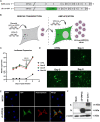
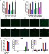
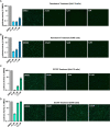
Research activities with infectious severe acute respiratory syndrome coronavirus 2 (SARS-CoV-2) are currently permitted only under biosafety level 3 (BSL3) containment. Here, we report the development of a single-cycle infectious SARS-CoV-2 virus replicon particle (VRP) system with a luciferase and green fluorescent protein (GFP) dual reporter that can be safely handled in BSL2 laboratories to study SARS-CoV-2 biology. The spike (S) gene of SARS-CoV-2 encodes the envelope glycoprotein, which is essential for mediating infection of new host cells. Through deletion and replacement of this essential S gene with a luciferase and GFP dual reporter, we have generated a conditional SARS-CoV-2 mutant (ΔS-VRP) that produces infectious particles only in cells expressing a viral envelope glycoprotein of choice. Interestingly, we observed more efficient production of infectious particles in cells expressing vesicular stomatitis virus (VSV) glycoprotein G [ΔS-VRP(G)] than in cells expressing other viral glycoproteins, including S. We confirmed that infection from ΔS-VRP(G) is limited to a single round and can be neutralized by anti-VSV serum. In our studies with ΔS-VRP(G), we observed robust expression of both luciferase and GFP reporters in various human and murine cell types, demonstrating that a broad variety of cells can support intracellular replication of SARS-CoV-2. In addition, treatment of ΔS-VRP(G)-infected cells with either of the anti-CoV drugs remdesivir (nucleoside analog) and GC376 (CoV 3CL protease inhibitor) resulted in a robust decrease in both luciferase and GFP expression in a drug dose- and cell-type-dependent manner. Taken together, our findings show that we have developed a single-cycle infectious SARS-CoV-2 VRP system that serves as a versatile platform to study SARS-CoV-2 intracellular biology and to perform high-throughput screening of antiviral drugs under BSL2 containment. IMPORTANCE Due to the highly contagious nature of SARS-CoV-2 and the lack of immunity in the human population, research on SARS-CoV-2 has been restricted to biosafety level 3 laboratories. This has greatly limited participation of the broader scientific community in SARS-CoV-2 research and thus has hindered the development of vaccines and antiviral drugs. By deleting the essential spike gene in the viral genome, we have developed a conditional mutant of SARS-CoV-2 with luciferase and fluorescent reporters, which can be safely used under biosafety level 2 conditions. Our single-cycle infectious SARS-CoV-2 virus replicon system can serve as a versatile platform to study SARS-CoV-2 intracellular biology and to perform high-throughput screening of antiviral drugs under BSL2 containment.
Keywords: SARS-CoV-2; replicon.
Conflict of interest statement
The authors declare no conflict of interest.
Figures


Similar articles
-
A BSL-2 compliant mouse model of SARS-CoV-2 infection for efficient and convenient antiviral evaluation.J Virol. 2024 Jul 23;98(7):e0050424. doi: 10.1128/jvi.00504-24. Epub 2024 Jun 20. J Virol. 2024. PMID: 38899934 Free PMC article.
-
Construction of a Noninfectious SARS-CoV-2 Replicon for Antiviral-Drug Testing and Gene Function Studies.J Virol. 2021 Aug 25;95(18):e0068721. doi: 10.1128/JVI.00687-21. Epub 2021 Aug 25. J Virol. 2021. PMID: 34191580 Free PMC article.
-
A Convenient and Biosafe Replicon with Accessory Genes of SARS-CoV-2 and Its Potential Application in Antiviral Drug Discovery.Virol Sin. 2021 Oct;36(5):913-923. doi: 10.1007/s12250-021-00385-9. Epub 2021 May 17. Virol Sin. 2021. PMID: 33999369 Free PMC article.
-
Dimming the corona: studying SARS-coronavirus-2 at reduced biocontainment level using replicons and virus-like particles.mBio. 2024 Dec 11;15(12):e0336823. doi: 10.1128/mbio.03368-23. Epub 2024 Nov 12. mBio. 2024. PMID: 39530689 Free PMC article. Review.
-
Reverse genetics systems for SARS-CoV-2.J Med Virol. 2022 Jul;94(7):3017-3031. doi: 10.1002/jmv.27738. Epub 2022 Apr 5. J Med Virol. 2022. PMID: 35324008 Free PMC article. Review.
Cited by
-
Boosting the detection performance of severe acute respiratory syndrome coronavirus 2 test through a sensitive optical biosensor with new superior antibody.Bioeng Transl Med. 2022 Sep 16;8(5):e10410. doi: 10.1002/btm2.10410. Online ahead of print. Bioeng Transl Med. 2022. PMID: 36248235 Free PMC article.
-
Reverse genetics systems for SARS-CoV-2: Development and applications.Virol Sin. 2023 Dec;38(6):837-850. doi: 10.1016/j.virs.2023.10.001. Epub 2023 Oct 11. Virol Sin. 2023. PMID: 37832720 Free PMC article. Review.
-
Single-dose mucosal replicon-particle vaccine protects against lethal Nipah virus infection up to 3 days after vaccination.Sci Adv. 2023 Aug 4;9(31):eadh4057. doi: 10.1126/sciadv.adh4057. Epub 2023 Aug 4. Sci Adv. 2023. PMID: 37540755 Free PMC article.
-
A BSL-2 compliant mouse model of SARS-CoV-2 infection for efficient and convenient antiviral evaluation.J Virol. 2024 Jul 23;98(7):e0050424. doi: 10.1128/jvi.00504-24. Epub 2024 Jun 20. J Virol. 2024. PMID: 38899934 Free PMC article.
-
Epitranscriptomic N6-Methyladenosine Profile of SARS-CoV-2-Infected Human Lung Epithelial Cells.Microbiol Spectr. 2023 Feb 14;11(1):e0394322. doi: 10.1128/spectrum.03943-22. Epub 2023 Jan 10. Microbiol Spectr. 2023. PMID: 36625663 Free PMC article.
References
-
- Friedrich BM, Scully CE, Brannan JM, Ogg MM, Johnston SC, Hensley LE, Olinger GG, Smith DR. 2011. Assessment of high-throughput screening (HTS) methods for high-consequence pathogens. J Bioterror Biodef S3:005.
-
- Federal Select Agent Program. Select agents and toxins exclusions: excluded attenuated strains of HHS select agents, section 73.3 (e). https://www.selectagents.gov/sat/exclusions/hhs.htm.
-
- Gangadharan D, Smith J, Weyant R, Centers for Disease Control and Prevention. 2013. Biosafety recommendations for work with influenza viruses containing a hemagglutinin from the A/goose/Guangdong/1/96 lineage. MMWR Recommend Rep 62:1–7. - PubMed
Publication types
MeSH terms
Substances
Grants and funding
LinkOut - more resources
Full Text Sources
Research Materials
Miscellaneous

