

Therapeutic potential of naturally derived carbon dots in sepsis-associated acute kidney injury
- PMID: 40217355
- PMCID: PMC11992765
- DOI: 10.1186/s13020-025-01103-3
Therapeutic potential of naturally derived carbon dots in sepsis-associated acute kidney injury
Abstract
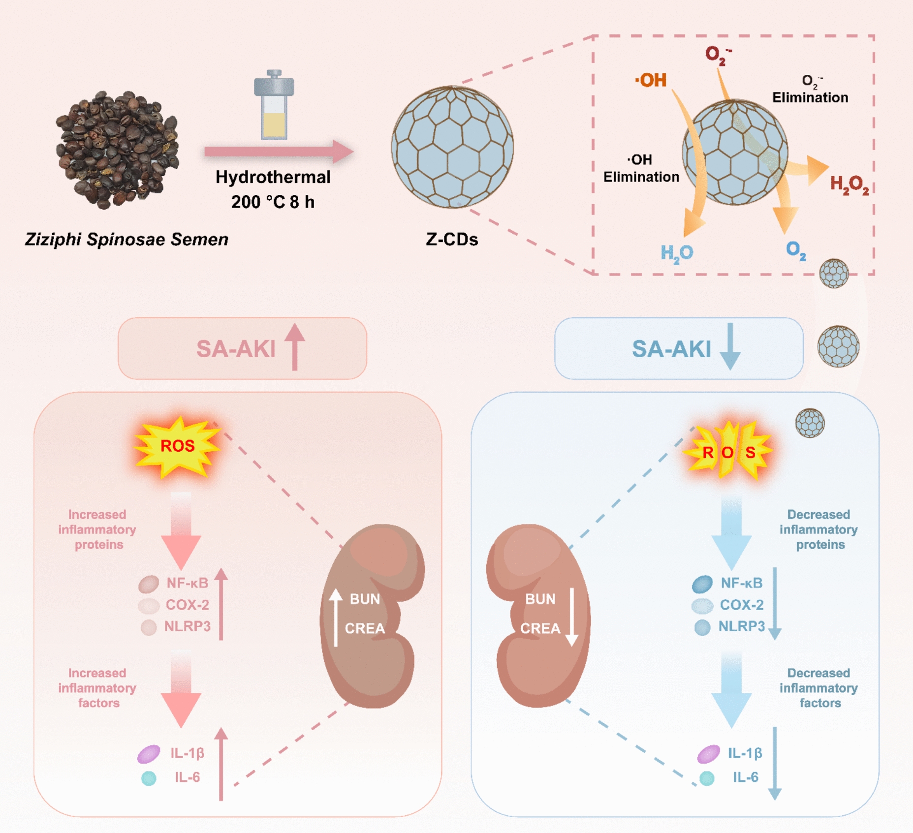
Background: Sepsis is a life-threatening infectious disease characterized by an uncontrolled inflammatory response and consequent multi-organ dysfunction. The kidneys, as primary excretory organs with high blood flow, are particularly susceptible to damage during sepsis. Nonetheless, the existing treatment options for sepsis-associated acute kidney injury (SA-AKI) are still restricted. Nanomedicine, especially carbon dots (CDs), has attracted considerable interest lately for outstanding biomedical characteristics.
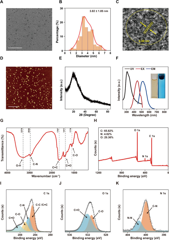
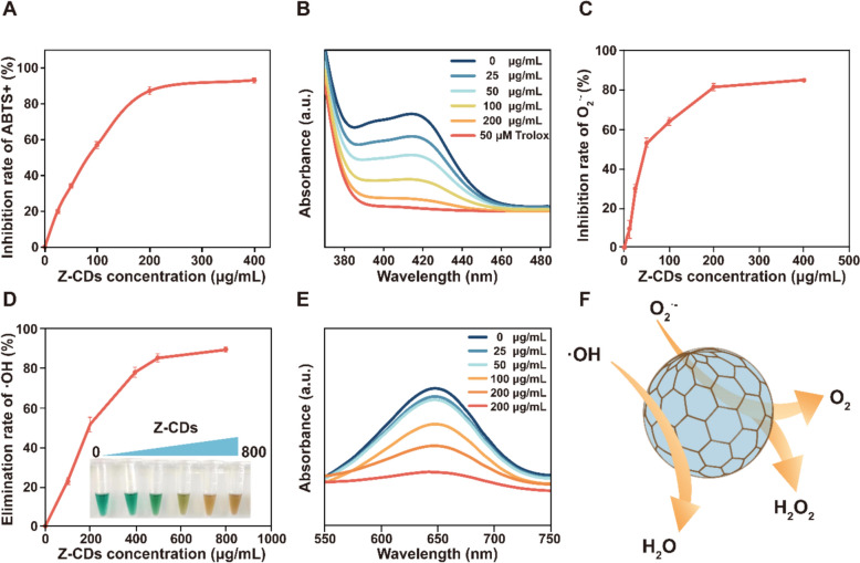
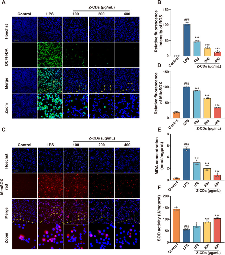
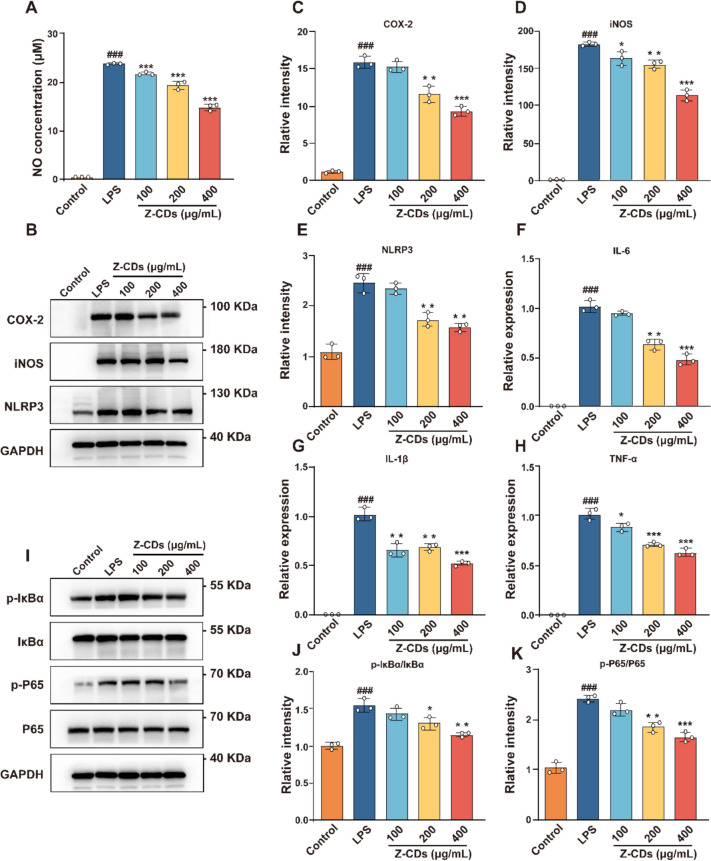
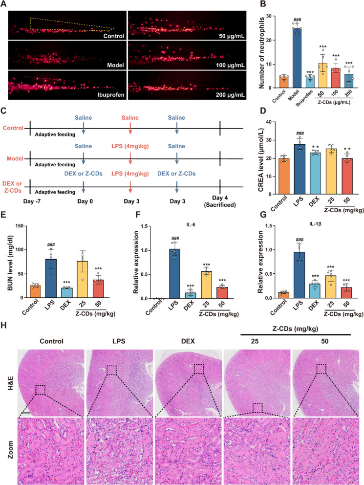
Methods: To avoid the generation of toxic effects, the natural CDs derived from Ziziphi Spinosae Semen (Z-CDs) were synthesized employing a hydrothermal method. The free radical scavenging capabilities of Z-CDs were evaluated by utilizing ABTS assay, NBT method, and Fenton reaction. A lipopolysaccharide (LPS)-stimulated RAW 264.7 cell model was used to explore the therapeutic potential of Z-CDs on cellular oxidative stress and inflammation. The CuSO4-induced zebrafish inflammation model and LPS-exposed SA-AKI mouse model were employed to assess the therapeutic efficacy of Z-CDs in vivo.
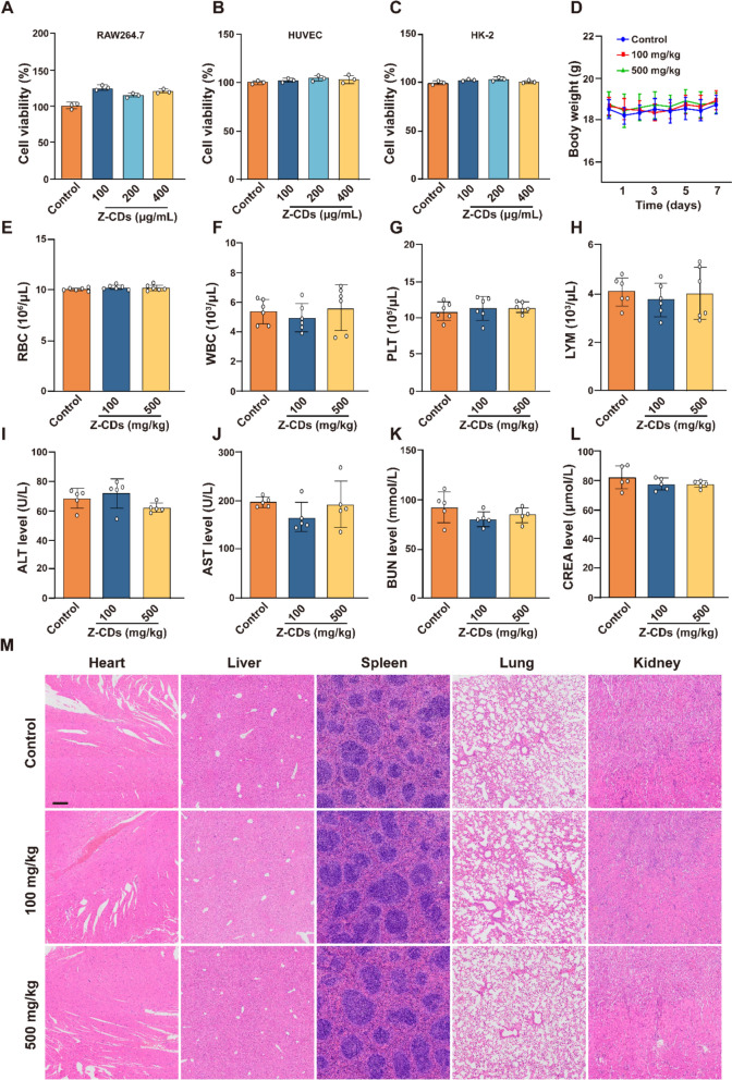
Results: The synthesized Z-CDs exhibited distinctive unsaturated surface functional groups, which confer exceptional biocompatibility and the ability to scavenge free radicals. Moreover, Z-CDs were particularly effective in eliminating excess reactive oxygen species (ROS) in cells, thus protecting mitochondrial function from oxidative damage. Notably, Z-CDs have demonstrated significant therapeutic benefits in protecting kidney tissue in SA-AKI mouse model with minimizing side effects. In mechanism, Z-CDs effectively reduced ROS production, thereby alleviating inflammatory responses in macrophages through the suppression of the NF-κB pathway.
Conclusions: This study developed a multifunctional nanomedicine derived from traditional medicinal herb, providing a promising pathway for the advancement of innovative drug therapies to treat SA-AKI.
Keywords: Acute kidney injury; Antioxidant; Carbon dots; NF-κB pathway; Phytochemicals; Sepsis.
© 2025. The Author(s).
Conflict of interest statement
Declarations. Ethics approval and consent to participate: Approval for all animal studies was granted by the Ethics Committee of the Experimental Animal Centre of Shandong First Medical University (No. W202410180685). Laboratory Animal Ethical and Welfare Committee at the Institute of Materia Medica, Shandong Academy of Medical Sciences (No. 202405). Consent for publication: All authors agreed with the content of the manuscript and approved the final version of the manuscript. Competing interests: We declare that we have no financial or personal relationships with other people or organizations that can inappropriately influence our work.
Figures
Similar articles
-
Melatonin-Derived Carbon Dots with Free Radical Scavenging Property for Effective Periodontitis Treatment via the Nrf2/HO-1 Pathway.ACS Nano. 2024 Mar 19;18(11):8307-8324. doi: 10.1021/acsnano.3c12580. Epub 2024 Mar 4. ACS Nano. 2024. PMID: 38437643
-
Hydrothermally Derived Green Carbon Dots from Broccoli Water Extracts: Decreased Toxicity, Enhanced Free-Radical Scavenging, and Anti-Inflammatory Performance.ACS Biomater Sci Eng. 2023 Mar 13;9(3):1307-1319. doi: 10.1021/acsbiomaterials.2c01537. Epub 2023 Feb 6. ACS Biomater Sci Eng. 2023. PMID: 36744996
-
Heteroatom doped carbon dots with nanoenzyme like properties as theranostic platforms for free radical scavenging, imaging, and chemotherapy.Acta Biomater. 2020 Sep 15;114:343-357. doi: 10.1016/j.actbio.2020.07.022. Epub 2020 Jul 15. Acta Biomater. 2020. PMID: 32682058
-
The future of plant based green carbon dots as cancer Nanomedicine: From current progress to future Perspectives and beyond.J Adv Res. 2025 Jan;67:133-159. doi: 10.1016/j.jare.2024.01.034. Epub 2024 Feb 5. J Adv Res. 2025. PMID: 38320729 Free PMC article. Review.
-
The role of inflammatory response and metabolic reprogramming in sepsis-associated acute kidney injury: mechanistic insights and therapeutic potential.Front Immunol. 2024 Oct 31;15:1487576. doi: 10.3389/fimmu.2024.1487576. eCollection 2024. Front Immunol. 2024. PMID: 39544947 Free PMC article. Review.
References
-
- Meyer NJ, Prescott HC. Sepsis and septic shock. N Engl J Med. 2024;391(22):2133–46. - PubMed
-
- Xiao YQ, Fang H, Wang X, Liu M, Shen T, Zhang M, et al. Modulation of unregulated inflammation-associated coagulopathy in sepsis using multifunctional nanosheets. Adv Func Mater. 2024;34(38):2402785.
-
- Desposito L, Bascara C. Review: sepsis guidelines and core measure bundles. Postgrad Med. 2024;136(7):702–11. - PubMed
Grants and funding
- ZR2023ZD25/Natural Science Foundation of Shandong Province
- ZR2023QH427/Natural Science Foundation of Shandong Province
- ZR2022LZY021/Natural Science Foundation of Shandong Province
- ZR2023MH361/Natural Science Foundation of Shandong Province
- Q-2023107/the Shandong Province Chinese Medicine Science and Technology Development Project
- 32300957/National Natural Science Foundation of China
- tstp20230633/Taishan Scholars Project in Shandong Province
- tsqn202408246/Taishan Scholars Project in Shandong Province
- L246029/Beijing Municipal Natural Science Foundation-Key Research Project of the Daxing
- 202401/WT_/Wellcome Trust/United Kingdom
- 202408/the Joint Innovation Team for Clinical & Basic Research
- 202228048/Jinan New 20 Policies for Higher Education Funding
- 2022KJ197/Youth Innovation Technology Project of Higher School in Shandong Province
- 202002040923/Project of Shandong Medical and Health Science and Technology
LinkOut - more resources
Full Text Sources
Molecular Biology Databases
