. 2006 Dec;2(12):e127.
doi: 10.1371/journal.ppat.0020127.

HIV-1 Vpr-induced apoptosis is cell cycle dependent and requires Bax but not ANT

Affiliations

HIV-1 Vpr-induced apoptosis is cell cycle dependent and requires Bax but not ANT

Joshua L Andersen et al. PLoS Pathog. 2006 Dec.

Abstract

The HIV-1 accessory protein viral protein R (Vpr) causes G2 arrest and apoptosis in infected cells. We previously identified the DNA damage-signaling protein ATR as the cellular factor that mediates Vpr-induced G2 arrest and apoptosis. Here, we examine the mechanism of induction of apoptosis by Vpr and how it relates to induction of G2 arrest. We find that entry into G2 is a requirement for Vpr to induce apoptosis. We investigated the role of the mitochondrial permeability transition pore by knockdown of its essential component, the adenine nucleotide translocator. We found that Vpr-induced apoptosis was unaffected by knockdown of ANT. Instead, apoptosis is triggered through a different mitochondrial pore protein, Bax. In support of the idea that checkpoint activation and apoptosis induction are functionally linked, we show that Bax activation by Vpr was ablated when ATR or GADD45alpha was knocked down. Certain mutants of Vpr, such as R77Q and I74A, identified in long-term nonprogressors, have been proposed to inefficiently induce apoptosis while activating the G2 checkpoint in a normal manner. We tested the in vitro phenotypes of these mutants and found that their abilities to induce apoptosis and G2 arrest are indistinguishable from those of HIV-1NL4-3 vpr, providing additional support to the idea that G2 arrest and apoptosis induction are mechanistically linked.


Conflict of interest statement

Competing interests. The authors have declared that no competing interests exist.

Figures

Figure 1
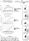
Figure 1. Vpr-Induced Caspase Activation and Smac Release from the Mitochondria Are Temporally Delayed in Relation to G2 Arrest
(A) pHR-VPR-G and pHR-VPR-R are bicistronic lentiviral vectors that encode HIV-1NL4–3 vpr, an internal ribosome entry site, and either the gene for GFP or that for mRFP, respectively; pHR-VPR(R80A) was derived from pHR-VPR (both the mRFP and the GFP versions) by site-directed mutagenesis; DHIV3 is an envelope-truncated (see gray box) version of HIV-1NL4–3; DHIV3-ΔVPR was derived from DHIV-3 by introducing a frameshift mutation in vpr. (B) SupT1 T lymphocytes were transduced by spin-infection in the presence of 10 μg/ml Polybrene with indicated vectors. Mock-infected cells were subjected to spin-infection in the presence of 10 μg/ml Polybrene without virus. Cells were collected at specified time points post-transduction, stained with propidium iodide, and analyzed for DNA content by flow cytometry to determine cell cycle profiles. The percentage of cells transduced with pHR vectors and DHIV3 viruses ranged between 70% to 80% and between 65% to 70%, respectively, as determined by mRFP or GFP expression (with pHR vectors) or intracellular p24 staining (with DHIV3 vectors). (C) Caspase activation was measured as an indication of apoptosis. SupT1 cells were infected with indicated vectors, harvested, and incubated with FITC-VAD-FMK. The percentage of caspase-active cells at each time point was measured by flow cytometry. (D) Infected SupT1 cells were lysed, and lysates were fractionated into mitochondrial (m) and cytoplasmic (c) fractions and then assayed by Western blot. Western blots were probed with antibodies specific to Smac to measure release from mitochondria and with anti-VDAC antibodies to measure mitochondrial contamination in the cytoplasmic fractions. As a positive control for apoptosis, SupT1 cells were treated with 0.8 μg/ml doxorubicin (dox) for 48 h. (E) Examples of flow cytometric analysis of caspase activation, corresponding to the 72-h time points from (C).
Figure 2
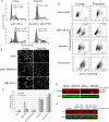
Figure 2. The Apoptotic Effect of Vpr Is Lost in Cells Synchronized in G1/S
(A) HeLa cells were transduced with pHR-VPR-G or mock-transduced and then incubated with 2 mM thymidine. After 24 h of incubation, cells were harvested, stained with PI, and analyzed for DNA content by flow cytometry to determine the percentage of cells in G1/S and G2/M. Transduction efficiency of pHR-VPR in both thymidine-treated and cycling cells was 70% to 75% as determined by analysis of GFP expression by flow cytometry (unpublished data). (B) Cells from experiments shown in (A) were stained for DAPI at 72 h postinfection, in order to evaluate apoptosis via chromatin morphology (C) Quantitation of apoptosis in DAPI-stained samples shown in (B); incubation with 25 μM etoposide for 48 h or 1 μM staurosporine for 8 h was included in both cycling and thymidine-treated cells, for comparative purposes. (D) Cycling or thymidine-synchronized HeLa cells were mock-transduced or transduced with either pHR-VPR-R or pHR-VPR(R80A) for apoptosis analysis, using FITC-VAD-FMK. As positive controls for apoptosis, cycling and synchronized cells were treated with etoposide or staurosporine as shown in (C). (E) Cycling or thymidine-synchronized HeLa cells expressing Vpr or treated with 25 μM etoposide for 48 h were lysed and analyzed by Western blot for PARP cleavage as a marker of apoptosis/caspase activity. The caspase-cleaved PARP band is observed at 89 kDa. (F) Cell lysates from pHR-VPR–transduced cells, with or without thymidine block, were harvested at 12 and 24 h post-transduction and analyzed by Western blot for Vpr expression with antibodies specific to the amino-terminal hemagglutinin tag.
Figure 3
Figure 3. Release of Vpr-Expressing Cells from Thymidine Block Leads to Reentry into the Cell Cycle and Vpr-Induced, G2-Dependent Apoptosis
(A) Thymidine-synchronized HeLa cells transduced with pHR-VPR-G or mock-transduced were released from thymidine block and harvested at specified time points postrelease. Cells from each time point were stained with PI and analyzed for DNA content by flow cytometry. (B) pHR-VPR–transduced HeLa cells from the same experiment were monitored by Western blot for PARP cleavage at specified time points. Time points labeled under “Thym + pHR-VPR” indicate hours following release from thymidine block; time points under “pHR-VPR” indicate hours following transduction, as these cells were not synchronized. (C) Cells treated as those in (A) and (B) were harvested at 24 and 48 h were analyzed for apoptosis by DAPI staining, and the results are quantified.
Figure 4
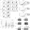
Figure 4. siRNA-Mediated Knockdown of ATR or Bax, but Not of ANT, Suppresses Vpr-Induced Apoptosis
(A) HeLa cells were transfected with nonspecific (NS) siRNA or siRNA targeted to ATR, or ATM as indicated. At 48 h post-transfection, cells were either treated with 25 μM etoposide, treated with 25 μM MNNG, mock-transduced, or transduced with pHR-VPR-R. Additionally, cells transfected with siRNAs targeted to Bax or ANT were transduced with pHR-VPR-R or mock-transduced (lower left dot plots). Cells from each treatment were assayed for caspase activity as in Figure 2B. (B) Cells treated as in (A) were harvested at specified time points post-transduction and assayed for caspase activity. (C) Cells treated as in (A) were stained with DAPI, and the results were quantified by microscopy. (D) Cells treated with the indicated siRNAs were lysed and analyzed by Western blot to verify knockdown efficiency. (E) Primary human CD4+ lymphocytes were infected with DHIV3, infected with DHIV3ΔVPR, or mock-infected. At 48 h postinfection, cells in each treatment were lysed and assayed for protein concentration. Equal amounts of protein from each treatment were incubated with Bax6A7 monoclonal antibody. Antibody–protein complexes were precipitated with agarose beads and boiled and then subjected to Western blot analysis with a polyclonal antibody to Bax. (F) HeLa cells treated with the indicated siRNAs and transduced with pHR-VPR or mock-transduced were lysed; reactivity to Bax6A7 antibody was assayed as described in (E).
Figure 5
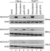
Figure 5. Vpr Induces Histone 3 Phosphorylation in HeLa but Not in SupT1 Cells
HeLa or Supt1 cells were transduced with pHR-VPR-G and at indicated time points, lysed, and analyzed by Western blot with a phospho-specific antibody that recognizes phosphorylation of H3 at serine-10. Nocodazole (250 ng/ml) and doxorubicin (1 μM) were used as positive and negative controls, respectively.
Figure 6
Figure 6. Functional Analysis of Vpr Mutants
(A) SupT1 cells were infected with pHR-VPR-R or indicated mutants, at an MOI of 0.5. At 48 h postinfection, cells were stained with hypotonic PI to determine the cell cycle profiles. At 72 h postinfection, cells were incubated with FITC-VAD-FMK and analyzed by flow cytometry to determine the percentage of cells with active caspases. (B) Cells from above treatments were lysed at 48 h postinfection, and Western blot was performed to assay for phosphorylation of BRCA1 at Ser1423 by the ATR kinase. To establish the role of ATR in BRCA1 phosphorylation, parallel infections were treated with caffeine (2 mM). (C) Induction of apoptosis by Vpr-GFP and Vpr(R80A)-GFP fusion proteins. HPB-ALL cells were transfected with indicated constructs or mock-transfected and 48 after transfection, phosphatidylserine exposure was analyzed by flow cytometry using phycoerythrin-conjugated annexin V.
Figure 7
Figure 7. Models to Explain the Activities of HIV-1 Vpr
(A). Activation of ATR by Vpr and downstream signaling consequences leading to both G2 arrest and apoptosis. The functional link between GADD45α and activation of Bax and the mechanism by which G2 arrest leads to Bax activation remain unknown (question marks). (B) Binding of Vpr to mitochondrial PTPC was proposed to trigger release of cytochrome c and induction of apoptosis, independently of cell cycle status; in this work, we propose that an alternative pore-forming mitochondrial protein, Bax, is the effector of Vpr-induced apoptosis, and that Bax activation requires upstream stress signals derived from ATR.

References

    1. Derdeyn CA, Silvestri G. Viral and host factors in the pathogenesis of HIV infection. Curr Opin Immunol. 2005;17:366–373. - PubMed
    1. Gougeon ML. Apoptosis as an HIV strategy to escape immune attack. Nat Rev Immunol. 2003;3:392–404. - PubMed
    1. Roshal M, Zhu Y, Planelles V. Apoptosis in AIDS. Apoptosis. 2001;6:103–116. - PubMed
    1. Haase AT. Population biology of HIV-1 infection: Viral and CD4+ T cell demographics and dynamics in lymphatic tissues. Annu Rev Immunol. 1999;17:625–656. - PubMed
    1. Perelson AS, Neumann AU, Markowitz M, Leonard JM, Ho DD. HIV-1 dynamics in vivo: Virion clearance rate, infected cell life-span, and viral generation time. Science. 1996;271:1582–1586. - PubMed

Publication types

MeSH terms