. 1999 Sep 14;96(19):10608-13.
doi: 10.1073/pnas.96.19.10608.

Molecular cloning of an apoptosis-inducing protein, pierisin, from cabbage butterfly: possible involvement of ADP-ribosylation in its activity

Affiliations

Molecular cloning of an apoptosis-inducing protein, pierisin, from cabbage butterfly: possible involvement of ADP-ribosylation in its activity

M Watanabe et al. Proc Natl Acad Sci U S A. .

Abstract

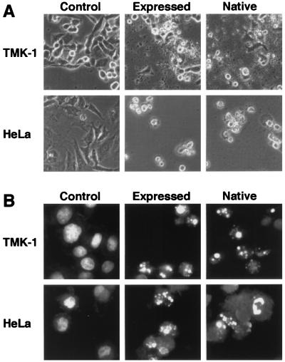
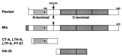
We have previously reported that the cabbage butterfly, Pieris rapae, contains a 98-kDa protein, named pierisin, that induces apoptosis in a variety of human cancer cell lines. In the present study, sequencing and cloning of a cDNA encoding pierisin was accomplished. PCR-direct sequencing showed that the gene encodes an 850-amino acid protein with a calculated molecular weight of 98,081. An intact clone at the amino acid level encompassing the entire coding region was obtained by recombination of two independent clones, and the molecular mass of its in vitro expressed protein was about 100 kDa on SDS/PAGE, the same as that of purified native pierisin. The expressed protein induced apoptosis in human gastric carcinoma TMK-1 and cervical carcinoma HeLa cells, like the native protein, indicating functional activity. The deduced amino acid sequence of pierisin showed 32% homology with a 100-kDa mosquitocidal toxin from Bacillus sphaericus SSII-1. In addition, pierisin showed regional sequence similarities with ADP-ribosylating toxins, such as the A subunit of cholera toxin. A glutamic acid residue at the putative NAD-binding site, conserved in all ADP-ribosylating toxins, was also found in pierisin. Substitution of another amino acid for glutamic acid 165 resulted in a great decrease in cytotoxicity and induction of apoptosis. Moreover, inhibitors of ADP-ribosylating enzymes reduced pierisin-induced apoptosis. These results suggest that the apoptosis-inducing protein pierisin might possess ADP-ribosylation activity that leads to apoptosis of the cells.


Figures

Figure 1
Figure 1
Nucleotide and deduced amino acid sequences of the cDNA for pierisin by PCR-direct sequencing. All determined internal amino acid sequences of purified native pierisin from pupae of P. rapae, including those used to make degenerate primers, existed in the deduced amino acid sequence of the gene, as underlined. Termination codons found at the 5′ end of the coding region are indicated by asterisks (∗). A possible polyadenylation signal, found near the 3′ end, is indicated by “#.”
Figure 2
Figure 2
SDS/PAGE of [35S]methionine-labeled proteins, expressed from the subclone in the pMAL-p2 plasmid. The arrow indicates the molecular size of the native pierisin.
Figure 3
Figure 3
Morphological analysis of TMK-1 and HeLa cells treated with the in vitro-expressed protein from the subclone. The cells were treated for 18 h with 3% (TMK-1) or 0.6% (HeLa) of rabbit reticulocyte lysate alone (control), or containing synthesized protein (expressed), or 2.5 ng/ml (TMK-1) or 0.5 ng/ml (HeLa) purified native pierisin (native). (A) Phase-contrast micrographs. (B) Fluorescent micrographs.
Figure 4
Figure 4
DNA fragmentation in TMK-1 and HeLa cells treated with the in vitro-expressed protein. The cells were treated with synthesized protein (expressed) or purified native pierisin (native) under the same conditions described in Fig. 3, and their DNA was extracted and run on agarose gels.
Figure 5
Figure 5
Sequence similarity of pierisin with Mtx and ADP-ribosylating toxins. The conserved glutamic acid residue is boxed. (A) Alignment of the deduced amino acid sequence of pierisin with that of Mtx (15). P, pierisin; M, Mtx. Identical amino acids are marked by asterisks (∗). (B) Homologous region of pierisin with ADP-ribosylating toxins. CT-A, the A subunit of cholera toxin (16); LTH-A and LTP-A, the A subunit of E. coli heat-labile toxin pathogenic for humans and pigs, respectively (–19); PT-S1, the S1 subunit of pertussis toxin (20). Completely conserved amino acids are marked by asterisks (∗).
Figure 6
Figure 6
Dose-dependent cytotoxic effects of nonmutated and mutated proteins on HeLa cells. Cells were incubated with various concentrations of rabbit reticulocyte lysate: control lysate (○); lysate containing nonmutated protein (●), E165D protein (■), or E165Q protein (▴). After incubation for 72 h at 37°C, MTT assay was carried out.
Figure 7
Figure 7
Structural similarity of pierisin with other proteins. Cross-hatched bar, possible signal sequence observed in Mtx; striped bars, homologous region among pierisin, Mtx, and ADP-ribosylating toxins; shaded bars, repeat unit of 150 amino acids. The conserved glutamic acid residue is indicated as “E”. The trypsin cleavage site of pierisin, indicated by an arrow, was determined by using a Shimadzu PSQ-1 protein sequencer after separation and blotting of the digested C-terminal 70-kDa fragment on an SDS/polyacrylamide gel. Pronase E and proteinase K also produced the 70-kDa fragment, suggesting that there is a protease-sensitive region close to the trypsin cleavage site. Mtx possesses a protease-sensitive region (29). The protease-sensitive regions in pierisin and Mtx are indicated by double-headed arrows. HA-33, hemagglutinin component of botulinus toxin; other protein abbreviations as in Fig. 5.

References

    1. Boman H G, Hultmark D. Annu Rev Microbiol. 1987;41:103–126. - PubMed
    1. Hultmark D. Trends Genet. 1993;9:178–183. - PubMed
    1. Hoffmann J A. Curr Opin Immunol. 1995;7:4–10. - PubMed
    1. Itoh A, Iizuka K, Natori S. Jpn J Cancer Res (GANN) 1985;76:1027–1033. - PubMed
    1. Moore A J, Devine D A, Bibby M C. Pept Res. 1994;7:265–269. - PubMed

Publication types

MeSH terms

Associated data