. 2001 Sep 11;98(19):10960-5.
doi: 10.1073/pnas.181331498.

Two types of MGDG synthase genes, found widely in both 16:3 and 18:3 plants, differentially mediate galactolipid syntheses in photosynthetic and nonphotosynthetic tissues in Arabidopsis thaliana

Affiliations

Two types of MGDG synthase genes, found widely in both 16:3 and 18:3 plants, differentially mediate galactolipid syntheses in photosynthetic and nonphotosynthetic tissues in Arabidopsis thaliana

K Awai et al. Proc Natl Acad Sci U S A. .

Abstract

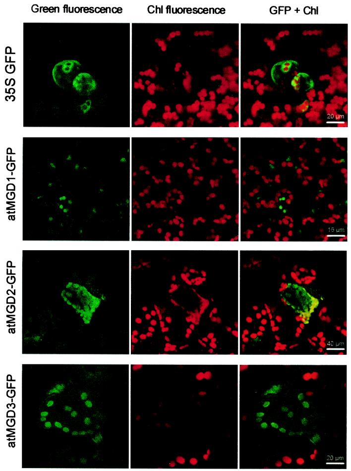
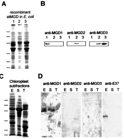
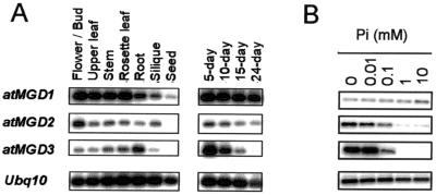
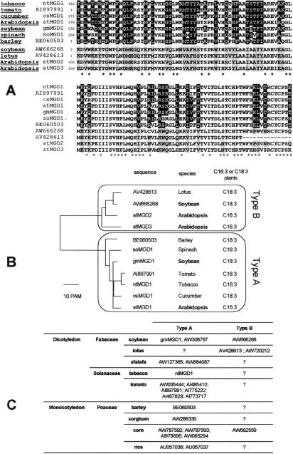
In Arabidopsis, monogalactosyldiacylglycerol (MGDG) is synthesized by a multigenic family of MGDG synthases consisting of two types of enzymes differing in their N-terminal portion: type A (atMGD1) and type B (atMGD2 and atMGD3). The present paper compares type B isoforms with the enzymes of type A that are known to sit in the inner membrane of plastid envelope. The occurrence of types A and B in 16:3 and 18:3 plants shows that both types are not specialized isoforms for the prokaryotic and eukaryotic glycerolipid biosynthetic pathways. Type A atMGD1 gene is abundantly expressed in green tissues and along plant development and encodes the most active enzyme. Its mature polypeptide is immunodetected in the envelope of chloroplasts from Arabidopsis leaves after cleavage of its transit peptide. atMGD1 is therefore likely devoted to the massive production of MGDG required to expand the inner envelope membrane and build up the thylakoids network. Transient expression of green fluorescent protein fusions in Arabidopsis leaves and in vitro import experiments show that type B precursors are targeted to plastids, owing to a different mechanism. Noncanonical addressing peptides, whose processing could not be assessed, are involved in the targeting of type B precursors, possibly to the outer envelope membrane where they might contribute to membrane expansion. Expression of type B enzymes was higher in nongreen tissues, i.e., in inflorescence (atMGD2) and roots (atMGD3), where they conceivably influence the eukaryotic structure prominence in MGDG. In addition, their expression of type B enzymes is enhanced under phosphate deprivation.


Figures

Figure 1
Figure 1
Chloroplast localization of atMGD-GFP fusion after transient expression in rosette leaves of Arabidopsis. Expression of four constructs was analyzed: N-terminus fusions of atMGD1 (atMGD1-GFP), atMGD2 (atMGD2-GFP), or atMGD3 (atMGD3-GFP) to the GFP sequence under the control of cauliflower mosaic virus 35S promoter and unfused GFP as a cytosolic control (35S GFP). GFP fluorescence (in green) was compared with chlorophyll autofluorescence (in red).
Figure 2
Figure 2
Immunolocalization of atMGD1. The specificity of polyclonal antibodies prepared against atMGD1, atMGD2, and atMGD3 was first determined (A and B) and used to analyze the content of chloroplast subfractions (C and D). (A) Inclusion bodies enriched in atMGDs. Twenty five micrograms of protein was loaded in each lane and separated by SDS/PAGE. * indicate the positions of the overexpressed proteins. (B) Western blot analysis using 1:5,000-diluted primary antibodies. Detection was obtained after incubation with an secondary antibody linked to alkaline phosphatase. Anti-atMGD1 and anti-atMGD2 exhibit a high specificity, and anti-atMGD3 reacts with both atMGD2 and atMGD3. (C) Subfractions of Arabidopsis chloroplasts prepared from rosette leaves. Proteins (30 μg) from each chloroplast subfraction (E: envelope, S: stroma, T: thylakoids) were separated by SDS/PAGE. (D) Immunodetection of atMGD1 in chloroplast envelope membranes. Western blot using antibodies against atMGD1 (dilution 1:500), atMGD2 (dilution 1:500), atMGD3 (dilution 1:500), or E37, an envelope membrane marker (dilution 1:10,000) was carried out on E, S, and T lanes transferred to nitrocellulose. A polypeptide having a size consistent with the processed atMGD1 protein (type A) was detected. Type B enzymes are not detected. Positions of molecular mass standards (29, 33, 49, 82, and 105 kDa) are shown on the left.
Figure 3
Figure 3
Analyses of atMGD gene expression. (A) Expression of atMGD genes in various organs and developmental stages. (B) Expression of atMGD genes in total extracts from plants grown on Murashige and Skoog media supplemented with phosphate as indicated. RT-PCR analyses for Arabidopsis MGD genes were carried out as described in Materials and Methods. Amounts of DNA loaded per gel were adjusted for each experiment so as not to saturate the image.
Figure 4
Figure 4
Type A and type B MGDG synthases in the plant kingdom. A basic blast screening (blosum 62 matrix) of the GenBank database dbEST, using atMGD2 as a probe, led to the identification of putative MGDG synthases in various plant species. (A) Example of a local alignment. Identical amino acids are indicated by *; amino acids that are common to cloned type A enzymes are highlighted in black whereas amino acids that are common to cloned type B enzymes (atMGD2 and atMGD3) are shown in gray. (B) Unrooted phenogram drawn by using the previous alignment. Distance is indicated in PAM (probability of accepted mutation). (C) Compiled results of local alignments of MGDG synthases ESTs exhibiting signatures of types A and B. Accession numbers of all ESTs are indicated.

References

    1. Joyard J, Maréchal E, Block M A, Douce R. In: Membranes: Specialized Functions in Plants. Smallwood M, Knox P, Bowles D J, editors. Oxford: BIOS Scientific; 1996. pp. 179–194.
    1. Heinz E. In: Lipids and Lipid Polymers. Tevini M, Lichtenthaler H K, editors. Berlin: Springer; 1977. pp. 102–120.
    1. Block M A, Dorne A-J, Joyard J, Douce R. J Biol Chem. 1983;258:13281–13286. - PubMed
    1. Cline K, Keegstra K. Plant Physiol. 1983;71:366–372. - PMC - PubMed
    1. Shimojima M, Ohta H, Iwamatsu A, Masuda T, Shioi Y, Takamiya K. Proc Natl Acad Sci USA. 1997;94:333–337. - PMC - PubMed

Publication types

MeSH terms