. 2006 Aug 8;103(32):12021-6.
doi: 10.1073/pnas.0600021103. Epub 2006 Jul 27.

Functional development of Src tyrosine kinases during evolution from a unicellular ancestor to multicellular animals

Affiliations

Functional development of Src tyrosine kinases during evolution from a unicellular ancestor to multicellular animals

Yuko Segawa et al. Proc Natl Acad Sci U S A. .

Abstract

The Src family of tyrosine kinases play pivotal roles in regulating cellular functions characteristic of multicellular animals, including cell-cell interactions, cell-substrate adhesion, and cell migration. To investigate the functional alteration of Src kinases during evolution from a unicellular ancestor to multicellular animals, we characterized Src orthologs from the unicellular choanoflagellate Monosiga ovata and the primitive multicellular sponge Ephydatia fluviatilis. Here, we show that the src gene family and its C-terminal Src kinase (Csk)-mediated regulatory system already were established in the unicellular M. ovata and that unicellular Src has unique features relative to multicellular Src: It can be phosphorylated by Csk at the negative regulatory site but still exhibits substantial activity even in the phosphorylated form. Analyses of chimera molecules between M. ovata and E. fluviatilis Src orthologs reveal that structural alterations in the kinase domain are responsible for the unstable negative regulation of M. ovata Src. When expressed in vertebrate fibroblasts, M. ovata Src can induce cell transformation irrespective of the presence of Csk. These findings suggest that a structure of Src required for the stable Csk-mediated negative regulation still is immature in the unicellular M. ovata and that the development of stable negative regulation of Src may correlate with the evolution of multicellularity in animals.


Conflict of interest statement

Conflict of interest statement: No conflicts declared.

Figures

Fig. 1.
Fig. 1.
Schematic of Src and Csk ortholog structures and C-terminal sequence alignments. (A) Structures of Src and Csk orthologs from E. fluviatilis and M. ovata. SH3, SH2, and kinase domains are boxed. Numbers in the boxes denote percent amino acid identity to chicken c-Src. An autophosphorylation site (Y416) and the C-terminal regulatory site (Y527) are indicated by Ys. Accession numbers of the sequences and alignments of the deduced amino acid sequences of Src and Csk orthologs are shown in Figs. 6 and 7, respectively. (B) C-terminal sequence alignments of Src relatives from the species indicated. Csk-phosphorylated tyrosine residues (Y) and adjacent Gln (Q) residues are shown in red.
Fig. 2.
Fig. 2.
EfCsk and MoCsk activity and regulation of EfSrc. (A) Expression vectors for rat Csk (rCsk), EfCsk, or MoCsk were transfected into Csk-deficient [Csk(−/−)] cells (35) at the concentrations indicated. Total cell lysate was analyzed by Western blotting with anti-Csk (for rCsk and EfCsk), anti-Myc (for MoCsk), anti-Src pY529, anti-Src pY418, or anti-Src. (B) Specificities of EfCsk and MoCsk. A kinase-deficient MoSrcFv (K271M), a double mutant MoSrcFv having an additional substitution at Tyr-503 (K271M-Y503F), a kinase-deficient EfSrc2 (K288M), and a double mutant EfSrc2 (K288M-Y523F) were expressed in S. cerevisiae with or without the Csk ortholog indicated. Total cell lysates were subjected to Western blotting by using anti-phosphotyrosine 4G10 (Top), anti-FLAG (for Src, Middle), or anti-Myc (for Csk, Bottom). Asterisks indicate autophosphorylated MoCsk. (C) EfSrc isoforms or their YF C-terminal mutants were transiently expressed in 293T cells, and total cell lysates were analyzed by Western blotting with 4G10, anti-Myc (for EfSrc), anti-Csk, and anti-Src. V, empty vector; C, Csk expression vector; Y, wild-type Src; F, YF mutant Src; 2, EfSrc2; 6, EfSrc6; 45, EfSrc45; 301, EfSrc301. (D) Increasing amounts (0, 0.25, or 0.5 μg) of vectors expressing hFyn and hFyn-F were transfected transiently into 293T cells. Total cell lysates were analyzed by Western blotting.
Fig. 3.
Fig. 3.
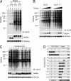
Regulatory features of MoSrc. (A) MoSrc isoforms or their YF C-terminal mutants were coexpressed with MoCsk in 293T cells, and total cell lysates were analyzed by Western blotting with 4G10, anti-Myc (for MoSrc), or anti-HA (for MoCsk). (B) Dose dependency of MoSrcFv activity. 293T cells were cotransfected transiently with a fixed amount of vector expressing MoCsk (0.5 μg) and increasing amounts of vectors (0–1.0 μg) expressing MoSrcFv or their YF mutants. Total cell lysates were analyzed by Western blotting. (C) The chimeric Src molecules consisting of domains from MoSrcFv and EfSrc301 were coexpressed transiently with MoCsk in 293T cells. Total cell lysates were analyzed by Western blotting for the proteins indicated. (D) Structures of the chimeric Src molecules. Construct numbers correspond to lane numbers in C. A, activation loop; N, N lobe of the kinase domain; αC, N-terminal half of the N lobe containing the αC helix. Detailed maps of the regions substituted in these chimeras are shown in Fig. 7.
Fig. 4.
Fig. 4.
Role of C-terminal tyrosine phosphorylation in the negative regulation of M. ovata Src. (A) MoSrcFv, the Fv/301 chimera or their YF C-terminal mutants were coexpressed with MoCsk in 293T cells. Total cell lysates were analyzed by Western blotting with the antibodies indicated. For the anti-pY421 competition assay, the antibody was preincubated with nonphosphorylated or phosphorylated peptides (20 μg/ml) corresponding to the C-terminal region of SrcFv. (B) MoSrc was immunoprecipitated from cell lysates by using anti-Myc and analyzed by Western blotting with the antibodies indicated. (C) The immunoprecipitates were subjected to in vitro kinase assays by using cortactin as a substrate. (C Upper) Tyrosine phosphorylation of cortactin and MoSrc levels were detected by Western blotting with anti-pY421 and anti-Myc, respectively (upper blot). Control immunorpecipitates gave no significant signals (data not shown). The results of triplicate assays are shown. The signal intensities for MoSrc (lower blot) and phosphorylated cortactin were quantified by using the Image J program (National Institutes of Health), and the relative specific activities were plotted (C Lower). Data represent means ± SD.
Fig. 5.
Fig. 5.
Functional features of M. ovata Src. (A) Cell morphologies were observed under phase-contrast microscopy at a magnification of ×400. (B) Colony formation assay of the REF-T transfectants. Cells (4 × 10⁴) were plated in soft agar, and after 5 d, colonies were stained with MTT reagent and photographed.

References

    1. Spector D. H., Varmus H. E., Bishop J. M. Proc. Natl. Acad. Sci. USA. 1978;75:4102–4106. - PMC - PubMed
    1. Hunter T., Sefton B. M. Proc. Natl. Acad. Sci. USA. 1980;77:1311–1315. - PMC - PubMed
    1. Brown M. T., Cooper J. A. Biochim. Biophys. Acta. 1996;1287:121–149. - PubMed
    1. Takeichi M. Annu. Rev. Biochem. 1990;59:237–252. - PubMed
    1. DeMali K. A., Wennerberg K., Burridge K. Curr. Opin. Cell Biol. 2003;15:572–582. - PubMed

Publication types

Substances

LinkOut - more resources