. 2001 Sep;69(9):5864-73.
doi: 10.1128/IAI.69.9.5864-5873.2001.

Gene cluster for assembly of pilus colonization factor antigen III of enterotoxigenic Escherichia coli

Affiliations

Gene cluster for assembly of pilus colonization factor antigen III of enterotoxigenic Escherichia coli

T Taniguchi et al. Infect Immun. 2001 Sep.

Abstract

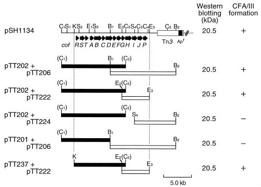
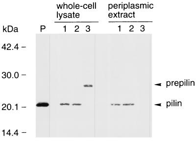
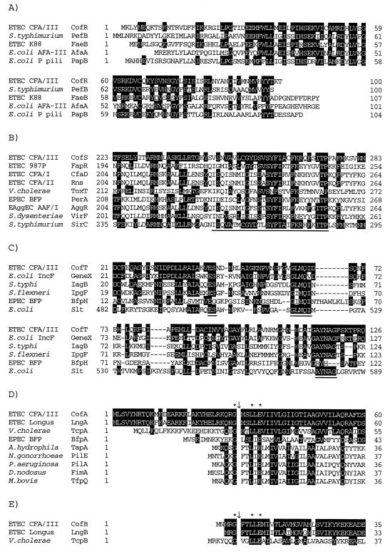
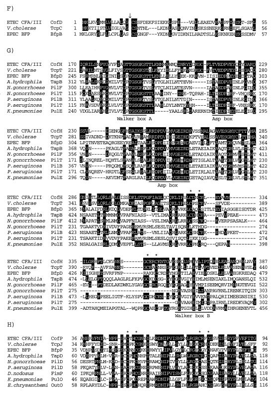
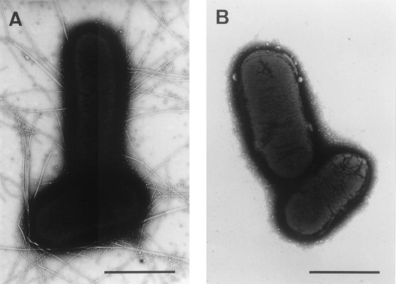
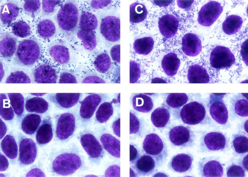
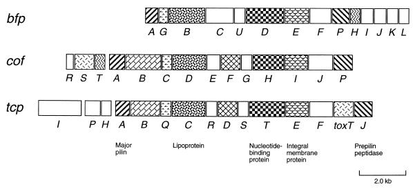
The assembly of pilus colonization factor antigen III (CFA/III) of enterotoxigenic Escherichia coli (ETEC) requires the processing of CFA/III major pilin (CofA) by a prepilin peptidase (CofP), similar to other type IV pilus formation systems. CofA is produced initially as a 26.5-kDa preform pilin (prepilin) and then processed to a 20.5-kDa mature pilin by CofP which is predicted to be localized in the inner membrane. In the present experiment, we determined the nucleotide sequence of the whole region for CFA/III formation and identified a cluster of 14 genes, including cofA and cofP. Several proteins encoded by cof genes were similar to previously described proteins, such as the toxin-coregulated pili of Vibrio cholerae and the bundle-forming pili of enteropathogenic E. coli. The G+C content of the cof gene cluster was 37%, which was significantly lower than the average for the E. coli genome (50%). The introduction of a recombinant plasmid containing the cof gene cluster into the E. coli K-12 strain conferred CFA/III biogenesis and the ability of adhesion to the human colon carcinoma cell line Caco-2. This is the first report of a complete nucleotide sequence of the type IV pili found in human ETEC, and our results provide a useful model for studying the molecular mechanism of CFA/III biogenesis and the role of CFA/III in ETEC infection.


Figures

FIG. 1
FIG. 1
Restriction maps of pSH1134 and CFA/III clones and their expression. pSH1134, pTT202, and pTT206 were digested with the appropriate restriction endonucleases and the fragments (solid and open boxes) were cloned into pMW119 and pACYC184, respectively. The cloned genes were expressed under the control of their own promoter or promoter on the cloning vector. The proposed organization of the cof gene cluster is illustrated in the upper part of the figure. The values represent the molecular mass (in kilodaltons) on Western blot analysis with anti-CFA/III antiserum. The symbols (+ and −) on the right side show the results of CFA/III formation. + and − represent the formation and the nonformation of CFA/III, respectively. Restriction endonuclease sites: B, BamHI; C, ClaI; E, EcoRI; K, KpnI; and S, SalI.
FIG. 2
FIG. 2
Western blot analysis of E. coli HB101 whole-cell lysates (left) and periplasmic extracts (right) using anti-CFA/III antiserum. Lane P, purified CFA/III; lane 1, E. coli HB101 harboring pTT202 and pTT224; lane 2, E. coli HB101 harboring pTT201 and pTT206; lane 3, E. coli HB101 harboring pTT202. The prepilin and processed pilin bands are indicated by arrowheads. Molecular mass markers are noted on the left side.
FIG. 3
FIG. 3
Partial alignment of the deduced amino acid sequences of the Cof proteins with those of known proteins. Identical amino acids are indicated by a black background. Gaps introduced for alignment are represented by dashes. (A) Alignment of the deduced amino acid sequences of CofR, Salmonella serovar Typhimurium PefB (GenBank accession no. L08613), ETEC FaeB (Z11709), E. coli AfaA (X76688), and E. coli PapB (X03391). (B) Alignment of the deduced amino acid sequences of CofS, ETEC FapR (X53494), ETEC CfaD (M55609), ETEC Rns (J04166), V. cholerae ToxT (X64098), EPEC PerA (Z48561), enteroaggregative E. coli (EAggEC) AggR (Z32523), S. dysenteriae VirF (X58464), and serovar Typhimurium SirC (AF134856) in the region containing the DNA-binding domain (underline). (C) Alignment of the deduced amino acid sequences of CofT, E. coli Gene X (X07264), Salmonella serovar Typhi IagB (X80892), S. flexneri IpgF (L04309), EPEC BfpH (Z68186), and E. coli Slt (M69185). Three motifs conserved in putative lytic transglycosylases are underlined. (D) Alignment of the deduced amino acid sequences of CofA, ETEC LngA (AF004308), V. cholerae TcpA (X64098), EPEC BfpA (Z68186), A. hydrophila TapA (U20255), N. gonorrhoeae PilE (X66144), P. aeruginosa PilA (M14849), Dichelobacter nodosus FimA (X52405), and Moraxella bovis TfpQ (M59712) in the N-terminal region. The cleavage site of type IV pilins is shown by a downward arrow. The conserved glycine, leucine, and glutamic acid residues are marked by asterisks. (E) Alignment of the deduced amino acid sequences of CofB, ETEC LngB, and V. cholerae TcpB (X64098) in the N-terminal region. The type IV pilin-like cleavage site is shown by a downward arrow. The conserved glycine, leucine, and glutamic acid residues are marked by asterisks. (F) Alignment of the deduced amino acid sequences of CofD, V. cholerae TcpC (X64098), and EPEC BfpB (Z68186) in the N-terminal region. The lipoprotein-cleavage site is shown by a downward arrow. (G) Alignment of the deduced amino acid sequences of CofH, V. cholerae TcpT (X64098), EPEC BfpD (Z68186), A. hydrophila TapB (U20255), N. gonorrhoeae PilF (U32588), N. gonorrhoeae PilT (S72391), P. aeruginosa PilB (M32066), P. aeruginosa PilT (M55524), and K. pneumoniae PulE (M32613) in the region containing the nucleotide-binding domain. The Walker box A, Asp boxes, and Walker box B are underlined. The conserved CXXC motifs are marked by asterisks. (H) Alignment of the deduced amino acid sequences of CofP, V. cholerae TcpJ (M74708), EPEC BfpP (Z68186), A. hydrophila TapD (U20255), N. gonorrhoeae PilD (U32588), P. aeruginosa PilD (M32066), D. nodosus FimP (U17138), K. pneumoniae PulO (M32613), and Erwinia chrysanthemi OutO (L02214) in the region containing the conserved CXXC motifs (asterisks).
FIG. 3
FIG. 3
Partial alignment of the deduced amino acid sequences of the Cof proteins with those of known proteins. Identical amino acids are indicated by a black background. Gaps introduced for alignment are represented by dashes. (A) Alignment of the deduced amino acid sequences of CofR, Salmonella serovar Typhimurium PefB (GenBank accession no. L08613), ETEC FaeB (Z11709), E. coli AfaA (X76688), and E. coli PapB (X03391). (B) Alignment of the deduced amino acid sequences of CofS, ETEC FapR (X53494), ETEC CfaD (M55609), ETEC Rns (J04166), V. cholerae ToxT (X64098), EPEC PerA (Z48561), enteroaggregative E. coli (EAggEC) AggR (Z32523), S. dysenteriae VirF (X58464), and serovar Typhimurium SirC (AF134856) in the region containing the DNA-binding domain (underline). (C) Alignment of the deduced amino acid sequences of CofT, E. coli Gene X (X07264), Salmonella serovar Typhi IagB (X80892), S. flexneri IpgF (L04309), EPEC BfpH (Z68186), and E. coli Slt (M69185). Three motifs conserved in putative lytic transglycosylases are underlined. (D) Alignment of the deduced amino acid sequences of CofA, ETEC LngA (AF004308), V. cholerae TcpA (X64098), EPEC BfpA (Z68186), A. hydrophila TapA (U20255), N. gonorrhoeae PilE (X66144), P. aeruginosa PilA (M14849), Dichelobacter nodosus FimA (X52405), and Moraxella bovis TfpQ (M59712) in the N-terminal region. The cleavage site of type IV pilins is shown by a downward arrow. The conserved glycine, leucine, and glutamic acid residues are marked by asterisks. (E) Alignment of the deduced amino acid sequences of CofB, ETEC LngB, and V. cholerae TcpB (X64098) in the N-terminal region. The type IV pilin-like cleavage site is shown by a downward arrow. The conserved glycine, leucine, and glutamic acid residues are marked by asterisks. (F) Alignment of the deduced amino acid sequences of CofD, V. cholerae TcpC (X64098), and EPEC BfpB (Z68186) in the N-terminal region. The lipoprotein-cleavage site is shown by a downward arrow. (G) Alignment of the deduced amino acid sequences of CofH, V. cholerae TcpT (X64098), EPEC BfpD (Z68186), A. hydrophila TapB (U20255), N. gonorrhoeae PilF (U32588), N. gonorrhoeae PilT (S72391), P. aeruginosa PilB (M32066), P. aeruginosa PilT (M55524), and K. pneumoniae PulE (M32613) in the region containing the nucleotide-binding domain. The Walker box A, Asp boxes, and Walker box B are underlined. The conserved CXXC motifs are marked by asterisks. (H) Alignment of the deduced amino acid sequences of CofP, V. cholerae TcpJ (M74708), EPEC BfpP (Z68186), A. hydrophila TapD (U20255), N. gonorrhoeae PilD (U32588), P. aeruginosa PilD (M32066), D. nodosus FimP (U17138), K. pneumoniae PulO (M32613), and Erwinia chrysanthemi OutO (L02214) in the region containing the conserved CXXC motifs (asterisks).
FIG. 4
FIG. 4
Electron micrographs of E. coli HB101 after growth on CFA agar plates at 37°C for 20 h. (A) E. coli HB101 harboring pTT237 and pTT222. (B) E. coli HB101 harboring pMW119 and pACYC184. Bar, 1 μm.
FIG. 5
FIG. 5
Micrographs showing adhesion of E. coli strains to Caco-2 cells. (A) ETEC 31-10 (CFA/III-positive strain). (B) ETEC 31-10P (CFA/III-negative strain). (C) E. coli HB101 harboring pTT237 and pTT222. (D) E. coli HB101 harboring pMW119 and pACYC184.
FIG. 6
FIG. 6
Genetic organizations of cof, bfp, and tcp gene clusters. All genes except tcpI are transcribed rightward. The homologous genes are indicated by the same shading patterns. Predicted similar functions are indicated in the bottom of the figure.

References

    1. Allaoui A, Ménard R, Sansonetti P J, Parsot C. Characterization of the Shigella flexneri ipgD and ipgF genes, which are located in the proximal part of the mxi locus. Infect Immun. 1993;61:1707–1714. - PMC - PubMed
    1. Båga M, Göransson M, Normark S, Uhlin B E. Transcriptional activation of a Pap pilus virulence operon from uropathogenic Escherichia coli. EMBO J. 1985;4:3887–3893. - PMC - PubMed
    1. Darfeulle-Michaud A, Aubel D, Chauviere G, Rich C, Bourges M, Servin A, Joly B. Adhesion of enterotoxigenic Escherichia coli to the human colon carcinoma cell line Caco-2 in culture. Infect Immun. 1990;58:893–902. - PMC - PubMed
    1. Dijkstra A J, Keck W. Peptidoglycan as a barrier to transenvelope transport. J Bacteriol. 1996;178:5555–5562. - PMC - PubMed
    1. Evans D G, Evans D J, Jr, Tjoa W. Hemagglutination of human group A erythrocytes by enterotoxigenic Escherichia coli isolated from adults with diarrhea: correlation with colonization factor. Infect Immun. 1977;18:330–337. - PMC - PubMed

Publication types

Associated data

LinkOut - more resources