. 2004 Nov;2(11):e327.
doi: 10.1371/journal.pbio.0020327. Epub 2004 Oct 5.

Sensitivity to oxidative stress in DJ-1-deficient dopamine neurons: an ES- derived cell model of primary Parkinsonism

Affiliations

Sensitivity to oxidative stress in DJ-1-deficient dopamine neurons: an ES- derived cell model of primary Parkinsonism

Cecile Martinat et al. PLoS Biol. 2004 Nov.

Abstract

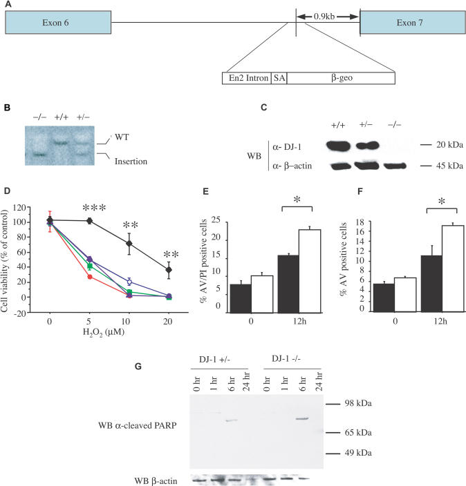
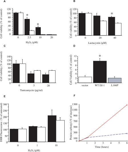
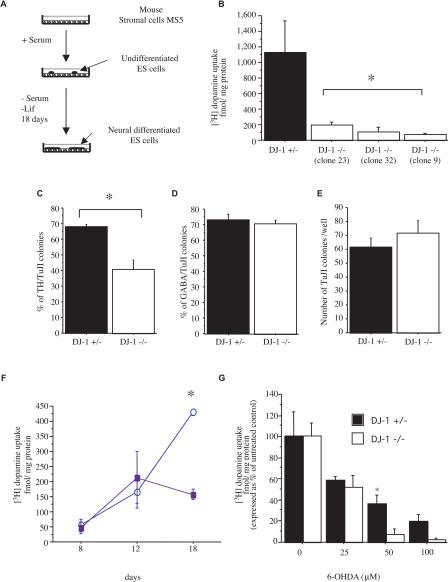
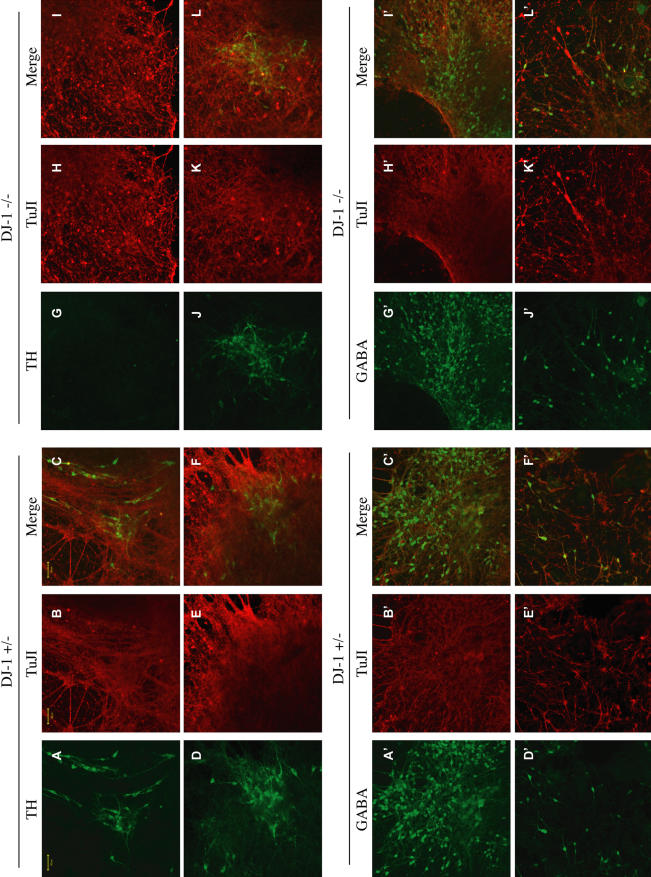
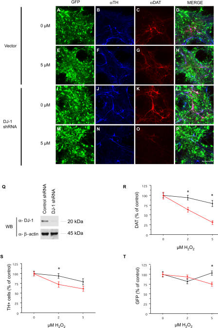
The hallmark of Parkinson's disease (PD) is the selective loss of dopamine neurons in the ventral midbrain. Although the cause of neurodegeneration in PD is unknown, a Mendelian inheritance pattern is observed in rare cases, indicating a genetic factor. Furthermore, pathological analyses of PD substantia nigra have correlated cellular oxidative stress and altered proteasomal function with PD. Homozygous mutations in DJ-1 were recently described in two families with autosomal recessive Parkinsonism, one of which is a large deletion that is likely to lead to loss of function. Here we show that embryonic stem cells deficient in DJ-1 display increased sensitivity to oxidative stress and proteasomal inhibition. The accumulation of reactive oxygen species in toxin-treated DJ-1-deficient cells initially appears normal, but these cells are unable to cope with the consequent damage that ultimately leads to apoptotic death. Furthermore, we find that dopamine neurons derived from in vitro-differentiated DJ-1-deficient embryonic stem cells display decreased survival and increased sensitivity to oxidative stress. These data are consistent with a protective role for DJ-1, and demonstrate the utility of genetically modified embryonic stem cell-derived neurons as cellular models of neuronal disorders.


Conflict of interest statement

The authors have declared that no conflicts of interest exist.

Figures

Figure 1
Figure 1. DJ-1-Deficient ES Cells Are Sensitized to Oxidative Stress
(A) Schematic map of the murine DJ-1 gene in clone F063A04. The retroviral insertion places the engrailed-2 (En2) intron, the splice acceptor (SA), and the β-galactosidase/neomycin resistance gene fusion (β-geo) between exons 6 and 7. (B) Southern blot analysis of KpnI-digested genomic DNA from DJ-1 homozygous mutant (–/–), WT (+/+), and heterozygous (+/–)cells, probed with murine DJ-1 cDNA. WT DNA shows a predicted 14-kb band (WT), whereas the mutant allele migrates as a 9-kb band (insertion). (C) Western blot (WB) of ES cell lysates from WT (+/+), DJ-1 heterozygous (+/–), and mutant homozygous (–/–) clones with antibodies to murine DJ-1 (α-DJ-1) or β-actin (α-β-actin). DJ-1 migrates at 20 kDa, β-actin at 45 kDa. (D) ES cells were exposed to 0, 5, 10, and 20 μM H2O2 for 15 h and viability was assayed by MTT. Responses of DJ-1 heterozygous cells (diamonds) and DJ-1 knockout clones 9 (open circles), 16 (solid circles), 23 (squares), and 32 (triangles) are shown. ** p ≤ 0.01; *** p ≤ 0.0001. (E and F) Cell death of DJ-1 heterozygous and DJ-1-deficient cells (clone 32) after exposure to H2O2 (10 μM) was quantified by staining with PI and an antibody to AV with subsequent FACS analysis. AV staining marks cells undergoing apoptosis, whereas PI staining indicates dead cells. * p ≤ 0.05. (G) DJ-1 heterozygous (+/–) and knockout (clone 32; –/–) cells were assayed at 1, 6, and 24 h after treatment with 10 μM H2O2 by Western blotting for cleaved PARP (89 kDa), which indicates apoptosis. No band is seen for cleaved PARP or β-actin for the DJ-1-deficient cells at 24 h due to cell death. Data represent means ± standard error of the mean (SEM) and were analyzed by ANOVA with Fisher's post-hoc test.
Figure 2
Figure 2. Specificity and Mechanism of Altered Toxin Sensitivity in DJ-1-Deficient Cells
(A–C) Cell viability of DJ-1 heterozygous cells (solid bar) and DJ-1-deficient knockout clone 32 cells (open bar) after 15 h exposure to H2O2 (A), lactacystin (B), or tunicamycin (C) as assayed by MTT reduction. * p ≤ 0.05. (D) DJ-1-deficient knockout cells (clone 32) were transiently transfected with plasmids containing WT human DJ-1 vector (solid bar) and PD-associated L166P mutant DJ-1 vector (gray bar); as a control, knockout cells were also transfected with vector alone (open bar). 48 h after transfection, cells were exposed to 10 μM H2O2 for 15 h and then assayed by MTT reduction. WT human DJ-1 significantly enhanced survival of the knockout cells, whereas the L166P mutant did not. Similar results were obtained at 20 μM H2O2 and with a second DJ-1-deficient clone (unpublished data). Transfection efficiency exceeded 90% in all cases and protein expression level was comparable for human WT and L166P mutant DJ-1 as determined by Western blotting (Figure S1). * p ≤ 0.05. (E) DJ-1-deficient cells (clone 32; open bar) and control heterozygous cells (solid bar) were assayed for intracellular formation of ROS in response to H2O2 treatment (15 min, 1 or 10 μM) using DHR and FACS analysis. (F) Protein carbonyl levels were measured by spectrophotometric analysis of DNP-conjugated lysates from DJ-1-deficient (clone 32; solid red line) and control heterozygous cells (dashed blue line). Data are shown as the mean ± SEM and were analyzed by ANOVA with Fisher's post-hoc test.
Figure 3
Figure 3. DJ-1-Deficient ES Cell Cultures Display Reduced DN Production
(A) The SDIA coculture method. DJ-1 knockout or control heterozygous ES cells are cocultured with mouse stromal cells (MS5) in the absence of serum and leukemia inhibitory factor for 18 DIV. (B) DN production was quantified at 18 DIV by 3H-dopamine uptake assay. DJ-1-deficient ES cell cultures were defective relative to heterozygous control cultures. (C–D) Neuron production was quantified by immunohistochemical analysis as a percent of TuJ1-positive colonies that express TH (C) or GABA (D). Quantification of TH and GABA immunostaining was performed on all colonies in each of three independent wells. Colonies were scored as positive if any immunostained cells were present. * p ≤ 0.05. (E) The absolute number of TuJ1-positive colonies was not significantly different between the two genotypes. (F) Kinetic analysis of DN differentiation in DJ-1-deficient cultures (clone 32, solid square) and heterozygous controls (open circle) as quantified by 3H-dopamine uptake assay. * p ≤ 0.05. (G) DJ-1-deficient (open bar) and heterozygous control (closed bar) cultures differentiated for 9 DIV and then exposed to 6-OHDA at the indicated dose for 72 h. DNs were quantified by 3H-dopamine uptake assay. Data represent the means ± SEM and were analyzed by ANOVA followed by Fisher's post-hoc test. * p ≤ 0.05.
Figure 4
Figure 4. Neuronal Differentiation of DJ-1-Deficient and Control Heterozygous ES Cell Cultures
(A–L) DJ-1 heterozygous (+/–; A–F) and knockout (–/– [clone 32]; G–L) cultures were differentiated by SDIA for 18 DIV and immunostained with antibodies to TH (green) and TuJ1 (red). Images of both (Merge) are also shown. (A′–L′) Immunostaining of DJ-1 heterozygous (+/–, A′–F′) and deficient (–/–, G′–L′) cultures with antibodies for GABA (green) and TuJ1 (red). Scale bar, 50 μm. Images of both (Merge) are also shown.
Figure 5
Figure 5. RNAi “Knockdown” of DJ-1 in Primary Embryonic Midbrain DNs Display Increased Sensitivity to Oxidative Stress
(A–P) Primary midbrain cultures from E13.5 embryos were infected with lentiviral vectors encoding DJ-1 shRNA (or vector alone) under the regulation of the control vector (A–H) or the U6 promoter (I–P). Cells were cultured for 1 wk after infection and then exposed to H2O2 (5 μM; E–H and M–P) for 24 h. Cultures were immunostained for TH (B, F, J, and N) or DAT (C, G, K, or O) and visualized by confocal microscopy. Images containing all stains are included (Merge; D, H, L, and P). Scale bar, 100 μm. (Q) Cell lysates prepared from midbrain primary cultures infected with DJ-1 shRNA lentivirus (or control vector) were analyzed by Western blotting for murine DJ-1 or β-actin. (R–T) Quantification of TH, DAT, and GFP signal was performed on ten randomly selected fields in each of three wells for each condition. Red triangles, DJ-1 shRNA treated; black circles, control vector. Data represent the means ± SEM and were analyzed by ANOVA followed by Fisher's post-hoc test. * p ≤ 0.05.

References

    1. Abeliovich A, Schmitz Y, Farinas I, Choi-Lundberg D, Ho WH, et al. Mice lacking α-synuclein display functional deficits in the nigrostriatal dopamine system. Neuron. 2000;25:239–252. - PubMed
    1. Barberi T, Klivenyi P, Calingasan NY, Lee H, Kawamata H, et al. Neural subtype specification of fertilization and nuclear transfer embryonic stem cells and application in parkinsonian mice. Nat Biotechnol. 2003;21:1200–1207. - PubMed
    1. Bian K, Gao Z, Weisbrodt N, Murad F. The nature of heme/iron-induced protein tyrosine nitration. Proc Natl Acad Sci U S A. 2003;100:5712–5717. - PMC - PubMed
    1. Bonifati V, Rizzu P, van Baren MJ, Schaap O, Breedveld GJ, et al. Mutations in the DJ-1 gene associated with autosomal recessive early-onset parkinsonism. Science. 2003;299:256–259. - PubMed
    1. Dauer W, Przedborski S. Parkinson's disease: Mechanisms and models. Neuron. 2003;39:889–909. - PubMed

Publication types

MeSH terms

Associated data